www.impactjournals.com/oncotarget/ Oncotarget, Vol. 6, No. 14
Molecular stratification of metastatic melanoma using gene
expression profiling : Prediction of survival outcome and benefit
from molecular targeted therapy
Helena Cirenajwis1, Henrik Ekedahl2, Martin Lauss1, Katja Harbst1, Ana Carneiro1,3, Jens Enoksson4, Frida Rosengren1, Linda Werner-Hartman1, Therese Törngren1, Anders Kvist1, Erik Fredlund5, Pär-Ola Bendahl1, Karin Jirström4, Lotta Lundgren1,3, Jillian Howlin1, Åke Borg1, Sofia K. Gruvberger-Saal1, Lao H. Saal1, Kari Nielsen6, Markus Ringnér1, Hensin Tsao7,8, Håkan Olsson1,3, Christian Ingvar2, Johan Staaf1, Göran Jönsson1
1 Department of Clinical Sciences, Division of Oncology and Pathology, Lund University, Lund, Sweden 2 Department of Clinical Sciences, Division of Surgery, Lund University, Lund, Sweden
3 Department of Oncology, Skåne University Hospital, Lund University, Lund, Sweden 4 Department of Clinical Pathology, Skåne University Hospital, Lund University, Lund, Sweden 5 Department of Oncology-Pathology, Karolinska Institute, Stockholm, Sweden
6 Department of Dermatology, Helsingborg General Hospital, Helsingborg, Sweden 7 Department of Dermatology, Harvard Medical School, Boston, USA
8 Wellman Center for Photomedicine, MGH Cancer Center, Massachusetts General Hospital, Boston, USA
Correspondence to: Göran Jönsson, email: goran_b.jonsson@med.lu.se
Keywords: gene expression, melanoma, BRAF, BRAF inhibitor, mutation
Received: January 09, 2015 Accepted: February 27, 2015 Published: March 26, 2015
This is an open-access article distributed under the terms of the Creative Commons Attribution License, which permits unrestricted use, distribution, and reproduction in any medium, provided the original author and source are credited.
AbstrAct
Melanoma is currently divided on a genetic level according to mutational status. However, this classification does not optimally predict prognosis. In prior studies, we have defined gene expression phenotypes (high-immune, pigmentation, proliferative and normal-like), which are predictive of survival outcome as well as informative of biology. Herein, we employed a population-based metastatic melanoma cohort and external cohorts to determine the prognostic and predictive significance of the gene expression phenotypes. We performed expression profiling on 214 cutaneous melanoma tumors and found an increased risk of developing distant metastases in the pigmentation (HR, 1.9; 95% CI, 1.05-3.28; P=0.03) and proliferative (HR, 2.8; 95% CI, 1.43-5.57; P=0.003) groups as compared to the high-immune response group. Further genetic characterization of melanomas using targeted deep-sequencing revealed similar mutational patterns across these phenotypes. We also used publicly available expression profiling data from melanoma patients treated with targeted or vaccine therapy in order to determine if our signatures predicted therapeutic response. In patients receiving targeted therapy, melanomas resistant to targeted therapy were enriched in the MITF-low proliferative subtype as compared to pre-treatment biopsies (P=0.02). In summary, the melanoma gene expression phenotypes are highly predictive of survival outcome and can further help to discriminate patients responding to targeted therapy.
IntroductIon
Cutaneous malignant melanoma (CMM) is the
provide clinicians and patients with accurate prognostic information about the disease, a correct staging system is fundamental. Since 1998, the American Joint Committee on Cancer (AJCC) melanoma staging system has served
as a foundation for clinical classification and it was
recently updated to the 7th edition after adding more tumor intrinsic factors with prognostic significance [1]. However,
clinical outcome of patients with similar or even identical
clinical and histological features varies considerably [2],
especially within the AJCC intermediate risk stages and in
patients with advanced disease [3].
Gene expression profiling may provide additional
information to the current prognostic assessment. Several attempts to introduce this approach as a step towards individualized patient management have been
made by defining new molecular biomarkers and gene
signatures correlating with clinical outcome. Since the initial search for prognostic signatures in melanoma by Winnepenninckx et al. [4], several signatures have been
proposed. These range from a single-gene signature of osteopontin in primary melanoma, to different multi-gene signatures in stage III and IV metastatic melanoma lesions
[5-8]. However, at present time there is still no extensively
validated prognostic molecular signature in melanoma. We have reported distinct gene expression
phenotypes significantly associated with survival outcome in stage IV metastatic melanoma [9]. The phenotypes,
mainly characterized by differential expression of immune
response, melanocyte-specific and proliferation genes,
have also been validated in cutaneous primary melanoma
[10].
In melanoma, molecular targeted anticancer therapy has received much focus lately and new emerging treatment approaches have shown dramatic clinical results. The prospective drugs include kinase inhibitors, targeting
oncogenic BRAFV600E or MEK [11]; immune system
activators, e.g. vaccines or immune checkpoint blockades, with the latter approach utilizing monoclonal antibodies
directed against the inhibitory immune receptors CTLA-4, PD-1 or PD-L1 [12-14]. Since treatment with these agents can be associated with significant morbidity and since
response is far from universal, there is a compelling need
to better identify patients who may benefit from the newer
generation therapies.
In the present work we aimed to further establish the clinical relevance and delineate the mutational landscape of our previously described gene expression phenotypes in
a population-based retrospective collection of 214 CMM
specimens obtained from a single clinical institution. This is, to our knowledge, the largest study correlating genome-wide molecular and mutation data to clinical patient information in metastatic melanoma. Our data
firmly validates the prognostic significance of the gene
expression phenotypes and provides novel evidence that
the gene expression phenotypes may predict benefit from
molecular targeted therapies in advanced stage melanoma
beyond BRAF status.
results
repeated observation of gene expression phenotypes in a population-based metastatic melanoma cohort
Gene expression profiling was performed on a total of 214 CMM tumor tissues representing a
population-based retrospective collection from a single institution. In this cohort, all previously reported phenotypes were
present: high-immune response (30%), normal-like (6%), pigmentation (44%) and proliferative (15%) (Figure 1A) [9]. An overview of the patient clinicopathological
characteristics and the gene expression phenotypes is
provided in Table 1. Specifically, the normal-like group
was more prevalent among the primary melanoma tumors,
comprising 50% (8/16), whereas only 2% (4/188) of the tumors in the metastatic setting were classified as
normal-like. When excluding the normal-like group (due to lower
number of cases), there was no significant association
between gene expression phenotype and metastasis type (P=0.1, Fisher’s exact test). Furthermore, when
analyzing primary melanoma tumor features we found that histological type and primary site varied between the phenotypes (P=0.01 or P=0.02, respectively, Fisher’s
exact test). In contrast, Breslow thickness, age at primary diagnosis and Clark´s level of invasion did not show any
significant differences (P > 0.05, Table 1). Analysis of
the median time from primary melanoma diagnosis to the diagnosis of the analysed metastatic lesion indicated
a significant difference between the groups, with longer durations for the proliferative-classified melanomas
(P=0.04, Kruskal-Wallis test).
To further describe the phenotypes and determine transcriptional programs in melanoma, we performed transcriptional network analysis of highly correlated
genes in the cohort, as previously described [15]. Based
on gene ontology analysis and published associations
with melanoma-specific tumor biology, we could extract five transcriptional modules defined herein as the
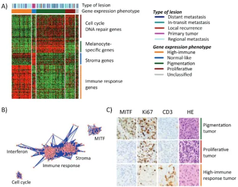
Figure 1: Gene expression phenotypes in melanoma. A) Heatmap of 299 genes (rows) included in the classification of 214 melanoma tumors (columns). Tumor descriptions are shown in the color bars including phenotype classification and tumor type. b) Network
analysis identified five clusters of genes reflecting biological mechanisms of relevance in melanoma and named thereafter. Each dot (pink)
represents a gene that is connected by lines (blue) representing correlations between the genes. c) Immunohistochemical staining of MITF,
Thus, the transcriptional modules reflect gene expression
phenotypes to a large extent.
To investigate whether gene expression
phenotypes are reflected on protein expression levels, we examined MITF (a melanocyte-specific marker), cluster of differentiation 3 (CD3, expressed by mature T-lymphocytes), and the proliferative marker Ki67 in a
subset of our tumors (n=59) by immunohistochemical
analysis. A striking agreement between protein and gene expression was observed in the phenotypes with strong
infiltration of CD3 positive T lymphocytes in the high-immune response classified tumors and a high prevalence of Ki67 positive melanoma cells in the proliferative tumors
with few, if any, cells staining positive for MITF (Figure
1C). The pigmentation-classified tumors comprised a high
fraction of MITF positive cells and a high prevalence of
Ki67 positive cells (Figure 1C).
Gene expression phenotypes and somatic mutation status
To further characterize the mutational landscape of the gene expression phenotypes, we used targeted deep sequencing to screen for somatic mutations in
1697 cancer-associated genes in tumors from 146 CMM
patients. Among these tumors, the mutation burden
demonstrated wide heterogeneity, ranging from 5 up
to 768 somatic mutations per tumor (Table 1). A small subset of acral lentiginous melanomas (ALMs, n=6)
had a significantly lower mutation burden (range: 6-51
mutations), as compared to metastases of unknown
origin, superficial spreading or nodular melanoma (P <
0.001, Kruskal-Wallis test). Moreover, the ALMs were all classified as pigmentation tumors. The mutation burden was not significantly different between the gene
expression phenotypes (P=0.5, Kruskal-Wallis test)
(Table 1). Furthermore, most melanomas harbored the UV-induced mutational signature C -> T preceded by a
pyrimidine (Figure 2).
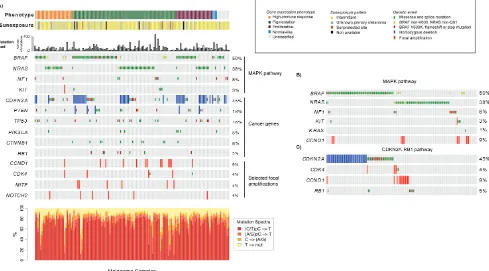
Next, we focused on a set of genes highly implicated in melanomagenesis, including BRAF, NRAS, KIT,
CDK4, CDKN2A, TP53, PTEN, CTNNB1, NF1, RB1 and
PIK3CA. To provide a more comprehensive context of the
mutational landscape we also included focal amplifications
and homozygous deletions of melanoma signature genes, like MITF, KIT, CDK4, NOTCH2 and CCND1,CDKN2A
and PTEN (Figure 2A). Analysis of the mutation spectrum
within the context of the gene expression phenotypes showed that: i) CDKN2A alterations were more prevalent
[image:5.612.63.552.379.650.2]in the proliferative-classified melanomas (P=0.05, Fisher’s exact test, consistent with previous reports [9]),
Figure 2: Analysis of the mutational landscape in melanoma tumors. A) Genetic events such as mutations, homozygous
deletions and focal amplifications in cancer genes within the context of the gene expression phenotypes. Tumors are ordered according to
the gene expression phenotypes and the genes of interest. The mutation frequency plot corresponds to the number of somatically acquired mutations observed in the 1697 investigated cancer-associated genes in each melanoma tumor. b) Mutations in genes involved in the
MAPK pathway. Tumors are ordered according to mutations in BRAF, NRAS, NF1, KIT, KRAS and CCND1. c). Genetic events in genes
ii) pigmentation-classified melanomas were enriched for
genetic events in CTNNB1, MITF or CCND1 (P=0.02, P=0.04 and P=0.04, respectively, Fisher’s exact test),
whereas iii) BRAF and NRAS mutations were equally
distributed across phenotypes (Supplementary Table 2). BRAF and NRAS mutations were mutually exclusive
in 96% of tumors, while six cases had co-occurring
mutations (P < 0.001, Fisher’s exact test). Interestingly,
the majority of the samples with concurrent BRAF and
NRAS mutations (4/6 tumors) seemed to have a NRAS
hotspot mutation and a non-hotspot mutation of BRAF (Figure 2B). The BRAF/NRAS wild-type melanomas,
i.e. samples negative for mutations in both BRAF and
NRAS, more often had genetic alterations in KIT or NF1
(P < 0.001, Fisher’s exact test) (Figure 2B). In total, only 9.6% of all cases in the cohort were negative for
either of the genetic changes in BRAF, NRAS, NF1 and
KIT, indicating that the majority of melanomas could
potentially have activated MAPK pathway through
these genetic alterations. In addition, one tumor negative for these four genes harbored a KRAS G13D mutation.
When considering hotspot mutations in BRAF and NRAS, alterations in KIT and loss of function mutations in NF1,
we found that the majority (96%) of the proliferative-classified tumors had an alteration. Furthermore, we also found mutually exclusive genetic events in the
CDKN2A-RB1 pathway where CDK4, CCND1 and RB1 alterations occurred mainly in CDKN2A wild-type tumors (Figure
2C). In summary, these results suggest that the most
Gene expression phenotypes are predictive of survival outcome in melanoma
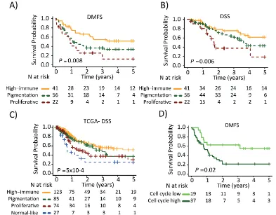
Next, we determined the association between the gene expression phenotypes and the survival outcome in the metastatic cohort. Among the patients with regional metastatic disease (n=125), we found an increased risk of having a distant metastasis (5-year distant metastasis-free survival, DMFS) in the pigmentation (HR, 1.9; 95% CI, 1.05-3.28; P=0.03) and proliferative (HR, 2.8; 95% CI, 1.43-5.57; P=0.003) phenotypes, as compared to the high-immune response phenotype (Table 2). In addition, an increased risk of death from melanoma (5-year disease-specific survival, DSS) was observed in the pigmentation (HR, 1.7; 95% CI, 0.83-3.28; P=0.2) and proliferative (HR, 3.5; 95% CI, 1.56-7.80; P=0.002) phenotypes, as
compared to the high-immune response phenotype (Table
2). The corresponding Kaplan-Meier analyzes are shown in Figures 3A and 3B, respectively. In a multivariable Cox
regression model (adjusting for age, gender and metastasis type), with the high-immune response phenotype as the reference group, the pigmentation and proliferative phenotypes exhibited an increased risk of distant metastases, whereas only the proliferative phenotype had
a significantly increased risk of death from melanoma (Table 2). The confounders were chosen based on their level of significance from the univariable analyzes with a
cutoff at P ≤ 0.05 (Supplementary Table 3). In the stage IV
melanoma cohort, we analyzed the distribution of the gene expression phenotypes among the distantly metastasized tumors (n=23). In total, there were two proliferative-classified melanomas and both had a poor survival (Figure S2). To further validate our findings we used the 309
regional and distant metastatic lesions from the TCGA
data set. Here, we could firmly validate an improved
survival in the high-immune response group as compared to the other groups (P=5x10⁻⁴, Figure 3C).
Based on the findings from the network analysis
described above, we divided the patients into low or high
activity groups for each of the five expression modules. Each module was analyzed separately and further linked
to the gene expression phenotypes (Figure S1C). Notably, the pigmentation phenotype could be split into low and high cell cycle activity groups with the high activity group
being associated with a poor 5-year DMFS (HR, 2.6; 95% CI, 1.16-5.70; P=0.02, Figure 3D). The other phenotypes could also be further stratified, however, the number
of patients included was too few to obtain statistical
[image:7.612.103.496.357.664.2]significance.
Figure 3: Survival analysis of metastatic melanomas stratified by gene expression phenotype using the Kaplan-Meier
estimator to determine. A) Distant metastasis free survival (DMFS) and b) and disease specific survival (DSS). c) Metastatic tumors
from the TCGA data were stratified and Kaplan-Meier analysis was performed. d) Pigmentation-classified tumors were stratified by the cell cycle module (low or high). Survival differences between low and high groups were estimated using Kaplan-Meier analysis. P-values
Gene expression phenotypes and prediction of
benefit from molecular targeted therapies
Since these molecular groupings exhibit an impact
on survival, it may be hypothesized that the profiles could
also correlate with therapeutic response. We thus evaluated whether the gene expression phenotypes were associated
with response to therapies (MAPK pathway inhibition or MAGE-A3 vaccine) using three publicly available gene
expression datasets.
In the first dataset (GSE50509, Rizos et al.), 21
patients with BRAF-mutant metastatic melanoma were treated with BRAF inhibitors (BRAFi, dabrafenib or vemurafenib) and evaluated for best objective response
(RECIST response, %) and progression-free survival (PFS) [16], followed by gene expression profiling of
tumor samples taken pre-treatment and post-relapse.
When we classified this dataset into the gene expression
phenotypes, we found no clear correlation between the clinical response and the pre-treatment phenotypes owing
to small numbers (Figure 4A, upper panel). However, the
single MITF-low proliferative sample responded poorly
to treatment (RECIST response, %) with only 15 weeks until the patient progressed (patient 30). In contrast, the high-immune response-classified samples had a superior response with more than 25 weeks before progression (except in one case, patient 18) (Figure 4A, upper panel).
[image:8.612.99.505.245.664.2]Interestingly, the phenotype distribution was different in post-relapse samples, with an increased prevalence of
Figure 4: Gene expression phenotypes and prediction of clinical benefit from molecular targeted therapies. A) Melanoma
tumors from patients treated with BRAFi [16] or b) BRAFi/MEKi [17] were classified into the gene expression phenotypes and further analyzed for objective response (RECIST response, %) (4A, upper panel; 4B left panel) and phenotype distribution in pre-treatment and post-relapse biopsies (4A, lower panel; 4B, middle and right panel). c) Gene expression phenotype distribution in patients treated with
MITF-low proliferative-classified cases (P = 0.06, Fisher’s exact test; proliferative versus non-proliferative cases, excluding unclassified cases) (Figure 4A, lower panel).
The second dataset (GSE61992, Long et al.) included 10 patients with BRAF-mutant metastatic melanoma that were treated with a combination of
dabrafenib and the MEK inhibitor (MEKi) trametinib and evaluated for RECIST response and PFS [17]. There were too few observations to draw any definitive conclusions,
however again, the single MITF-low proliferative sample
responded poorly to treatment (RECIST response, %) with only 2.8 months until the patient progressed (patient 1) (Figure 4B, left panel). In line with the results from the
Rizos et al. dataset, there was an increased prevalence of
MITF-low proliferative-classified cases after treatment, although not reaching significance (P = 0.3, Fisher’s exact test; proliferative versus non-proliferative cases, excluding unclassified cases) (Figure 4B, middle and right panel). When combining the gene expression-based classification
results from the datasets by Rizos et al. and Long et al.
[16, 17], in total including 31 patients, the prevalence of MITF-low proliferative-classified cases increased
after resistance emerged to either BRAFi alone or in a
combination with BRAFi and MEKi (P = 0.02, Fisher’s exact test; proliferative versus non-proliferative cases, excluding unclassified cases).
In the third study, we analyzed 56 patients that had been evaluated for response to MAGE-A3 immunotherapeutic treatment (22 with clinical benefit and 34 with no benefit) [14]. When we classified these
samples into the gene expression phenotypes and analyzed
the patients’ treatment response to MAGE-A3, only 2/11 patients in the MITF-low proliferative phenotype had clinical benefit from treatment, while the highest
proportion of responders was found in the high-immune
response group (6/10) although not reaching statistical significance (P=0.2, Fisher’s exact test of proliferative versus non-proliferative cases) (Figure 4C).
In summary, we provide initial indications that gene
expression based classification may predict clinical benefit
from targeted therapy.
dIscussIon
It is well established that melanoma can be
genetically classified according to somatic gene mutation status. Although this stratification is relevant, controversy exists regarding the prognostic significance of classifying
melanomas based only on BRAF and NRAS mutations [18, 19]. Gene expression profiling may provide additional
information to current prognostic assessment in melanoma and several studies have associated an immune response
gene signature with improved prognosis [5-7, 9].
Prognostic assessment of stage III melanoma is currently performed by histopathological characterization, determining the number of involved lymph nodes and the
size of nodal metastatic disease. However these features
do not capture the extent of heterogeneity present in
this group of patients. Previously, we identified gene expression phenotypes reflecting biological mechanisms
relevant in melanoma such as melanocyte differentiation,
DNA repair and immunological responses [9]. The
existence of the reported gene expression phenotypes was further supported by analysis of a large cohort of primary
melanomas [10]. Moreover, Nsengimana et al. recently confirmed the independent prognostic significance of the
gene expression phenotypes in a population-based British cohort of melanoma patients (Nsengimana et al. Accepted
for publication in Oncotarget, 2015). In the current study,
we demonstrate that the gene expression phenotypes hold prognostic information also in a regional metastatic setting
and that patients with tumors classified as high-immune
response have an improved survival outcome as compared
to patients with pigmentation or proliferative classified tumors. The TCGA data further supported the significance
of the gene expression phenotypes in metastatic
melanoma. Importantly, gene expression classification
adds prognostic information to conventional markers such as gender and age in regional metastatic melanoma patients. In all, this highlights that gene expression-based
classification may improve prognostic stratification in
metastatic melanoma.
Previously, large screening efforts have uncovered novel melanoma driver genes using whole-exome
sequencing [20, 21]. Confirming these studies, we
found BRAF and NRAS mutations as almost mutually exclusive genetic events and enrichment of NF1 and KIT
alterations in melanomas wild-type for BRAF and NRAS. Furthermore, we found alterations in CDKN2A, CDK4,
CCND1 and RB1 to be almost mutually exclusive genetic events. In all, this suggests that there may be multiple ways of activating or inactivating certain pathways in melanoma. In this study, we investigated known cancer genes previously reported as mutated in melanoma within the context of the gene expression phenotypes. Overall, the most frequently mutated genes in our study (BRAF, NRAS,
TP53 and PTEN) were mutated at similar frequencies
across the gene expression phenotypes. However, CTNNB1 mutation was preferentially mutated in the
pigmentation phenotype supporting the role of the Wnt/ beta-catenin in activating MITF [22]. Thus, integrating mutation profiles with gene expression classification may
contribute to understanding of the molecular composition of individual melanomas.
Importantly, BRAF mutation (V600E) is a predictive
marker of BRAF inhibitor treatment and the majority of patients receiving such therapy have a dramatic initial
response [23]. However, resistance eventually develops in
a substantial fraction of the patients and several molecular mechanisms explaining this have emerged during the
melanoma tumors in the resistant fraction obtained after
BRAFi or BRAFi/MEKi treatment. These results are in
line with two recent studies showing that the MITF-low
state is associated with an intrinsic resistance to MAPK
pathway inhibition, as well as with an acquired resistance
observed later in initially responding melanomas [26, 27]. The underlying mechanism for the development of
resistance is not fully known, although melanoma tumors with low levels or absence of MITF have proliferative
and invasive capacity that is independent of the MAPK
signaling pathway. MITF is a melanocyte differentiation transcription factor considered to be the master regulator in pigmentation, but has also been described as a
lineage-specific oncogene in melanoma [28, 29]. In all, this
highlights the complexity of MITF function and the need for further studies on melanoma tumor specimens
obtained from MAPK pathway inhibitor treated patients
to fully investigate the role of melanocyte differentiation gene programs (including that of MITF) in resistance development.
The observation that immune response gene signatures may be associated with improved survival outcome is intriguing when considering novel
immunotherapies, such as anti-CTLA4 and anti-PD1
antibodies in melanoma. Such gene signatures may be
correlated with benefit from immunotherapy and thus of direct clinical relevance. Indeed, we found that 60% of the patients with tumors classified as high-immune response exhibited clinical benefit from MAGE-A3 immunotherapy, while only 18% of the MITF-low proliferative classified tumors had clinical benefit from the vaccine treatment.
Ulloa-Montoya et al. presented similar results in their own study suggesting a predictive immunogenic gene
signature for MAGE-A3 immunotherapy [14]. Similar
observations have been found in ipilimumab treated patients further suggesting an important role of the tumor microenvironment for improved immunotherapy response
[30]. In contrast, Snyder et al. elegantly described a study on genomic prediction of response to CTLA-4 blockade [31]. In detail, mutation load based on somatic neoepitopes were able to discriminate patients benefitting from CTLA-4 blockade and those not benefitting. Thus, it is likely that this may also influence response to MAGE-A3 vaccine and
more extensive prospective studies on
immunotherapy-treated patients are needed to define a clinically useful test.
In summary, we demonstrate that melanoma gene expression phenotypes are highly prognostic for survival outcome. Our data also provide evidence that the MITF-low proliferative phenotype is more common in post-relapse cases suggesting that these cells may be
selected for during the course of MAPK pathway inhibitor
treatment. Furthermore, we delineated the mutational landscape in the gene expression phenotypes providing support that integration of molecular data contributes to
the understanding of melanoma. Gene expression profiling
as well as targeting deep sequencing is easily performed,
and therefore these approaches provide novel and promising ways to improve prediction of patient prognosis as well as prediction of treatment response to molecular targeted therapies in melanoma.
MaterialS and MethodS
Patients
This study was approved by the Regional Ethics Committee at Lund University (Dnr. 191/2007 and 101/2013). The sample cohort, representing a
population-based retrospective collection (n=219), was obtained at the Department of Surgery at Skåne University Hospital (Figure S3).
The CMM (n=214) cohort comprised a minor
subset of primary melanoma tumors (n=16) and a larger fraction of metastases including regional metastases (n=139), distant metastases (n=23), local recurrences
(n=11) and in-transit metastases (n=15). In a small subset
of the samples we were lacking associated stage data and these samples were further entitled as not available (NA) (n=10). In general, local metastases were either cutaneous or subcutaneous (10/11), in-transit metastases were subcutaneous (13/15), regional metastases were typically found in lymph nodes (124/139), and distant metastases were found either subcutaneously (10/23) or in visceral areas (10/23). A summary of the patient characteristics is
provided in Table 1. This is an independent study without sample overlap with earlier studies performed by our
group [9, 10].
Gene expression and somatic mutation profiling
Genome-wide expression profiling was performed using Illumina Human-HT12v4.0 BeadChip arrays
by standard methods. Data from this study have been
submitted to the NCBI Gene Expression Omnibus (GEO) database (GSE65904). Detailed descriptions of the
procedures and data analysis steps are provided in the
Supplementary Data. The centroids from Harbst et al. were used to classify the samples into the four identified melanoma phenotypes [10]. The data were analyzed for
technical variations using principal component analysis
(PCA), (Figure S4) [32]. In order to further describe the phenotypes and find highly connected genes in the cohort,
we created a melanoma network as previously described
[15]. A subset of the samples was further analyzed using
immunohistochemistry. In addition, we performed targeted
deep sequencing of 1697 cancer associated genes in 146 patients (having a matched blood sample) out of the 214 CMM patients, as previously described [33]. Mutation data was visualized using Oncoprinter [34, 35]. The gene
evaluated in three independent external datasets obtained
from GEO (GSE50509 [16]; GSE61992 [17]; GSE35640 [14]). Before we performed the classification of the
external samples, we combined our dataset with the above external datasets (pairwise merging) and applied distance
weighted discrimination (DWD), (Figure S5) [36]. TCGA RNAseqv2 level 3 data (release 3.1.14.0, 2015-01-28), comprising 20,501 genes from 472 primary
and metastatic samples was downloaded in the form
of normalized RSEM count estimates (‘*rsem.genes. normalized_results’ files) from the TCGA data portal (https://tcga-data.nci.nih.gov/tcga/).
acKnowledGMentS
The study was supported by the Swedish Cancer
Society, the Swedish Research Council, BioCARE, the Berta Kamprad Foundation, the King Gustaf V Jubilee
foundation, the Gunnar Nilsson Cancer foundation, Mats
Paulsson’s foundation, Stefan Paulsson’s foundation and
the governmental funding for healthcare research (ALF).
The United States NIH (K24 CA149202 to HT). HO was supported by the European council ERC-2011-294576. We wish to thank Kristina Lövgren, Inger Remse and Björn
Nodin for their assistance with the immunohistochemical staining.
conFlIcts oF Interest
There is no conflict of interest that we should
disclose.
reFerences
1. Balch CM, Gershenwald JE, Soong SJ, Thompson JF,
Atkins MB, Byrd DR, Buzaid AC, Cochran AJ, Coit
DG, Ding S, Eggermont AM, Flaherty KT, Gimotty PA, Kirkwood JM, McMasters KM, Mihm MC, Jr., et al. Final version of 2009 AJCC melanoma staging and classification. J Clin Oncol. 2009; 27:6199-6206.
2. Thompson JF, Scolyer RA and Kefford RF. Cutaneous melanoma. Lancet. 2005; 365:687-701.
3. Brunner G, Reitz M, Heinecke A, Lippold A, Berking C,
Suter L and Atzpodien J. A nine-gene signature predicting clinical outcome in cutaneous melanoma. J Cancer Res Clin
Oncol. 2013; 139:249-258.
4. Winnepenninckx V, Lazar V, Michiels S, Dessen P, Stas
M, Alonso SR, Avril MF, Ortiz Romero PL, Robert T,
Balacescu O, Eggermont AM, Lenoir G, Sarasin A, Tursz T, van den Oord JJ and Spatz A. Gene expression profiling
of primary cutaneous melanoma and clinical outcome. J
Natl Cancer Inst. 2006; 98:472-482.
5. Mann GJ, Pupo GM, Campain AE, Carter CD, Schramm SJ, Pianova S, Gerega SK, De Silva C, Lai K, Wilmott JS, Synnott M, Hersey P, Kefford RF, Thompson JF, Yang YH
and Scolyer RA. BRAF mutation, NRAS mutation, and
the absence of an immune-related expressed gene profile
predict poor outcome in patients with stage III melanoma. J
Invest Dermatol. 2013; 133:509-517.
6. Bogunovic D, O’Neill DW, Belitskaya-Levy I, Vacic V, Yu YL, Adams S, Darvishian F, Berman R, Shapiro R, Pavlick
AC, Lonardi S, Zavadil J, Osman I and Bhardwaj N.
Immune profile and mitotic index of metastatic melanoma
lesions enhance clinical staging in predicting patient
survival. Proc Natl Acad Sci U S A. 2009; 106:20429-20434.
7. John T, Black MA, Toro TT, Leader D, Gedye CA, Davis ID, Guilford PJ and Cebon JS. Predicting clinical outcome
through molecular profiling in stage III melanoma. Clin Cancer Res. 2008; 14:5173-5180.
8. Conway C, Mitra A, Jewell R, Randerson-Moor J, Lobo S,
Nsengimana J, Edward S, Sanders DS, Cook M, Powell B, Boon A, Elliott F, de Kort F, Knowles MA, Bishop DT and Newton-Bishop J. Gene expression profiling of
paraffin-embedded primary melanoma using the DASL assay
identifies increased osteopontin expression as predictive of reduced relapse-free survival. Clin Cancer Res. 2009; 15:6939-6946.
9. Jonsson G, Busch C, Knappskog S, Geisler J, Miletic H, Ringner M, Lillehaug JR, Borg A and Lonning PE. Gene expression profiling-based identification of molecular
subtypes in stage IV melanomas with different clinical
outcome. Clin Cancer Res. 2010; 16:3356-3367.
10. Harbst K, Staaf J, Lauss M, Karlsson A, Masback A,
Johansson I, Bendahl PO, Vallon-Christersson J, Torngren
T, Ekedahl H, Geisler J, Hoglund M, Ringner M, Lundgren L, Jirstrom K, Olsson H, et al. Molecular profiling reveals
low- and high-grade forms of primary melanoma. Clin
Cancer Res. 2012; 18:4026-4036.
11. Johnson DB, Flaherty KT, Weber JS, Infante JR, Kim KB, Kefford RF, Hamid O, Schuchter L, Cebon J, Sharfman WH, McWilliams RR, Sznol M, Lawrence DP, Gibney GT, Burris HA, 3rd, Falchook GS, et al. Combined BRAF (Dabrafenib) and MEK Inhibition (Trametinib) in Patients With BRAFV600-Mutant Melanoma Experiencing
Progression With Single-Agent BRAF Inhibitor. J Clin
Oncol. 2014; 32:3697-3704.
12. Hodi FS, O’Day SJ, McDermott DF, Weber RW, Sosman JA, Haanen JB, Gonzalez R, Robert C, Schadendorf D, Hassel JC, Akerley W, van den Eertwegh AJ, Lutzky J,
Lorigan P, Vaubel JM, Linette GP, et al. Improved survival with ipilimumab in patients with metastatic melanoma. N
Engl J Med. 2010; 363:711-723.
13. Topalian SL, Sznol M, McDermott DF, Kluger HM, Carvajal RD, Sharfman WH, Brahmer JR, Lawrence DP, Atkins MB, Powderly JD, Leming PD, Lipson EJ, Puzanov
I, Smith DC, Taube JM, Wigginton JM, et al. Survival, durable tumor remission, and long-term safety in patients with advanced melanoma receiving nivolumab. J Clin
14. Ulloa-Montoya F, Louahed J, Dizier B, Gruselle O, Spiessens B, Lehmann FF, Suciu S, Kruit WH, Eggermont
AM, Vansteenkiste J and Brichard VG. Predictive
gene signature in MAGE-A3 antigen-specific cancer immunotherapy. J Clin Oncol. 2013; 31:2388-2395. 15. Fredlund E, Staaf J, Rantala JK, Kallioniemi O, Borg A
and Ringner M. The gene expression landscape of breast
cancer is shaped by tumor protein p53 status and epithelial-mesenchymal transition. Breast Cancer Res. 2012; 14:R113. 16. Rizos H, Menzies AM, Pupo GM, Carlino MS, Fung C,
Hyman J, Haydu LE, Mijatov B, Becker TM, Boyd SC, Howle J, Saw R, Thompson JF, Kefford RF, Scolyer RA
and Long GV. BRAF inhibitor resistance mechanisms in metastatic melanoma: spectrum and clinical impact. Clin
Cancer Res. 2014; 20:1965-1977.
17. Long GV, Fung C, Menzies AM, Pupo GM, Carlino MS,
Hyman J, Shahheydari H, Tembe V, Thompson JF, Saw RP, Howle J, Hayward NK, Johansson P, Scolyer RA, Kefford RF and Rizos H. Increased MAPK reactivation in early resistance to dabrafenib/trametinib combination
therapy of BRAF-mutant metastatic melanoma. Nature
communications. 2014; 5:5694.
18. Carlino MS, Haydu LE, Kakavand H, Menzies AM, Hamilton AL, Yu B, Ng CC, Cooper WA, Thompson JF, Kefford RF, O’Toole SA, Scolyer RA and Long
GV. Correlation of BRAF and NRAS mutation status with outcome, site of distant metastasis and response to
chemotherapy in metastatic melanoma. Br J Cancer. 2014. 19. Ekedahl H, Cirenajwis H, Harbst K, Carneiro A, Nielsen
K, Olsson H, Lundgren L, Ingvar C and Jonsson G. The clinical significance of BRAF and NRAS mutations in a
clinic-based metastatic melanoma cohort. Br J Dermatol.
2013; 169:1049-1055.
20. Hodis E, Watson IR, Kryukov GV, Arold ST, Imielinski M, Theurillat JP, Nickerson E, Auclair D, Li L, Place C, Dicara D, Ramos AH, Lawrence MS, Cibulskis K, Sivachenko A,
Voet D, et al. A landscape of driver mutations in melanoma.
Cell. 2012; 150:251-263.
21. Krauthammer M, Kong Y, Ha BH, Evans P, Bacchiocchi A, McCusker JP, Cheng E, Davis MJ, Goh G, Choi M, Ariyan S, Narayan D, Dutton-Regester K, Capatana A, Holman EC, Bosenberg M, et al. Exome sequencing identifies recurrent
somatic RAC1 mutations in melanoma. Nature genetics.
2012; 44:1006-1014.
22. Liu J, Fukunaga-Kalabis M, Li L and Herlyn M.
Developmental pathways activated in melanocytes and
melanoma. Archives of biochemistry and biophysics. 2014. 23. Chapman PB, Hauschild A, Robert C, Haanen JB, Ascierto
P, Larkin J, Dummer R, Garbe C, Testori A, Maio M,
Hogg D, Lorigan P, Lebbe C, Jouary T, Schadendorf D,
Ribas A, et al. Improved survival with vemurafenib in
melanoma with BRAF V600E mutation. N Engl J Med. 2011; 364:2507-2516.
24. Holderfield M, Deuker MM, McCormick F and McMahon
M. Targeting RAF kinases for cancer therapy:
BRAF-mutated melanoma and beyond. Nat Rev Cancer. 2014; 14:455-467.
25. Van Allen EM, Wagle N, Sucker A, Treacy DJ, Johannessen CM, Goetz EM, Place CS, Taylor-Weiner A, Whittaker S, Kryukov GV, Hodis E, Rosenberg M, McKenna A, Cibulskis K, Farlow D, Zimmer L, et al. The
genetic landscape of clinical resistance to RAF inhibition in
metastatic melanoma. Cancer Discov. 2014; 4:94-109. 26. Konieczkowski DJ, Johannessen CM, Abudayyeh O, Kim
JW, Cooper ZA, Piris A, Frederick DT, Barzily-Rokni M,
Straussman R, Haq R, Fisher DE, Mesirov JP, Hahn WC, Flaherty KT, Wargo JA, Tamayo P, et al. A Melanoma Cell State Distinction Influences Sensitivity to MAPK Pathway Inhibitors. Cancer Discov. 2014.
27. Muller J, Krijgsman O, Tsoi J, Robert L, Hugo W, Song C, Kong X, Possik PA, Cornelissen-Steijger PD, Foppen MH, Kemper K, Goding CR, McDermott U, Blank C, Haanen J, Graeber TG, et al. Low MITF/AXL ratio predicts early
resistance to multiple targeted drugs in melanoma. Nature
communications. 2014; 5:5712.
28. Steingrimsson E, Copeland NG and Jenkins NA.
Melanocytes and the microphthalmia transcription factor
network. Annual review of genetics. 2004; 38:365-411. 29. Garraway LA, Widlund HR, Rubin MA, Getz G, Berger
AJ, Ramaswamy S, Beroukhim R, Milner DA, Granter SR, Du J, Lee C, Wagner SN, Li C, Golub TR, Rimm DL, Meyerson ML, et al. Integrative genomic analyses identify
MITF as a lineage survival oncogene amplified in malignant melanoma. Nature. 2005; 436:117-122.
30. Ji RR, Chasalow SD, Wang L, Hamid O, Schmidt H, Cogswell J, Alaparthy S, Berman D, Jure-Kunkel M,
Siemers NO, Jackson JR and Shahabi V. An immune-active tumor microenvironment favors clinical response to
ipilimumab. Cancer Immunol Immunother. 2012; 61:1019-1031.
31. Snyder A, Makarov V, Merghoub T, Yuan J, Zaretsky JM, Desrichard A, Walsh LA, Postow MA, Wong P, Ho TS, Hollmann TJ, Bruggeman C, Kannan K, Li Y, Elipenahli
C, Liu C, et al. Genetic basis for clinical response to
CTLA-4 blockade in melanoma. The New England journal of medicine. 2014; 371:2189-2199.
32. Lauss M, Visne I, Kriegner A, Ringner M, Jonsson G and Hoglund M. Monitoring of technical variation in
quantitative high-throughput datasets. Cancer informatics.
2013; 12:193-201.
33. Harbst K, Lauss M, Cirenajwis H, Winter C, Howlin J, Torngren T, Kvist A, Nodin B, Olsson E, Hakkinen J, Jirstrom K, Staaf J, Lundgren L, Olsson H, Ingvar C, Gruvberger-Saal SK, et al. Molecular and genetic diversity
in the metastatic process of melanoma. The Journal of
pathology. 2014; 233:39-50.
34. Cerami E, Gao J, Dogrusoz U, Gross BE, Sumer SO, Aksoy BA, Jacobsen A, Byrne CJ, Heuer ML, Larsson E, Antipin Y, Reva B, Goldberg AP, Sander C and Schultz
exploring multidimensional cancer genomics data. Cancer
discovery. 2012; 2:401-404.
35. Gao J, Aksoy BA, Dogrusoz U, Dresdner G, Gross B, Sumer SO, Sun Y, Jacobsen A, Sinha R, Larsson E, Cerami E, Sander C and Schultz N. Integrative analysis of complex cancer genomics and clinical profiles using the cBioPortal. Science signaling. 2013; 6:pl1.
36. Benito M, Parker J, Du Q, Wu J, Xiang D, Perou CM and
Marron JS. Adjustment of systematic microarray data