0095-1137/08/$08.00⫹0 doi:10.1128/JCM.01459-07
Copyright © 2008, American Society for Microbiology. All Rights Reserved.
Sequential Outbreaks of Infections by Distinct
Acinetobacter baumannii
Strains in a Public Teaching Hospital in Houston, Texas
䌤
Samuel A. Shelburne III,
1* Kavindra V. Singh,
3A. Clinton White, Jr.,
1† Laura Byrne,
1Alexis Carmer,
1Celest Austin,
2Edward Graviss,
2Charles Stager,
2Barbara E. Murray,
3and Robert L. Atmar
1Section of Infectious Diseases, Department of Medicine,1and Department of Pathology,2Baylor College of Medicine, Houston, Texas, and Division of Infectious Diseases, Department of Medicine, University of
Texas Health Science Center at Houston, Houston, Texas3
Received 19 July 2007/Returned for modification 24 October 2007/Accepted 30 October 2007
Invasive disease due toAcinetobacter baumanniiis an increasing problem in health care settings worldwide. Whether certain clones ofA. baumanniiare more likely to cause invasive disease in hospitalized patients is unknown. We studied all patients at a public teaching hospital in Houston, Texas, from whom theAcinetobacter
calcoaceticus-Acinetobacter baumannii complex was isolated over a 14-month period in 2005 to 2006. One
hundred seven unique patient isolates were identified, with 87 of the strains classified as beingA. baumannii, the majority of which were multidrug resistant. The A. baumannii isolates were comprised of 18 unique pulsed-field types, with strains of clone A and clone B accounting for 66 of the 87 isolates. Epidemiologic analysis showed the predominance of the twoA. baumanniiclones at distinct time periods, with the remainder of theA. baumanniiand non-A. baumanniistrains being evenly distributed. Patients from whom clone A strains were isolated were more likely to be bacteremic than were patients with otherA.baumanniiisolates. Conversely, clone B strains were more likely to be isolated from patients with tertiary peritonitis. Patients from whom clone A was isolated had a significantly higher rate of mortality. Multilocus sequence typing demonstrated that clones A and B are related to each other and toA. baumanniistrains previously isolated in Western Europe, sharing five of seven alleles. Taken together, we conclude that the outbreak of theA. calcoaceticus-A. baumannii complex in our institution was due to two distinctA. baumanniiclones that were associated with significantly different patient outcomes.
Infections due to antimicrobial-resistant organisms are a major public health issue (10, 14, 35).Acinetobacter baumannii
is a gram-negative bacterium that is increasingly being identi-fied as a significant cause of serious antibiotic-resistant noso-comial infections, especially in the intensive care unit (ICU) setting (12, 15, 31, 34). Moreover, the presence of multidrug-resistantA. baumanniiinfections in personnel returning from the war in Iraq has drawn additional attention to this organism (4, 7, 17, 27, 30).
Our understanding of the clinical characteristics ofA. bau-manniiinfection and the molecular basis of its pathogenesis is hampered by several issues (2, 12). First, standard laboratory techniques do not reliably distinguishA. baumanniifrom other species of theA. calcoaceticus-A. baumanniicomplex (26). The
A. calcoaceticus-A. baumanniicomplex consists of the pheno-typically similar but genetically diverseA. calcoaceticusandA. baumanniistrains,Acinetobactergenomic species 3, and Acin-etobactergenomic species 13TU (33). Thus, studies that do not use molecular techniques to distinguish A. baumannii from relatedAcinetobacterspecies may generate inaccurate data re-garding clinical epidemiology and disease outcomes. Second,
A. baumannii often colonizes rather than infects patients,
thereby obscuring the true significance of organism isolation (25). Finally, given that patients are often critically ill at the time of infection with A. baumannii, whether A. baumannii
infection actually increases patient mortality has been debated (11, 18, 20). These difficulties have left major gaps in our understanding of the epidemiology and clinical relevance ofA. baumanniiinfections, which in turn limits the effectiveness of preventive and therapeutic strategies.
To date, the virulence of specificA. baumanniistrains has been attributed mainly to the presence or absence of drug resistance elements (12, 20). However, factors other than drug resistance are likely to be involved in the pathogenesis ofA. baumannii infection in a fashion similar to those of other bacteria (6). To this end, we investigated a recentA. calcoace-ticus-A. baumanniicomplex outbreak at a public teaching hos-pital in Houston, Texas. We used a combination of epidemio-logic and molecular techniques, including the newly described
A. baumanniimultilocus sequence typing (MLST), to investi-gate whether patient acquisition of a particular A. calcoace-ticus-A. baumanniicomplex clone resulted in a difference in clinical outcome. The outbreak investigation showed thatA. baumanniiclone A strains, which are related to invasive strains previously isolated in Europe by MLST, caused high rates of bacteremia and were associated with increased patient mortal-ity (1).
MATERIALS AND METHODS
Hospital description and infection control practices.The study was conducted at Ben Taub General Hospital, a 588-bed teaching facility that is the primary public hospital in Houston. There are three separate adult ICUs at Ben Taub
* Corresponding author. Mailing address: Section of Infectious Dis-eases, Department of Medicine, Baylor College of Medicine, Room 535E, 1 Baylor Plaza, Houston, TX 77030. Phone: (713) 798-2079. Fax: (713) 798-8895. E-mail: samuels@bcm.tmc.edu.
† Present address: Section of Infectious Diseases, Department of Medicine, University of Texas Medical Branch, Galveston, TX.
䌤Published ahead of print on 14 November 2007.
198
on May 16, 2020 by guest
http://jcm.asm.org/
General Hospital: (i) a 30-bed open unit and six-isolation-room surgical ICU, (ii) a 14-bed open unit and two-isolation-room neurosurgical ICU, and (iii) a 16-bed individual-room medical ICU located two floors above the surgical and
neuro-surgical ICUs. Before a perceived increase in seriousA. calcoaceticus-A.
bau-manniicomplex infections in the winter of 2004 to 2005, standard infection control interventions included infection control audits and education, alcohol gel dispensers, and cohorting and contact isolation procedures for patients with multidrug-resistant isolates. Additional responses to the outbreak included in-creasing nursing and house staff education, intensive environmental cleaning, dedicated equipment for patients with multidrug-resistant isolates, and environ-mental sampling.
Sample and data collection.To gain insight into whether an outbreak was
occurring, we compared the annual numbers ofA. calcoaceticus-A. baumannii
complex isolates from 1995 to 2004. Having determined that an outbreak was occurring, we sought to characterize the molecular epidemiology of the outbreak
by saving allA. calcoaceticus-A. baumanniicomplex isolates beginning on 1 May
2005. This report details isolates collected between 1 May 2005 and 30 June 2006.
The date of first isolation of anA. calcoaceticus-A. baumanniiisolate and the
isolate’s antibiotic resistance profile were recorded for all patients. Subsequent reviews of the patients’ medical records were utilized to extract epidemiologic data and clinical information including age, sex, hospital location, duration of ICU and hospital stay, date and type of antimicrobial therapy received, mechan-ical ventilation, renal replacement therapy, date of insertion and duration of central catheters, surgical intervention, and laboratory data. The severity of patient illness was calculated using the acute physiology and chronic health evaluation II (APACHE II) score at the time of admission (21). Data for calculating the Glasgow coma score were not consistently available retrospec-tively; therefore, the APACHE II score was modified to exclude the Glasgow coma score. A cluster of patients was defined as patients located within three
beds of one another from whom anA. calcoaceticus-A. baumanniiisolate with
identical drug susceptibility patterns was isolated. The study was performed under a protocol approved by the Baylor College of Medicine Institutional Review Board.
Infection definitions.Infection was defined as the isolation of anA. calcoace-ticus-A. baumanniicomplex isolate from a normally sterile site with signs and symptoms compatible with infection or as noted below. Criteria for defining catheter-related bloodstream infection were not consistently available (i.e., quan-titative cultures from a catheter segment, simultaneous quanquan-titative blood cul-tures from the catheter and peripheral blood, or differential period to positivity of catheter versus peripheral cultures), so we identified catheter-associated bloodstream infections according to Centers for Disease Control and Prevention guidelines: a primary bloodstream infection (i.e., the isolate was not related to an infection present at another site) that occurred in a patient with a central catheter that was in use in the 48 h prior to the development of the bacteremia (24). Ventilator-associated pneumonia was diagnosed using standard definitions, i.e., a patient on mechanical ventilation in whom a new or progressive pulmonary infiltrate developed on chest radiography, and when the organism was isolated from a lower respiratory tract sample or from either pleural fluid or blood (3).
Tertiary peritonitis was defined as the isolation of anA. calcoaceticus-A.
bau-manniicomplex isolate from peritoneal fluid⬎48 h after treatment for either primary or secondary peritonitis (3). Colonization was defined as the isolation of anA. calcoaceticus-A. baumannii complex isolate from at least one clinical specimen in the absence of clinical symptoms consistent with infection.
Species-level identification and susceptibility testing.Organisms were
identi-fied as belonging to theA. calcoaceticus-A. baumanniicomplex by using standard
microbiologic methods. Antimicrobial susceptibility was determined by broth automated microdilution testing and/or Kirby-Bauer disk diffusion testing. Or-ganisms were defined as multidrug resistant if there was resistance to two or more of the following: expanded-spectrum cephalosporins or extended-spectrum
-lactams, quinolones, carbapenems, trimethoprim-sulfamethoxazole, or
amin-oglycosides. Species-level identification of organisms in theA. calcoaceticus-A.
baumanniicomplex was performed using amplified ribosomal DNA restriction analysis (8).
Genotyping.Genotyping of all organisms identified as beingA. baumannii
isolates was performed using digestion with ApaI followed by pulsed-field gel electrophoresis (PFGE) according to previously reported standardized tech-niques (28). Isolates were assigned to clonal groups according to the criteria described previously by Tenover et al. (29). For each of the major clonal types of
A. baumanniiidentified by PFGE, five strains from unique patients that were not geographically or temporally related were selected for MLST, which was per-formed as previously described (1; http://pubmlst.org/abaumannii/).
Statistical analysis.Statistical analysis was performed using the NCSS
statis-tical package, 2004 version (NCSS Statisstatis-tical Software). A Student’sttest was
used for analysis of continuous data with normal distribution (e.g., age), whereas the Wilcoxon rank sum test was used for analysis of nonparametric data (e.g., time from ICU admission to organism isolation). Categorical data were analyzed
using the2test. Stepwise logistic regression was employed for multivariate
analysis of variables found to have aPvalue ofⱕ0.20 by univariate analysis. All
test results with a two-sidedPvalue of⬍0.05 were considered to be significant.
RESULTS
Outbreak description.From 1995 to 2003, the total number ofA. calcoaceticus-A. baumanniiisolates in the ICUs was, on average, 55⫾12. Without significant changes in ICU patient load or institution of new surveillance practices, the total num-ber ofA. calcoaceticus-A. baumanniicomplex isolates from the ICUs increased to 177 in 2004 and 193 in 2005. In response to a perceived increase in severe A. calcoaceticus-A. baumannii
complex infections in the surgical ICU in the winter of 2004 to 2005, a hospital-wide prospective collection of A. calcoace-ticus-A. baumanniicomplex isolates was begun on 1 May 2005. Over the 14 months of active surveillance, we identified 107 unique patients from whom A. calcoaceticus-A. baumannii
complex isolates were isolated (Table 1). Ninety-nine patients (93%) met the definition of nosocomial acquisition. Ninety-five patients (89%) had anA. calcoaceticus-A. baumannii com-plex isolate initially isolated while in the ICU or within 48 h of leaving the ICU. The breakdown by ICU was as follows: 56 patients (52%) in the surgical ICU, 25 patients (23%) in the medical ICU, and 14 patients (13%) in the neurosurgical ICU. The number of isolates peaked during the summer of 2005, declined in the winter of 2005 to 2006, and then increased again as the summer of 2006 began (Fig. 1A). Increased envi-ronmental cleaning and patient cohorting began in spring 2005 and was reemphasized in the fall of 2005, with a subsequent decrease inA. calcoaceticus-A. baumannii complex isolation that was not sustained (Fig. 1A). Environmental sampling in the surgical ICU resulted in the detection of multidrug-resis-tant A. calcoaceticus-A. baumannii complex isolates on two separate occasions (Fig. 1A). Temporal and geographical clus-terings were noted on 15 occasions, with 12 cases occurring in the open-ward surgical ICU. When clusterings were noted, intensive environmental cleaning and focused health care worker education were undertaken.
Cohort characteristics. Sixty-three patients (58%) met the definition forA. calcoaceticus-A. baumanniiinfection, with a breakdown as follows: 30 patients with catheter-associated bloodstream infection, 25 patients with ventilator-associated pneumonia, 7 patients with tertiary peritonitis, and one patient with brain abscess (Table 2). Overall, 43 patients (40.1%) were bacteremic due to either a catheter or another focus, usually pneumonia. The patients who were deemed to be colonized by
A. calcoaceticus-A. baumanniihad isolates obtained from non-sterile sites (e.g., sputum, urine, and surgical wounds) without objective evidence of infection. We note two unusual aspects of our cohort. First, more than 40% of our patients had blood-stream infection, which is higher than the typical 5 to 10% reported previously for other A. calcoaceticus-A. baumannii
complex cohorts (25, 31). Moreover, the recovery of A. cal-coaceticus-A. baumannii complex isolates from an intra-ab-dominal abscess, as occurred in seven of our patients, is dis-tinctly unusual (16).
on May 16, 2020 by guest
http://jcm.asm.org/
Comparison of infected versus colonized patients.Patients may or may not develop invasive disease following initial col-onization withA. calcoaceticus-A. baumanniicomplex isolates, possibly depending on coexisting illnesses or perhaps due to characteristics of the A. calcoaceticus-A. baumannii complex isolate (25). We investigated the possibility that sicker patients were more likely to develop infection by comparing modified APACHE II scores at the time of hospital admission for col-onized versus infected patients (Table 1). Interestingly, we found no statistical difference in the initial modified APACHE II scores among infected versus colonized patients (12.4⫾7.7 for colonized patients versus 14.2⫾8.5 for infected patients;
P⫽0.30). Although the baseline APACHE II scores were not significantly different, the mortality rate for patients infected with A. calcoaceticus-A. baumannii was significantly higher than that for colonized patients (36.5% versus 13.6%; P ⫽
0.009). The initial modified APACHE II levels were signifi-cantly higher in patients who subsequently died than in pa-tients who did not die, confirming that APACHE II scores in our cohort did provide prognostic information (17.2 ⫾ 6.9
versus 12.1⫾8.5, respectively;P⫽0.002). Given the similar clinical characteristics of the colonized versus infected pa-tients, these data suggest that variability in the virulence of the
A. calcoaceticus-A. baumanniicomplex isolates may have con-tributed to the development of invasive disease.
Characteristics ofA. calcoaceticus-A. baumanniicomplex iso-lates.Standard automated microbiology techniques can assign organisms to theA. calcoaceticus-A. baumanniicomplex only, which comprises at least four distinct species (20). Using am-plified ribosomal DNA restriction analysis, we found that 87 of the 107A. calcoaceticus-A. baumanniicomplex isolates (81%) wereA. baumannii(33). The remaining 20 isolates were either
Acinetobacter genomic species 3 (15 isolates), Acinetobacter
[image:3.585.52.539.79.451.2]genomic species 13 (three isolates), or not identified (two iso-lates). Sixty-one of the 63 cases of infection were due toA. baumanniiisolates, whereas 18 of the 44 colonizing isolates were non-A. baumannii strains (P ⬍0.001). Moreover, more than 90% of theA. baumanniiisolates were multidrug resis-tant, compared to 15% of the non-A. baumanniiisolates (Table 2) (P⬍0.001). When the outbreak epidemiologic data were
TABLE 1. Comparison ofA. calcoaceticus-A. baumanniicomplex-colonized and -infected patientsa
Patient characteristic
Value for group:
Colonized (n⫽44) Infected (n⫽63) Total (n⫽107)
Mean age (yr) (SD) 47.8 (18.3) 44.7 (15.2) 46.0 (16.8)
No. of males (%) 34 (77.3) 44 (69.8) 78 (75.7)
Race [no. (%)]
African-American 17 (38.7) 25 (39.7) 42 (39.2)
White, non-Hispanic 9 (20.5) 16 (25.4) 25 (23.3)
Hispanic, any race 16 (30.4) 18 (28.6) 34 (31.7)
Asian 2 (4.5) 4 (6.3) 6 (5.6)
Hospital acquired [no. (%)] 37 (84.1) 62 (98.4) 99 (92.5)b
In ICU when organism was isolated [no. (%)] 33 (75.0) 62 (98.4) 95 (88.8)b
Mean time from ICU admission to isolate isolation (days) (SD)
15.3 (10.7) 18.7 (13.0) 17.4 (12.3)
Mechanically ventilated when organism isolated [no. (%)] 24 (54.5) 47 (74.6) 71 (66.4)b
Surgical procedure during hospitalization [no. (%)] 31 (70.5) 55 (87.3) 86 (80.4)b
Central bloodstream catheter present when organism isolated [no. (%)]
28 (63.6) 48 (76.2) 76 (71.0)
Renal replacement [no. (%)] 11 (25.0) 20 (31.7) 31 (28.9)
Received expanded-spectrum cephalosporin 30 days prior to initial organism isolation [no. (%)]
26 (59.1) 54 (85.7) 80 (74.8)b
Received quinolone 30 days prior to initial organism isolation [no. (%)]
9 (20.5) 25 (39.7) 34 (31.8)b
Mean modified APACHE II score (SD) 12.4 (7.7) 14.2 (8.5) 13.5 (8.2)
Isolation of multidrug-resistant organism [no. (%)] 25 (29.8) 59 (93.7) 84 (78.5)b
Died during hospitalization [no. (%)] 6 (13.6) 23 (36.5) 29 (25.2)b
aCharacteristics of infected versus colonized patients were compared with a Student’sttest or Wilcoxon rank sum test for continuous variables and with2tests for
categorical variables.
bP⬍0.05 for the colonized versus infected patients.
on May 16, 2020 by guest
http://jcm.asm.org/
stratified by species, the variability in the rates ofA. calcoace-ticus-A. baumanniiisolation was found to be due to A. bau-mannii; non-A. baumannii strain isolation rates remained steady (Fig. 1B). These data support previous observations that
A. baumanniiisolates cause the majority of serious infections among isolates of theA. calcoaceticus-A. baumannii complex and are more likely to be multidrug resistant than otherA. calcoaceticus-A. baumanniicomplex species (2, 19, 22).
Genetic characterization ofA. baumanniiisolates.Next, we used PFGE to determine genetic relatednesses among theA. baumanniistrains in our cohort (28). Of the 87 strains studied, there were 18 distinct pulsed-field types. We identified two major clonal types, with clone A comprising 44 strains and clone B consisting of 20 strains (Fig. 2). The remaining 23 strains belonged to one of 16 clonal types or were nonresolv-able by PFGE (four strains). Over the 14 months of the study, the two major clones showed distinct temporal trends, with clone A predominating for several months before gradually being replaced by clone B (Fig. 1C). The otherA. baumannii
isolates (not clone A or B) were evenly distributed over the 14-month period (Fig. 1C). Clone A was identified in all adult ICUs (25 isolates in the surgical ICU, 13 isolates in the medical ICU, and 6 isolates in the neurosurgical ICU), whereas clone B isolates were found predominantly in the surgical ICU (17 of 20 isolates). Of the 15 clusters of patients withA. calcoace-ticus-A. baumanniiisolates, 7 were due to clone A strains and 5 were due to clone B strains. The isolates in the remaining three clusters had no clear genetic relationship. Given that the majority ofA. baumanniiisolates were multidrug resistant, we could discern no statistically significant differences in the per-centages of strains that were multidrug resistant for a partic-ular clonal type. Although all clone A and clone B strains were resistant to amikacin, clone A strains were universally resistant to gentamicin, whereas 19 of 20 clone B strains were gentami-cin susceptible, suggesting differing mechanisms of amino-glycoside resistance between the two major clones (23). We conclude that in our cohort, there were two major multidrug-resistant clones that made up the majority of theA. baumannii
isolates.
[image:4.585.103.225.67.409.2]Site specificity and virulence of the two majorA. baumannii clones.In light of the large numbers of cases of bacteremia in
FIG. 1. Temporal distribution ofAcinetobacter calcoacetius-A. bau-manniicomplex isolates. (A) Number ofA. calcoaceticus-A. baumannii isolates by month. Arrows refer to times of increased infection control initiatives as described in Materials and Methods. Stars refer to time of environmental sampling of the surgical ICU forA. calcoaceticus-A. baumanniiisolates. (B) Number ofA. calcoaceticus-A. baumannii iso-lates by month broken down by species identification as noted in the legend. (C) Number ofA. baumanniiisolates broken down by PFGE type as noted in the legend.
[image:4.585.301.542.93.261.2]FIG. 2. Representative PFGE profiles ofA. baumanniiisolates di-gested with ApaI. Lanes 1 to 4 contain clone A strains. Lanes 5 and 6 contain clone B strains. Lanes 7 and 8 containA. baumanniistrains other than clone A or B.
TABLE 2. Antimicrobial susceptibility patterns of Acinetobacter calcoaceticus-A. baumannii
complex isolates
Antibiotic
No. (%) of sensitive isolates
A. baumannii
(n⫽87)
Non-A. baumannii
(n⫽20)
Imipenem 71 (81.6) 20 (100)
Meropenem 16 (18.3) 18 (90.0)
Ampicillin-sulbactam 79 (90.1) 20 (100) Ticarcillin-clavulanate 13 (14.9) 18 (90.0)
Ceftriaxone 12 (13.7) 17 (85.0)
Ceftazidine 15 (17.2) 18 (90.0)
Cefepime 23 (26.4) 19 (95.0)
Levofloxacin 11 (12.6) 18 (90.0)
Ciprofloxacin 10 (11.4) 18 (90.0)
Trimethoprim-sulfamethoxazole 8 (9.2) 17 (85.0)
Gentamicin 32 (42.5) 19 (95.0)
Tobramycin 56 (64.3) 19 (95.0)
Amikacin 24 (27.5) 18 (90.0)
on May 16, 2020 by guest
http://jcm.asm.org/
[image:4.585.354.487.553.686.2]our cohort, we next investigated whether a particularA. bau-manniiclone was responsible for a disproportionate number of bacteremias. Indeed, 36 of the 43 cases (83.7%) of bacteremia were caused by clone A, although clone A accounted for only 41% of allA. calcoaceticus-A. baumanniiisolates and 50% of
A. baumanniiisolates (P⬍0.001 for clone A causing a dispro-portionate number of bacteremias). When looked at in a dif-ferent manner, 36 of the 44 patients (81.8%) who had clone A isolates developed bacteremia, compared to only 7 patients (15.9%) who had otherA. baumanniistrains isolated. To eval-uate whether clone A was found preferentially in patients at high risk of developing bacteremia, we compared the clinical characteristics of patients from whom clone A was isolated to those of patients from whom otherA. baumanniistrains were isolated (Table 3). No statistically significant differences in measured clinical criteria including modified APACHE II scores upon admission to the ICU and the presence or dura-tion of use of an indwelling central venous catheter were ob-served between the two groups of patients (Table 3). However, the mortality rate for patients from whom clone A was isolated
was 45.5%, compared with 18.6% for patients from whom otherA. baumanniistrains were isolated (P⫽0.007).
In addition to the large numbers cases of bloodstream infection, our cohort was also characterized by a number of intra-abdominal abscesses. Although clone A accounted for the vast majority of bloodstream infections in our cohort, there was only one case of intra-abdominal abscess involving this particular clone. The remaining intra-abdominal infec-tions were all caused by clone B. Although clone B ac-counted for only 22% of theA. baumanniiisolates, it caused 85% of the intra-abdominal abscesses in this cohort (P ⬍
0.001). In contrast, clone B accounted for only 7% of the bacteremias due toA. baumanniistrains and was not asso-ciated with increased mortality compared to otherA. bau-mannii strains (P ⫽ 0.910). Taken together, we conclude that clone A caused a disproportionate number of blood-stream infections and was associated with increased mortal-ity, whereas clone B caused a high percentage of intra-abdominal abscesses and was not associated with increased mortality. These data suggest both a site-specific
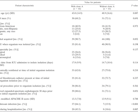
predispo-TABLE 3. Comparison of patients with and those withoutA. baumanniiclone Aa
Patient characteristic
Value for patients:
Pvalue
With clone A
(n⫽44)
Without clone A
(n⫽43)
Mean age (yr) (SD) 43.0 (14.9) 48.9 (16.6) 0.087
No. of men (%) 30 (68.2) 31 (72.1) 0.691
Race [no. (%)]
African-American 18 (40.9) 18 (41.9) 0.971
White, non-Hispanic 11 (25.0) 9 (20.9)
Hispanic, any race 12 (27.3) 13 (30.2)
Asian 3 (6.8) 3 (7.0)
Hospital acquired [no. (%)] 39 (90.7) 44 (100) 0.052
In ICU when organism was isolated [no. (%)] 35 (81.4) 40 (90.9) 0.198
ICU specialty [no. (%)] 0.137
Surgical 25 (56.8) 28 (56.0)
Medical 13 (29.5) 5 (11.6)
Neurosurgical 6 (13.6) 3 (7.0)
Mean time from ICU admission to isolate isolation (days) (SD)
17.8 (9.9) 14.7 (10.4) 0.114
[image:5.585.62.540.80.468.2]Mechanically ventilated at time of initial organism isolation [no. (%)]
33 (62.8) 27 (75.0) 0.210
Central bloodstream catheter present at time of initial organism isolation [no. (%)]
35 (81.4) 32 (72.7) 0.337
Surgical procedure prior to organism isolation [no. (%)] 38 (86.4) 34 (79.1) 0.361
Received expanded-spectrum cephalosporin 30 days prior to initial organism isolation [no. (%)]
39 (88.6) 31 (72.1) 0.051
Mean modified APACHE II score (SD) 13.3 (7.0) 13.9 (9.1) 0.780
Bloodstream infection [no. (%)] 37 (84.1) 7 (15.9) ⬍0.001
Died during hospitalization [no. (%)] 20 (45.5) 8 (18.6) 0.007
a
Characteristics of infected versus colonized patients were compared with a Student’sttest or Wilcoxon rank sum test for continuous variables and with2
tests for categorical variables.
on May 16, 2020 by guest
http://jcm.asm.org/
sition for the two clonal lineages and a difference in viru-lence.
Multivariate analysis of mortality associated with clone A.
To determine whether the isolation of clone A was indepen-dently associated with increased mortality, we performed a multivariate analysis using logistic regression for all patients from whomA. baumanniistrains were isolated. The variables entered into the model included all covariates associated with mortality at a P value of 0.20 or less in univariate analysis, which included isolation of clone A, modified APACHE II score, renal replacement therapy, and presence of a central bloodstream catheter. In the multivariate model, the odds ratio of death after acquisition of clone A was 3.31 (95% confidence interval, 1.27, 9.15;P ⫽0.024). Therefore, we conclude that isolation of clone A was associated with increased mortality independent of other risk factors, suggesting increased viru-lence compared with those of otherA. baumanniistrains iso-lated in this cohort.
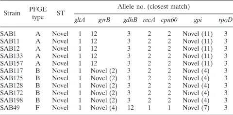
MLST. We next determined whether strains identified as belonging to clone A by PFGE would have the same MLST profile by analyzing five clone A isolates from unique patients. Each of the five clone A strains had the same MLST profile (Table 4). We also performed MLST on five clone B isolates from unique patients and found that all five clone B isolates had the same MLST profile, which was distinct from that of clone A (Table 4). The clone A and clone B strains each shared five alleles (gltA,gdhB,recA,cpn60, andrpoD) but were distinct at thegyrB locus (4-bp difference) and thegpi locus (11-bp difference). In contrast, anA. baumanniiPFGE type F strain sharedgltAandrpoDalleles with clone A and clone B but had distinctgyrB, gdhB, recA, cpn60, and gpi alleles (Table 4). A comparison of clone A and clone B data to published MLST data found that each strain shares five alleles (gltA,gdhB,recA,
cpn60, and rpoD) with sequence types 6, 10, and 19, which include invasiveA. baumannii isolates from Spain and Ger-many (1). Therefore, we conclude that clone A and clone B are related to each other and to certainA. baumanniistrains pre-viously isolated in Europe. Finally, we performed MLST onA. baumannii strains isolated from environmental samples and found that clone A was isolated in December 2005, whereas clone B was isolated in May 2006 (Fig. 1A). These data dem-onstrate that the presence of the twoA. baumanniiclones in
the environmental samples was temporally associated with their isolation from patients.
DISCUSSION
In this cohort of all patients from whom the A. calcoace-ticus-A. baumanniicomplex was isolated over a 14-month pe-riod, we found that the isolation of a specific A. baumannii
clone (clone A) was associated with a high level of bacteremia and increased mortality compared to other strains ofA. bau-manniiand to strains of the broaderA. calcoaceticus-A. bau-mannii complex. We were unable to discern patient-related factors that explained these findings, suggesting that some in-trinsic property of clone A itself may be responsible. Although significant efforts were made to control for confounding vari-ables, it is important to note that there are limitations inherent in the retrospective design and single-center nature of our study. Our data have parallels with data from a recent report noting an increased rate of bacteremia for a particular methi-cillin-resistantStaphylococcus aureusclone and, as such, add to our understanding of serious nosocomial infections by multi-drug-resistant bacteria (9).
At present, colonization withA. baumanniiin a susceptible host is thought to be the major risk factor for the subsequent development of invasive disease (12, 20). However, there is tremendous genetic diversity amongA. baumanniistrains, and it seems reasonable to speculate that the development of in-vasive disease following colonization is not the same for all strains (13). Indeed, one of the key findings of our investigation was that clone A caused a disproportionate number of bac-teremias compared to other clones ofA. baumannii. Whereas clone A comprised about 50% of theA. baumanniiisolates, it accounted for 84% of the bloodstream infections. Our data suggest that clone A may possess some inherent property, such as an ability to produce biofilm or to adhere to epithelial cells, that provides it with a capacity to produce bacteremia.
An unexpected finding of our study was the presence ofA. baumanniistrains in abdominal abscesses. Reviews of descrip-tive studies ofA. baumanniicolonization/infection show that the majority of isolates are recovered from sputum, urine, blood, wounds, and catheters (2, 20, 25). Most previously re-ported cases of peritonitis involving A. baumannii have in-volved patients receiving chronic ambulatory peritoneal dialy-sis (32). However, one study found that multidrug-redialy-sistantA. baumanniiisolates could be recovered from the stool of pa-tients in the ICU (5). As our papa-tients with abdominal abscesses had gastrointestinal tract disruption secondary to various pro-cesses, it does not seem unexpected thatA. baumanniimight be recovered from patients with nosocomial peritonitis. Impor-tantly, however, we found that all but one of the patients with an intra-abdominal abscess were infected withA. baumannii
[image:6.585.43.283.81.200.2]clone B. Although we cannot rule out the possibility that the finding of clone B in abdominal abscesses was related to its prevalence in the surgical ICU, we note that of the 36 patients in the surgical ICU that hadA. baumanniistrains other than clone B isolated, only 1 had an isolate from an abdominal abscess. This finding suggests that clone B may have a partic-ular predilection for the gastrointestinal tract in a fashion sim-ilar to that of clone A, which seems predisposed to caused bloodstream infections.
TABLE 4. Multilocus sequence typing ofA. baumanniiisolatesa
Strain PFGE
type ST
Allele no. (closest match)
gltA gyrB gdhB recA cpn60 gpi rpoD
SAB1 A Novel 1 12 3 2 2 Novel (11) 3
SAB11 A Novel 1 12 3 2 2 Novel (11) 3
SAB12 A Novel 1 12 3 2 2 Novel (11) 3
SAB133 A Novel 1 12 3 2 2 Novel (11) 3
SAB157 A Novel 1 12 3 2 2 Novel (11) 3
SAB117 B Novel 1 Novel (2) 3 2 2 Novel (4) 3
SAB125 B Novel 1 Novel (2) 3 2 2 Novel (4) 3
SAB128 B Novel 1 Novel (2) 3 2 2 Novel (4) 3
SAB172 B Novel 1 Novel (2) 3 2 2 Novel (4) 3
SAB198 B Novel 1 Novel (2) 3 2 2 Novel (4) 3
SAB49 F Novel 1 Novel (4) 12 1 1 Novel (7) 3
a
Sequences of amplified genes were compared with sequences at theA.
baumannii MLST website (http://pubmlst.org/abaumannii/). ST, sequence type.
on May 16, 2020 by guest
http://jcm.asm.org/
Our data highlight the need for microbiology laboratories to provide clinicians and infection control practitioners with more discriminative information than reporting an isolate as belonging to the A. calcoaceticus-A. baumannii complex. Given the vastly different outcomes between patients in-fected with A. baumannii and those infected with non-A. baumanniistrains, practitioners need to know whatA. cal-coaceticus-A. baumanniicomplex species has been isolated and perhaps be provided with molecular data regardingA. baumannii strains. Without molecular investigation, we would not have discovered that our two peaks of infection were actually caused by the dissemination of two genetically distinct A. baumannii strains (Fig. 1). MLST provides re-searchers with a tool for geographically diverse investiga-tions and also allows the rapid molecular identification of strains in individual patients. Such information may help clinicians with therapeutic decisions and infection control departments with outbreak containment strategies.
In conclusion, we found that a particular clone ofA. bau-mannii was significantly associated with the development of bacteremia and adverse patient outcomes compared to other
A. baumanniistrains and non-A. baumanniistrains belonging to theA. calcoaceticus-A. baumanniicomplex. We present the first MLST data for A. baumannii strains isolated in North America and demonstrate that the dominant clones ofA. bau-manniiat our facility are related to strains previously isolated in Western Europe. Our findings provide a framework for a more detailed investigation ofA. baumanniipathogenesis and improved understanding ofA. baumanniiepidemiology.
ACKNOWLEDGMENTS
This study was supported by National Institutes of Allergy and Infectious Disease career development grant K08 RR17665-04 (S.A.S.) and by the Harris County Hospital District.
REFERENCES
1.Bartual, S. G., H. Seifert, C. Hippler, M. A. Luzon, H. Wisplinghoff, and F. Rodriguez-Valera. 2005. Development of a multilocus sequence typing
scheme for characterization of clinical isolates ofAcinetobacter baumannii.
J. Clin. Microbiol.43:4382–4390.
2.Bergogne-Berezin, E., and K. J. Towner.1996.Acinetobacterspp. as noso-comial pathogens: microbiological, clinical, and epidemiological features.
Clin. Microbiol. Rev.9:148–165.
3.Calandra, T., and J. Cohen.2005. The international sepsis forum consensus conference on definitions of infection in the intensive care unit. Crit. Care
Med.33:1538–1548.
4.Centers for Disease Control and Prevention.2004.Acinetobacter baumannii
infections among patients at military medical facilities treating injured U.S.
service members, 2002–2004. MMWR Morb. Mortal. Wkly. Rep.53:1063–
1066.
5.Corbella, X., M. Pujol, J. Ayats, M. Sendra, C. Ardanuy, M. A. Dominguez, J. Linares, J. Ariza, and F. Gudiol.1996. Relevance of digestive tract colo-nization in the epidemiology of nosocomial infections due to multiresistant
Acinetobacter baumannii. Clin. Infect. Dis.23:329–334.
6.Cunningham, M. W.2000. Pathogenesis of group A streptococcal infections.
Clin. Microbiol. Rev.13:470–511.
7.Davis, K. A., K. A. Moran, C. K. McAllister, and P. J. Gray.2005.
Multidrug-resistantAcinetobacterextremity infections in soldiers. Emerg. Infect. Dis.
11:1218–1224.
8.Dolzani, L., E. Tonin, C. Lagatolla, L. Prandin, and C. Monti-Bragadin.
1995. Identification ofAcinetobacterisolates in theA. calcoaceticus-A.
bau-manniicomplex by restriction analysis of the 16S-23S rRNA
intergenic-spacer sequences. J. Clin. Microbiol.33:1108–1113.
9.Edgeworth, J. D., G. Yadegarfar, S. Pathak, R. Batra, J. D. Cockfield, D. Wyncoll, R. Beale, and J. A. Lindsay.2007. An outbreak in an intensive care
unit of a strain of methicillin-resistantStaphylococcus aureussequence type
239 associated with an increased rate of vascular access device-related
bac-teremia. Clin. Infect. Dis.44:493–501.
10.Espinal, M. A., A. Laszlo, L. Simonsen, F. Boulahbal, S. J. Kim, A. Reniero,
S. Hoffner, H. L. Rieder, N. Binkin, C. Dye, R. Williams, M. C. Raviglione, et al.2001. Global trends in resistance to antituberculosis drugs. N. Engl.
J. Med.344:1294–1303.
11.Falagas, M. E., and P. I. Rafailidis.2007. Attributable mortality of Acineto-bacter baumannii: no longer a controversial issue. Crit. Care11:134. 12.Fournier, P. E., and H. Richet. 2006. The epidemiology and control of
Acinetobacter baumanniiin health care facilities. Clin. Infect. Dis.42:692– 699.
13.Fournier, P. E., D. Vallenet, V. Barbe, S. Audic, H. Ogata, L. Poirel, H. Richet, C. Robert, S. Mangenot, C. Abergel, P. Nordmann, J. Weissenbach, D. Raoult, and J. M. Claverie.2006. Comparative genomics of multidrug
resistance inAcinetobacter baumannii. PLoS Genet.2:e7.
14.Fridkin, S. K., J. C. Hageman, M. Morrison, L. T. Sanza, K. Como-Sabetti, J. A. Jernigan, K. Harriman, L. H. Harrison, R. Lynfield, and M. M. Farley.
2005. Methicillin-resistantStaphylococcus aureusdisease in three
communi-ties. N. Engl. J. Med.352:1436–1444.
15.Gaynes, R., and J. R. Edwards.2005. Overview of nosocomial infections
caused by gram-negative bacilli. Clin. Infect. Dis.41:848–854.
16.Goh, B. K., G. Alkouder, T. K. Lama, and C. E. Tan.2005.
Multi-drug-resistant Acinetobacter baumannii intra-abdominal abscess. Surg. Infect.
6:345–347.
17.Griffith, M. E., D. R. Lazarus, P. B. Mann, J. A. Boger, D. R. Hospenthal, and C. K. Murray.2007.Acinetobacterskin carriage among US army soldiers
deployed in Iraq. Infect. Control Hosp. Epidemiol.28:720–722.
18.Grupper, M., H. Sprecher, T. Mashiach, and R. Finkelstein.2007.
Attrib-utable mortality of nosocomialAcinetobacterbacteremia. Infect. Control
Hosp. Epidemiol.28:293–298.
19.Houang, E. T., Y. W. Chu, K. Y. Chu, K. C. Ng, C. M. Leung, and A. F. Cheng.2003. Significance of genomic DNA group delineation in
compara-tive studies of antimicrobial susceptibility ofAcinetobacterspp. Antimicrob.
Agents Chemother.47:1472–1475.
20.Joly-Guillou, M. L.2005. Clinical impact and pathogenicity ofAcinetobacter.
Clin. Microbiol. Infect.11:868–873.
21.Knaus, W. A., E. A. Draper, D. P. Wagner, and J. E. Zimmerman.1985. APACHE II: a severity of disease classification system. Crit. Care Med.
13:818–829.
22.Lee, J. H., C. H. Choi, H. Y. Kang, J. Y. Lee, J. Kim, Y. C. Lee, S. Y. Seol, D. T. Cho, K. W. Kim, D. Y. Song, and J. C. Lee.2007. Differences in
phenotypic and genotypic traits against antimicrobial agents between
Acin-etobacter baumanniiandAcinetobacter genomicspecies 13TU. J. Antimicrob.
Chemother.59:633–639.
23.Murray, B. E., and R. C. Moellering, Jr.1979. Aminoglycoside-modifying
enzymes among clinical isolates ofAcinetobacter calcoaceticussubsp.
anitra-tus (Herellea vaginicola): explanation for high-level aminoglycoside
resis-tance. Antimicrob. Agents Chemother.15:190–199.
24.O’Grady, N. P., M. Alexander, E. P. Dellinger, J. L. Gerberding, S. O. Heard, D. G. Maki, H. Masur, R. D. McCormick, L. A. Mermel, M. L. Pearson, I. I. Raad, A. Randolph, R. A. Weinstein, et al.2002. Guidelines for the preven-tion of intravascular catheter-related infecpreven-tions. MMWR Recomm. Rep.
51:1–29.
25.Rodriguez-Bano, J., J. M. Cisneros, F. Fernandez-Cuenca, A. Ribera, J. Vila, A. Pascual, L. Martinez-Martinez, G. Bou, and J. Pachon.2004.
Clinical features and epidemiology ofAcinetobacter baumannii
coloniza-tion and infeccoloniza-tion in Spanish hospitals. Infect. Control Hosp. Epidemiol.
25:819–824.
26.Schreckenberger, P., M. Daneshvar, R. S. Weyant, and D. Hollis.2003.
Acinetobacter,Achromobacter,Chryseobacterium,Moraxella, and other
non-fermentative gram-negative rods, p. 749–779.InP. R. Murray, E. J. Baron,
J. H. Jorgensen, M. A. Pfaller, and R. H. Yolken (ed.), Manual of clinical microbiology, 8th ed. ASM Press, Washington, DC.
27.Scott, P., G. Deye, A. Srinivasan, C. Murray, K. Moran, E. Hulten, J. Fishbain, D. Craft, S. Riddell, L. Lindler, J. Mancuso, E. Milstrey, C. T. Bautista, J. Patel, A. Ewell, T. Hamilton, C. Gaddy, M. Tenney, G. Chris-topher, K. Petersen, T. Endy, and B. Petruccelli.2007. An outbreak of
multidrug-resistantAcinetobacter baumannii-calcoaceticuscomplex infection
in the US military health care system associated with military operations in
Iraq. Clin. Infect. Dis.44:1577–1584.
28.Seifert, H., L. Dolzani, R. Bressan, T. van der Reijden, B. van Strijen, D. Stefanik, H. Heersma, and L. Dijkshoorn.2005. Standardization and inter-laboratory reproducibility assessment of pulsed-field gel
electrophoresis-generated fingerprints ofAcinetobacter baumannii. J. Clin. Microbiol.43:
4328–4335.
29.Tenover, F. C., R. D. Arbeit, R. V. Goering, P. A. Mickelsen, B. E. Murray, D. H. Persing, and B. Swaminathan.1995. Interpreting chromosomal DNA restriction patterns produced by pulsed-field gel electrophoresis: criteria for
bacterial strain typing. J. Clin. Microbiol.33:2233–2239.
30.Turton, J. F., M. E. Kaufmann, M. J. Gill, R. Pike, P. T. Scott, J. Fishbain, D. Craft, G. Deye, S. Riddell, L. E. Lindler, and T. L. Pitt.2006. Comparison of Acinetobacter baumanniiisolates from the United Kingdom and the United States that were associated with repatriated casualties of the Iraq
conflict. J. Clin. Microbiol.44:2630–2634.
31.Turton, J. F., M. E. Kaufmann, M. Warner, J. Coelho, L. Dijkshoorn, T. van
on May 16, 2020 by guest
http://jcm.asm.org/
der Reijden, and T. L. Pitt.2004. A prevalent, multiresistant clone of Acin-etobacter baumanniiin Southeast England. J. Hosp. Infect.58:170–179. 32.Valdez, J. M., M. O. Asperilla, and R. A. Smego, Jr.1991.Acinetobacter
peritonitis in patients receiving continuous ambulatory peritoneal dialysis.
South. Med. J.84:607–610.
33.Vaneechoutte, M., L. Dijkshoorn, I. Tjernberg, A. Elaichouni, P. de Vos, G. Claeys, and G. Verschraegen.1995. Identification ofAcinetobactergenomic
species by amplified ribosomal DNA restriction analysis. J. Clin. Microbiol.
33:11–15.
34.Villegas, M. V., and A. I. Hartstein.2003.Acinetobacteroutbreaks, 1977–
2000. Infect. Control Hosp. Epidemiol.24:284–295.
35.Wootton, J. C., X. Feng, M. T. Ferdig, R. A. Cooper, J. Mu, D. I. Baruch, A. J. Magill, and X. Z. Su.2002. Genetic diversity and chloroquine selective
sweeps inPlasmodium falciparum. Nature418:320–323.