0095-1137/05/$08.00⫹0 doi:10.1128/JCM.43.1.156–162.2005
Copyright © 2005, American Society for Microbiology. All Rights Reserved.
Rapid Multiplex Assay for Serotyping Pneumococci with Monoclonal
and Polyclonal Antibodies
Jigui Yu,
1Jisheng Lin,
1William H. Benjamin, Jr.,
1,2Ken B. Waites,
1,2Che-hung Lee,
3and
Moon H. Nahm
1,2*
Departments of Pathology1and Microbiology,2University of Alabama at Birmingham, Birmingham, Alabama, and
U.S. Food and Drug Administration, Bethesda, Maryland3
Received 20 July 2004/Returned for modification 7 September 2004/Accepted 14 September 2004
We have developed and characterized a rapid semiautomated pneumococcal serotyping system incorporating a pneumococcal lysate preparation protocol and a multiplex serotyping assay. The lysate preparation incor-porates a bile solubility test to confirm pneumococcal identification that also enhances assay specificity. The multiplex serotyping assay consists of 24 assays specific for 36 serotypes: serotypes 1, 2, 3, 4, 5, 6A, 6B, 7A/7F, 8, 9L/9N, 9V, 10A/10B/39/(33C), 11A/11D/11F, 12A/12B/12F, 14, 15B/(15C), 17F, 18C, 19A, 19F, 20, 22A/22F, 23F, and 33A/33F. The multiplex assay requires a flow cytometer, two sets of latex particles coated with pneumococcal polysaccharides, and serotype-specific antibodies. Fourteen newly developed monoclonal anti-bodies specific for common serotypes and a pool of polyclonal rabbit sera for some of the less-common serotypes are used. The two monoclonal antibodies specific for serotypes 18C and 23F recognize serotype-specific epitopes that have not been previously described. These monoclonal antibodies make the identification of the 14 common serotypes invariant. The specificity of the serotyping assay is fully characterized with pneumococci of all known (i.e., 90) serotypes. The assay is sensitive enough to use bacterial lysates diluted 20 fold. Our serotyping system can identify not only all the serotypes in pneumococcal vaccines but also most (>90%) of clinical isolates. This system should be very useful in serotyping clinical isolates for evaluating pneumococcal vaccine efficacy.
Streptococcus pneumoniaeis a well-known human pathogen and a major etiologic agent for pneumonia, meningitis, otitis media, and sepsis, primarily among young children and older
adults (3).S. pneumoniaeis divided into 90 serotypes, based on
its expression of serologically distinct carbohydrate capsules (6). Antibodies to a capsular polysaccharide (PS) provide pro-tection against pneumococci expressing the same capsular se-rotype. Currently available pneumococcal vaccines contain a mixture of capsular PS of multiple serotypes. One pneumococ-cal vaccine (pneumococ-called PS vaccine) contains capsular PS from 23 commonly found serotypes (17). The most recently developed type of vaccine (called conjugate vaccine) contains capsular PS from 7 to 11 serotypes that are conjugated to a protein mole-cule (26). A 7-valent conjugate vaccine was introduced in 2000 for routine immunization in the United States and has reduced the incidence of invasive pneumococcal diseases in children (25).
Accurate, efficient serotyping of pneumococcal isolates is important for measuring the efficacy of pneumococcal vac-cines. Since the vaccine-induced antibodies provide serotype-specific protection, following the introduction of a new pneu-mococcal vaccine, pneumococci expressing the serotypes included in the vaccine would become less common while the prevalence of the pneumococci expressing nonvaccine sero-types would stay the same. As a result, a reduction in vaccine serotypes reflects the vaccine efficacy. In some cases, pneumo-cocci expressing the nonvaccine types replace those expressing
the vaccine serotypes and the prevalence of nonvaccine types may become higher (16). Further, the prevalence of serotypes can change over time for unknown reasons (5). As these changes would influence the clinical effectiveness of a vaccine, serotyping a large number of pneumococcal isolates is required as a part of monitoring pneumococcal vaccines.
Although there are numerous serotyping methods (2, 4, 6, 9–11, 13, 18–21), the ones that are currently used are inade-quate. The assays use polyclonal antibodies, which may not be able to distinguish closely related serotypes easily. The assays are not quantitative and require considerable experience to correctly interpret the results. Because over 20 serotypes are common among clinical isolates, an isolate must be tested for a large number of serotypes. Yet the available methods are manual and slow. To overcome these problems, we previously described a multiplex assay based on flow cytometry for a small number of serotypes (15). We now describe 14 monoclonal antibodies and a multiplex assay for the serotypes included in the 23-valent PS vaccine.
MATERIALS AND METHODS
Pneumococci, pneumococcal lysates, rabbit serogrouping sera, and monoclo-nal antibodies.Pneumococci used for the study were selected from clinical isolates used in a previous study (15) and from the clinical isolate collection at the University of Alabama at Birmingham. Also used were 90 isolates (purchased from Statens Serum Institut, Copenhagen, Denmark) representing all 90 known
serotypes. As negative controls, we usedStaphylococcus aureus(ATCC 6538),
Streptococcus pyogenes(ATCC 19615),Escherichia coli(ATCC 12014), and Strep-tococcus agalactiae(strain M781 from Carol Baker, Houston, Tex.). Pneumococ-cal lysates were obtained as follows. A microtube containing 0.3 ml of Todd-Hewitt broth with 0.5% yeast extract was inoculated with one or several colonies
of pneumococci. After a 6-h incubation at 37°C, 50l of lysis buffer (0.2%
sodium deoxycholate, 0.02% sodium dodecyl sulfate, and 0.3 M sodium citrate)
* Corresponding author. Mailing address: 845 19th St. South (BBRB 614), Birmingham, AL 35249-7331. Phone: (205) 934-0163. Fax: (205) 975-2149. E mail: E-mail: Nahm@uab.edu.
156
on May 16, 2020 by guest
http://jcm.asm.org/
was added to the microtube, and the tube was incubated for an additional hour at 37°C.
Serogrouping rabbit antisera S, T, E, and F were purchased from the Statens Serum Institut. Mouse hybridomas were produced as described previously (22). Briefly, BALB/c mice were immunized twice subcutaneously with PS-protein conjugate (days 0 and 21) and once intraperitoneally on day 59. The immunogen for seven serotypes (4, 6B, 9V, 14, 18C, 19F, and 23F) was Prevnar (Wyeth Lederle Vaccines, Pearl River, N.Y.). Conjugates used for serotypes 5 and 7F were prepared at the U.S. Food and Drug Administration (Bethesda, Md.), the 6A conjugate was a gift of Porter Anderson (Rochester, N.Y.), and conjugates of serotypes 1, 3, and 9N to ovalbumin were prepared in our laboratory as follows. Cyanogen bromide-activated PS was coupled to ovalbumin during an overnight incubation. The PS-protein conjugate was purified from the reaction mixture
with a molecular weight sizing column. Each dose contained 1g of PS for
serotypes 4, 9V, 18C, 19F, and 23F; 2g for serotypes 3 and 6B; and 10g for
serotypes 1, 5, 6A, 7F, and 9N. The primary and secondary immunogens
con-tained 10g of Quil A (Sigma Chemical, St. Louis, Mo.).
Three days after the last immunization, the spleen was harvested, and spleno-cytes were fused with SP2/0 Ag-14 as previously described (14). Primary culture wells were screened for the production of desirable antibodies, and the wells producing desirable monoclonal antibodies were cloned twice by limiting dilu-tion. A human-mouse hybridoma, Dob9, was produced by hybridizing peripheral blood lymphocytes from a person immunized with a 23-valent PS vaccine, as described previously (23).
Preparation of latex beads coated with different pneumococcal PS serotypes.
Fifteen different types of latex beads were produced by staining latex beads of
five different sizes (2 to 4.8m in diameter) to three different levels (none, low,
and high) of red fluorescence with DiD oil (1,1⬘-dioctadecyl-3,3,3⬘,3⬘
-tetrameth-ylindodicarbocyanine perchlorate) from Molecular Probes (Eugene, Oreg.). One of 14 different pneumococcal PSs (serotypes 1, 3, 4, 5, 6A, 6B, 7F, 9N, 9V, 14, 18C, 19A, 19F, and 23F) was adsorbed onto each of 14 types of beads. The 14 different bead preparations were then mixed together and used as bead set 1 in the assay. Bead set 2 was created by coating each of 10 bead types with one of 10 different pneumococcal PS (serotypes 2, 8, 10A, 11A, 12F, 15B, 17F, 20, 22F, and 33F) and then by mixing the 10 types of PS-coated beads together. All the beads were coated with capsular PS from the American Type Culture Collection (Ma-nassas, Va.), except for the beads coated with serotypes 3, 18C, 6B, 9V, and 19F. Those five beads were coated with PS-protein conjugate to increase the stability of PS coating and the shelf life of the beads.
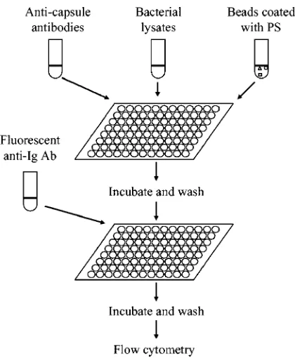
Multiplex serotype assay.The assay was performed as described in detail
previously (15) and summarized in Fig. 1. In brief, 30l of appropriately diluted
pneumococcal lysate, 40 to 56l of a mixture of latex beads (4l per each
serotype), and 30l of serotype-specific antibodies were added to a microwell of
a microtiter plate. The buffer used for dilutions was phosphate-buffered saline containing 1% bovine serum albumin, 0.05% Tween 20, and 0.05% sodium azide. As serotype-specific antibodies, a mixture of 14 monoclonal antibodies was used for bead set 1. For bead set 2, the type-specific antibodies were an equal volume mixture of serogrouping rabbit antisera S, T, E, and F that were diluted 600 fold
in the dilution buffer containing 40g of cell wall PS (Statens Serum Institut)/ml.
After 30 min of incubation at room temperature, the beads were washed in the
microtiter plate with 200l of normal saline containing 0.05% Tween 20 and
0.05% sodium azide, and the mixture of fluorescein-conjugated anti-immuno-globulin (Ig) antibodies was added. For bead set 1, a mixture of fluorescein-conjugated human (Sigma Chemical) and fluorescein-fluorescein-conjugated anti-mouse Ig was used. For bead set 2, a fluorescein-conjugated anti-rabbit Ig antiserum (Southern Biotechnology Assoc., Birmingham, Ala.) was used.
After 30 min of incubation, the beads were washed as described above, and the fluorescence of the beads was determined with a flow cytometer (FACSCalibur; Becton Dickinson, San Jose, Calif.). At least 300 beads were examined for each bead type. Bead types were recognized with forward scatter and red fluorescence. The geometric mean of green fluorescence was obtained for each type of bead. Normalized fluorescence was calculated as follows: (geometric mean
fluores-cence of a sample⫺geometric mean of background)/(geometric mean
fluores-cence of negative sample⫺geometric mean of background). Fluorescence of the
beads without any primary antibody was used as the background fluorescence. The geometric mean of the fluorescence obtained with the lysate of a noncap-sulated strain (R36A) or a strain expressing an unrelated serotype was used as the maximal fluorescence (100%).
RESULTS
Assay characteristics. (i) Production of monoclonal anti-bodies and assay development.To produce a set of fully char-acterized reagents that are invariant, we have produced mono-clonal antibodies to 14 commonly isolated serotypes (Table 1). One hybridoma (Dob9) is a human-mouse hybridoma, which produces human antibody specific for 19A and 19F. All others were obtained by hybridizing splenocytes of mice, which were immunized with a protein-PS conjugate. All hybridomas pro-duced either IgM or IgG antibodies except for one, which produced IgA antibody (Table 1). A mixture of 14 hybridoma culture supernatants was used for the multiplex serotyping assay.
(ii) Determination of assay sensitivity with purified PS and bacterial lysates.Once the multiplex assay format was devel-oped, the assay was first tested for assay sensitivity with
puri-fied capsular PS. For most serotypes, about 0.1g of PS/ml was
[image:2.585.58.268.65.320.2]sufficient to reduce 50% of the normalized fluorescence. For
FIG. 1. Diagram of the assay procedure.
TABLE 1. Monoclonal antibodies used for the assay
Name Specificity Isotype
Hyp1G4 1 G
Hyp3M6 3 M
Hyp4M3 4 M
Hyp5M3 5 M
Hyp6AM3 6A M
Hyp6BM7 6B M
Hyp7FM1 7F/7A M
Hyp9NA1 9N/9L A
Hyp9VG13 9V G
Hyp14M11 14 M
Hyp18CM1 18C M
Dob9 19A/19F M
Hyp19FM3 19F M
Hyp23FG3 23F G
on May 16, 2020 by guest
http://jcm.asm.org/
[image:2.585.302.541.79.228.2]serotypes 9N and 7F, about 0.7g/ml was required to reduce the normalized fluorescence by half.
To investigate whether the assay has an adequate sensitivity for actual bacterial isolates, we determined the dilution of pneumococcal lysates that reduces the normalized fluores-cence signal by 66.7% (Fig. 2). For the 66.7% inhibition, bac-terial lysates could be diluted more than 20 fold: pneumococcal lysates of serotypes 9V, 9N, 20, and 33F could be diluted 25 to 50 fold, and the lysates of the other serotypes could be diluted
more than 250 fold. Since 0.1 to 0.7g of purified PS/ml is
required for 50% inhibition, these observations suggest that pneumococcal lysates usually contain more than several micro-grams per milliliter of capsular PS.
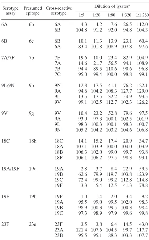
(iii) Determination of assay specificity with pneumococci expressing related serotypes.Once the assay sensitivity was determined, the specificity of the assay was evaluated with bacterial lysates of pneumococci expressing related serotypes (Table 2). When the assays using monoclonal antibodies were examined, they were highly specific for a single serotype, ex-cept for three serotypes (7F, 9N, and 19A). For instance, 9V lysate could inhibit the fluorescence signal of 9V assay by 50% even after an 80-fold dilution, but the lysates of related sero-types (9A, 9L, and 9N) inhibited the signal by less than 10% even when the lysates were diluted only 5 fold. The three cross-reactive serotypes (7F, 9N, and 19A) are known to ex-press cross-reactive epitopes (7b, 9b, and 19d, respectively) (6), and the recognition of these epitopes by the monoclonal anti-bodies could fully explain the observed cross-reaction patterns. For instance, 9L lysates inhibited the 9N assay as well as the 9N lysate, but 9A and 9V lysates did not inhibit at all. The 9b epitope was expressed on serotypes 9L and 9N but not on 9A and 9V. Also, the 19A assay reacted with 19F as well as 19A. This cross-reaction did not limit our ability to distinguish 19F and 19A strains: 19F strains inhibited both 19A and 19F assays, but 19A strains inhibited only the 19A assay. Since these cross-reactions did not limit the assays’ usefulness, the assays for the three serotypes were not modified any further. However, the
assays were labeled as 9N/9L, 7F/7A, and 19A/19F assays to avoid confusion.
An interesting finding was that our assays for 18C and 23F were type specific. So far, there are no serotype-specific epitopes identified for these two serotypes (6). Thus, our monoclonal antibodies for 18C and 23F may recognize new epitopes, and we provisionally labeled them 18h and 23e epitopes (Table 2).
[image:3.585.56.273.66.236.2]The assays based on the rabbit antisera were also evaluated for specificity with the lysates of related serotypes (Table 2). The assay did not show any cross-reaction with unrelated se-rotypes. Lysates of non-cross-reactive serotypes, even at a five-fold dilution, inhibited the fluorescence signal less than 33.3%. Among the related serotypes, the rabbit serum assays displayed
FIG. 2. Normalized fluorescence versus bacterial lysate dilutions is shown for bead set 1. Each line represents one serotype, and the serotype is identified. Lines for all others (serotypes 1, 4, 5, 6B, 14, 18C, 19A, 19F, and 23F) lie in the shaded area between the two lines produced by serotypes 6A and 3.
TABLE 2. Normalized fluorescence of each serotype assay in the presence of various dilutions of pneumococcal lysates of
cross-reactive serotypes
Serotype assay
Presumed epitope
Cross-reactive serotype
Dilution of lysatesa
1:5 1:20 1:80 1:320 1:1,280
6A 6b 6A 4.3 4.2 7.6 26.5 112.0
6B 104.8 91.2 92.0 94.8 104.3
6B 6c 6B 10.1 11.3 13.9 23.1 60.4
6A 83.4 101.8 108.9 107.8 97.6
7A/7F 7b 7F 19.6 10.0 23.4 82.9 104.9
7A 14.6 21.7 56.5 94.1 108.9 7B 94.4 89.5 110.6 90.6 96.8 7C 95.0 99.4 100.0 98.8 99.1
9L/9N 9b 9N 12.8 17.5 41.1 76.2 122.1
9A 94.6 104.2 108.3 127.7 129.0 9L 13.5 17.5 32.2 54.9 93.5 9V 99.1 102.5 112.7 102.3 126.2
9V 9g 9V 10.4 23.2 52.8 79.6 97.5
9A 93.0 97.3 100.1 102.5 101.9 9L 98.3 100.3 100.1 98.3 100.7 9N 105.2 104.2 103.2 104.6 106.8
18C 18h 18C 14.1 15.2 17.4 20.9 34.7
18A 107.1 103.9 100.0 104.0 103.9 18B 106.3 102.0 99.0 99.7 93.8 18F 106.1 106.2 97.5 98.3 93.1
19A/19F 19d 19A 2.8 3.7 8.4 22.9 59.5
19B 62.6 79.9 119.7 103.8 123.9 19C 72.4 99.0 99.2 112.8 114.8 19F 3.3 5.4 12.5 41.3 78.8
19F 19b 19F 1.0 1.4 2.0 3.4 9.2
19A 95.5 99.0 99.5 102.0 98.3 19B 98.9 100.3 99.5 100.3 98.4 19C 97.3 98.9 97.9 99.6 99.8
23F 23e 23F 3.5 3.8 6.4 14.5 43.0
23A 121.4 107.6 104.5 99.7 117.7 23B 95.5 95.1 88.3 103.3 107.7
aThe numbers in the table indicate normalized fluorescence, which was
cal-culated as (geometric mean fluorescence of a sample⫺geometric mean of
background)/(geometric mean fluorescence of negative sample⫺Geometric
mean of background). Fluorescence of the beads without any primary antibody was used as the background fluorescence. The fluorescence obtained with the lysate of a noncapsulated strain (R36A) or a strain expressing an unrelated serotype was used as the fluorescence of negative sample.
on May 16, 2020 by guest
http://jcm.asm.org/
[image:3.585.299.541.96.485.2]various cross-reaction patterns. The assay for 11A serotype fully cross-reacted with 11D and 11F serotypes but not with 11B and 11C serotypes. Also, the assay for 12F serotype rec-ognized both serotypes 12A and 12B. 12A lysate was less in-hibitory than the 12B or 12F lysates, this may be a result of the 12A strain producing less capsular PS than 12B or 12F strains. Consequently, the 12A serotype could not be distinguished from 12B or 12F serotypes. As with the three assays using monoclonal antibodies, we labeled these 11A/11D/11F and 12A/12B/12F assays. A similar rationale was used to name assays 10A/10B, 22A/22F, and 33A/33F. These cross-reaction patterns were consistent with assays recognizing cross-reactive epitopes indicated in Table 3.
In the case of the 15B assay, the assay cross-reacted with 15C but not with 15A and 15F. The cross-reaction with 15C was partial, shown by the fact that the normalized fluorescence remained at about 40% even though the dilution of the 15C lysate was reduced from 20 to 5 fold (Table 3). This cross-reactivity can be explained if our antisera contained antibodies recognizing the 15e epitope present in serotypes 15B and 15C and 15h epitope that was expressed only on the 15B serotype. Thus, the assay was labeled as the 15B/(15C) assay to reflect only the partial cross-reaction with 15C.
(iv) Operational characteristics of the test. Following the above studies, we examined the operational characteristics of all 24 assays together. Due to the use of microtiter plate sys-tems, we could easily handle a large number of samples. Also, the bacterial lysis step was specific for pneumococci. All pneu-mococcal isolates we examined so far were lysed, but other
bacterial species includingS. pyogenes, group B streptococcus,
staphylococcus, and E. coli were not lysed. Based on pilot
studies, bacterial lysates diluted 1:5 and 1:20 were analyzed in duplicate for the routine serotyping assay. Normalized fluores-cence signals greater than 66.7% were determined to be neg-ative, signals from 33.3 to 66.6% were indeterminate, and signals of less than 33.3% were positive.
Our 14 new monoclonal antibodies may react with capsular PS belonging to only a subset of a serotype. We therefore tested 104 pneumococcal strains representing the 14 serotypes identified by our monoclonal antibodies (Table 4) under the operational test conditions described above. While most strains were found to have the expected serotype, there were a few exceptions. All the strains displayed only one serotype, except one strain. The isolate previously reported to be sero-type 1 was contaminated with two other strains expressing serotype 9V and serotype 23F. One serotype 3 strain was found to produce no capsular PS, and it was found to be a laboratory-induced non-capsule-producing mutant. Two strains produced marginal amounts of capsular PS, and their capsular PS could be detectable only at 1:5 dilutions. Since the two isolates have been maintained in the laboratory for 10 and 30 years, respec-tively, variants producing small amounts of capsule may have been selected. Serotypes of three strains were clearly discrep-ant. One 6A isolate was found to express the 6B serotype. Another isolate was labeled 6B but expressed the 6A capsule. One 23F isolate was found to be serotype 22F. In view of the pattern of these discrepancies, the differences may have arisen from human errors in labeling the strains. Taken together, we concluded that the new monoclonal antibodies do not subdi-vide a serotype into subsets and that the operational test con-ditions (i.e., dilutions and decision criteria) were adequate.
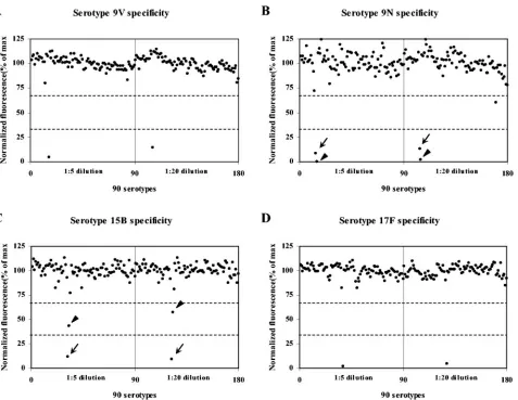
[image:4.585.302.541.89.263.2]As the second operational test of the assay, we examined all 90 serotypes with the assay. This also fully characterized the
TABLE 3. Normalized fluorescence of each serotype assay in the presence of various dilutions of pneumococcal lysates of
cross-reactive serotypes
Assay serotype Presumedepitope Cross-reactiveserotype
Dilution of lysatesa
1:5 1:20 1:80 1:320 1:1,280
10A/10B 10d 10A 16.4 18.7 20.3 31.6 37.9
10B 31.1 42.4 50.7 71.0 86.6
10C 90.9 98.7 103.2 101.6 105.6
10F 87.5 95.0 107.0 104.9 108.7
11A/11D/11F 11e 11A 1.0 1.9 6.6 16.1 34.7
11B 124.3 121.2 119.3 109.6 88.2
11C 86.5 114.9 106.5 95.6 92.8
11D 1.4 3.5 7.1 18.2 42.2
11F 7.9 12.2 22.9 38.8 54.9
12A/12B/12F 12a 12A 19.5 27.9 37.6 56.4 64.8
12B 12.0 17.1 27.1 43.9 62.8
12F 1.3 2.4 5.8 12.5 26.8
15B 15h 15A 81.3 88.3 99.2 93.4 97.9
15B 6.0 8.8 17.8 38.0 56.8
15C 39.9 60.2 67.0 87.5 85.4
15F 64.1 79.6 101.1 93.5 109.3
17F 17b 17A 82.5 93.7 85.5 88.9 85.9
17F 1.7 4.8 11.8 26.4 51.2
22A/22F 22a 22A 26.7 32.9 49.8 59.6 84.0
22F 4.2 13.6 22.3 44.4 66.4
33A/33F 33b 33A 14.7 21.7 34.5 50.3 68.3
33B 79.8 91.1 91.8 97.2 100.8
33C 99.6 106.3 119.6 113.4 106.6
33D 90.5 92.7 99.1 94.3 93.2
33F 16.4 23.6 28.6 42.9 60.7
aThe numbers in the table indicate normalized fluorescence, which was
cal-culated as (geometric mean fluorescence of a sample⫺geometric mean of
background)/(geometric mean fluorescence of negative sample⫺geometric
mean of background). Fluorescence of the beads without any primary antibody was used as the background fluorescence. The fluorescence obtained with the lysate of a noncapsulated strain (R36A) or a strain expressing an unrelated serotype was used as the fluorescence of negative sample.
TABLE 4. Study of pneumococcal strains with known serotypes with monoclonal antibodies
Serotype No. of isolates examined
No. of isolates confirmed
Comments
1 5 5 One strain is contaminated with 22F and 9V strains
3 6 5 One strain has no detectable signal 4 8 8 One strain is reactive at 1:5 only 5 5 5 One strain is reactive at 1:5 only 6A 7 6 One strain is 6B serotype 6B 9 8 One strain is 6A serotype
7F 5 5
9N 5 5
9V 8 8
14 11 11
18C 9 9
19A 7 7
19F 8 8
23F 11 10 One strain is 22F serotype
on May 16, 2020 by guest
http://jcm.asm.org/
[image:4.585.43.284.98.390.2]serotype specificity of the assay. As shown in Fig. 3 for sero-types 9V, 9N, 15B, and 17F, the assay was highly specific for the given serotype. Fluctuations of fluorescence signal around 100% were higher for some serotypes (e.g., serotype 9N) than others. The amount of fluctuation was inversely correlated with the magnitude of the fluorescence signal with no inhibitors. We found that contamination by the neighboring wells was a sig-nificant problem. Since the contamination occurred among neighboring wells and was not reproducible, the cross-contam-ination was easily recognized. Cross-reactions were observed for the serotypes that we had found to occur discussed above. For the 15B serotype assay, a 15B strain inhibited more than 67%; 15C inhibited less than 67% but more than 33%, even at the fivefold dilutions.
An unexpected cross-reaction was observed when the assay for serotype 10A was tested with serotypes 33C and 39. The reaction with serotype 39 was complete, and the cross-reaction was confirmed with another isolate of serotype 39. Serotype 39 is known to express epitope 10d, which is shared with serotypes 10A and 10B (6). Thus, our assay for serotype
10A detected this epitope. The cross-reaction with serotype 33C was partial (inhibition was between 33 and 67% for both 1:5 and 1:20 dilutions). Serum pools E and S had antibodies to 10A, and the cross-reaction was traceable to serum E. This partial cross-reaction may suggest the presence of an as-yet-unidentified epitope that is found in both serotype 33C and 10A. However, serotype 33C is extremely rare, and we cur-rently have no other strains expressing 33C; the explanation for this partial cross-reaction could not be further characterized.
DISCUSSION
[image:5.585.59.536.65.434.2]We describe a rapid, semiautomated multiplex serotyping assay system for pneumococci with several improvements over the conventional typing. A major advantage is that it uses well-characterized serotyping reagents. For the common sero-types included in the 7- to 11-valent conjugate vaccines, our assay uses monoclonal antibodies that are highly specific and that will be available indefinitely. These monoclonal antibodies could become the permanent reference serotyping reagents. In
FIG. 3. Normalized fluorescence for different serotypes is shown for assays of serotypes 9V (A), 9N (B), 15B (C), and 17F (D). The pneumococcal lysates used were diluted 1:5 (left half of each panel) or 1:20 (right half of each panel). Each dot represents normalized fluorescence for one serotype. Dotted lines indicate 33 and 67% of normalized fluorescence. Low normalized fluorescence (less than 33%) was produced with 9V lysate alone (A), 9L (arrow) and 9N (arrowhead) lysates (B), 15B (arrow) lysate (C), and 17F lysate (D). 15C (arrowhead) lysate produced intermediate normalized fluorescence (between 33 and 67%) (C). All other serotypes produced normalized fluorescence of greater than 67%.
on May 16, 2020 by guest
http://jcm.asm.org/
addition, this assay uses the standardized quellung reaction polyclonal antisera at a dilution of 100 to 1,000 fold. Thus, it is practical to fully characterize a batch of rabbit antisera with our rapid multiplex assay system and thus to use it for a long period of time worldwide.
Our monoclonal antibodies may have identified two new epitopes on pneumococcal PS. So far, no epitopes have been defined that are specific for 18C and 23F (6). Yet, our mono-clonal antibodies Hyp18CM1 and Hyp23FG3 are specific for 18C and 23F, respectively, and must recognize new epitopes. We have provisionally labeled the epitopes as 18h and 23e. In addition to the present findings, we had previously reported a new epitope for 6A PS with a mouse monoclonal antibody (22). Epitopes of pneumococcal PS have been identified so far with polyclonal rabbit antisera and mice and rabbits may rec-ognize different epitopes. It is likely that the use of mouse monoclonal antibodies will reveal many new epitopes in pneu-mococcal PS.
Another advantage is the use of the bile solubility test for producing the lysates. In addition to yielding pneumococcal lysates, this step functions as a confirmation test for pneumo-coccal identification. This step was found to be useful because not all bacterial colonies growing on gentamicin blood agar plates are pneumococci. Indeed, even during the testing we encountered several nonpneumococcal isolates that were given to us as pneumococci; their insolubility in bile alerted us their misidentification. Since rare pneumococcal isolates are bile insoluble, we plan to examine these strains for the presence of pneumococcal surface protein A, pneumococcal surface adhe-sin A, or pneumolyadhe-sin. An unused bead type in this assay may be used for pneumococcal surface protein A or pneumolysin assay detection.
Another advantage is that our present assay is very rapid due to the use of a multiplex assay for capsular PS. We have previously showed the feasibility of using a multiplex assay method for serotyping with polyclonal antibodies for 15 sero-types (15). We have now increased the serotype coverage and have fully characterized the assay specificity with all 90 known serotypes of pneumococci. The present assay is sensitive enough to allow about 20-fold dilutions of most bacterial ly-sates. The dilution significantly reduced random effects of dif-ferent strains and made the assay more robust. Further, our present method gives numerical outputs and printed records, which allow objective and reproducible interpretations. As a result, the present assay is a reliable and practical assay for serotyping pneumococci.
Our assay is not only practical but also compares favorably to alternative serotyping assays currently available such as the quellung reaction (6, 13, 20), bacterial agglutination (2, 10), latex agglutination (9, 19, 21), and enzyme-linked immunosor-bent assay for capsular PS (4, 11). Although bacterial aggluti-nation, latex agglutiaggluti-nation, and enzyme-linked immunosorbent assay methods are less laborious and have an end point easier to recognize than the quellung reaction, the three assays are manual, are not multiplexed, and have a limited repertoire of identifiable serotypes. Further, their assay specificity is not fully characterized with all 90 serotypes. Thus, our serotyping assay should be preferable to these methods.
Historically, the quellung reaction was used to define pneu-mococci serotypes (6, 13). The quellung reaction is now used in
many laboratories to type a pneumococcal isolate for common serogroups with specially designed antiserum pools (20). This limited use of the quellung reaction does not identify sero-types, and even this limited typing is laborious and requires expertise. When a large number of samples are analyzed with the quellung method, human errors are unavoidable. Our assay should be preferable to this limited use of quellung reactions. In a few reference laboratories, pneumococcal isolates are tested with a complete panel of serotyping antisera to deter-mine their exact serotypes (18, 25). The quellung reaction is well suited for this purpose, as the reagents exist. As a refer-ence laboratory tries to determine the exact serotype of a large number of clinical isolates, the effort needed for serotyping itself, as well as for the production and characterization of serotyping reagents, becomes very large. Perhaps, in these sit-uations, our method may be used to identify serotypes for most (about 90%) of isolates; the classical quellung reaction could then be used for the remainders that are at present not typed by our method.
A promising approach under development is to type pneumo-cocci by utilizing the DNA sequences of the capsule-producing genes (1) that are being produced by the Sanger Institute in England (http://www.sanger.ac.uk/Projects/S_pneumoniae/), as well as other laboratories. This approach is attractive, since ge-netic materials can be analyzed with various multiplex analytical tools and increasingly with reduced efforts and costs (12). To be able to serotype by DNA, all the capsule-producing genes of 90 serotypes are being sequenced (http://www.sanger.ac.uk/Projects /S_pneumoniae/). Also, the entire stretch of DNA containing all the capsule genes may be amplified from nearly all serotypes with one set of primers by PCR (8), although many of the amplicons are much too long for the routine use of PCR. Nevertheless, this approach still has several fundamental problems. Some genetic differences are extremely small. For instance, the difference be-tween the genes for serotypes 15B and 15C is a frameshift (24). Also, it is yet unknown how much genetic variability exists among different isolates expressing the same serotype. Furthermore, at this time our method is simpler, faster, and much cheaper than available DNA approaches. Perhaps DNA assay methods can be used for the rare and unusual serotypes our assay cannot identify. Our assay is designed for studies of pneumococcal vaccines. Bead set 1 is designed to evaluate conjugate vaccines with monoclonal antibodies. Bead set 2, along with set 1, is designed to evaluate the 23-valent PS vaccine. However, the advantages of our method, such as high throughput and the quantitative nature of the assay, should allow new applications. For in-stance, the frequency with which a person carries multiple pneumococcal serotypes in the nasopharyx is not clear, be-cause in the past, only a few isolates from one person could be studied (7). Because of the high throughput of our assay, one can obtain many isolates from nasopharyngeal cultures and serotype them all. Also, the quantitative nature of the assay may be useful in modifying this assay to detect capsular PS antigens in the urine of a patient. Such a test may be useful in establishing the diagnosis of pneumococcal pneumonia.
Lastly, our multiplex assay has several characteristics useful for a reference assay used for bacterial serotyping. Our assay can handle a large number of bacteria, produce quantitative results, and produce printed records. Also, our method uses monoclonal antibodies that can ensure serotype specificity
on May 16, 2020 by guest
http://jcm.asm.org/
definitely. In view of this, we plan to produce more monoclonal antibodies for additional pneumococcal serotypes and evaluate this assay technology for serotyping other bacteria such as
Haemophilus influenzaeandNeisseria meningitidis.
ACKNOWLEDGMENTS
The work was supported by National Institutes of Health AI-30021. We thank C. Frasch for careful reading of the manuscript and S. Hollingshead and D. Briles for their advice and encouragement.
REFERENCES
1.Brito, D. A., M. Ramirez, and H. de Lencastre.2003. Serotyping Streptococ-cus pneumoniaeby multiplex PCR. J. Clin. Microbiol.41:2378–2384. 2.Converse, G. M., III, and H. C. Dillon, Jr.1977. Epidemiological studies of
Streptococcus pneumoniaein infants: methods of isolating pneumococci.
J. Clin. Microbiol.5:293–296.
3.Fedson, D. S., and D. M. Musher.1994. Pneumococcal vaccine, p. 517–564.
InS. A. Plotkin and E. A. Mortimer (ed.), Vaccines, 2nd ed. W.B. Saunders
Co., Philadelphia, Pa.
4.Fenoll, A., A. Jado, D. Vicioso, and J. Casal.1997. Dot blot assay for the
serotyping of pneumococci. J. Clin. Microbiol.35:764–766.
5.Finland, M., and M. W. Barnes.1977. Changes in occurrence of capsular
serotypes ofStreptococcus pneumoniaeat Boston City Hospital during
se-lected years between 1935 and 1974. J. Clin. Microbiol.5:154–166.
6.Henrichsen, J. 1995. Six newly recognized types of Streptococcus pneu-moniae. J. Clin. Microbiol.33:2759–2762.
7.Huebner, R. E., R. Dagan, N. Porath, A. D. Wasas, and K. P. Klugman.2000. Lack of utility of serotyping multiple colonies for detection of simultaneous nasopharyngeal carriage of different pneumococcal serotypes. Pediatr.
In-fect. Dis. J.19:1017–1020.
8.Jiang, S. M., L. Wang, and P. R. Reeves.2001. Molecular characterization of Streptococcus pneumoniaetype 4, 6B, 8, and 18C capsular polysaccharide
gene clusters. Infect. Immun.69:1244–1255.
9.Lafong, A. C., and E. Crothers.1988. Simple latex agglutination method for
typing pneumococci. J. Clin. Pathol.41:230–231.
10.Lalitha, M. K., R. Pai, T. J. John, K. Thomas, M. V. Jesudason, K. N. Brahmadathan, G. Sridharan, and M. C. Steinhoff.1996. Serotyping of Streptococcus pneumoniae by agglutination assays: a cost-effective technique
for developing countries. Bull. W. H. O.74:387–390.
11.Lankinen, K. S., S. Rintamaki, R. Syrjanen, T. Kilpi, P. Ruutu, and M. Leinonen.2004. Type-specific enzyme immunoassay for detection of pneu-mococcal capsular polysaccharide antigens in nasopharyngeal specimens. J.
Microbiol. Methods56:193–199.
12.Lawrence, E. R., D. B. Griffiths, S. A. Martin, R. C. George, and L. M. Hall.
2003. Evaluation of semiautomated multiplex PCR assay for determination
ofStreptococcus pneumoniaeserotypes and serogroups. J. Clin. Microbiol.
41:601–607.
13.Lund, E. 1960. Laboratory diagnosis of Pneumococcus infections. Bull.
W. H. O.23:5–13.
14.Nahm, M. H., B. L. Clevinger, and J. M. Davie.1982. Monoclonal antibodies to streptococcal group A carbohydrate. I. A dominant idiotypic determinant
is located on Vk. J. Immunol.129:1513–1518.
15.Park, M. K., D. E. Briles, and M. H. Nahm.2000. A latex bead-based flow cytometric immunoassay capable of simultaneous typing of multiple
pneu-mococcal serotypes (multibead assay). Clin. Diagn. Lab. Immunol.7:486–
489.
16.Pelton, S. I.2000. Acute otitis media in the era of effective pneumococcal
conjugate vaccine: will new pathogens emerge? Vaccine19(Suppl. 1):S96–
S99.
17.Robbins, J. B., R. Austrian, C. J. Lee, S. C. Rastogi, G. Schiffman, J. Hen-richsen, P. H. Makela, C. V. Broome, R. R. Facklam, R. H. Tiesjema, and J. C. Parke, Jr.1983. Considerations for formulating the second-generation pneumococcal capsular polysaccharide vaccine with emphasis on the
cross-reactive types within groups. J. Infect. Dis.148:1136–1159.
18.Sandgren, A., K. Sjostrom, B. Olsson-Liljequist, B. Christensson, A. Sam-uelsson, G. Kronvall, and B. Henriques Normark.2004. Effect of clonal and serotype-specific properties on the invasive capacity of Streptococcus
pneu-moniae. J. Infect. Dis.189:785–796.
19.Slotved, H. C., M. Kaltoft, I. C. Skovsted, M. B. Kerrn, and F. Espersen.
2004. Simple, rapid latex agglutination test for serotyping of pneumococci
(Pneumotest-Latex). J. Clin. Microbiol.42:2518–2522.
20.Sorensen, U. B. S.1993. Typing of pneumococci by using 12 pooled antisera.
J. Clin. Microbiol.31:2097–2100.
21.Sridharan, G., T. J. John, M. K. Lalitha, L. H. Harrison, and M. C. Stein-hoff.1994. Serotypes of Streptococcus pneumoniae causing meningitis in southern India. Use of new direct latex agglutination antigen detection tests
in cerebrospinal fluid. Diagn. Microbiol. Infect. Dis.18:211–214.
22.Sun, Y., Y. Hwang, and M. H. Nahm.2001. Avidity, potency, and cross-reactivity of monoclonal antibodies to pneumococcal capsular
polysaccha-ride serotype 6B. Infect. Immun.69:336–344.
23.Sun, Y., M. K. Park, J. Kim, B. Diamond, A. Solomon, and M. H. Nahm.
1999. Repertoire of human antibodies against the polysaccharide capsule of
Streptococcus pneumoniae serotype 6B. Infect. Immun.67:1172–1179.
24.van Selm, S., L. M. van Cann, M. A. Kolkman, B. A. van der Zeijst, and J. P. van Putten.2003. Genetic basis for the structural difference between Strep-tococcus pneumoniaeserotype 15B and 15C capsular polysaccharides. Infect.
Immun.71:6192–6198.
25.Whitney, C. G., M. M. Farley, J. Hadler, L. H. Harrison, N. M. Bennett, R. Lynfield, A. Reingold, P. R. Cieslak, T. Pilishvili, D. Jackson, R. R. Facklam, J. H. Jorgensen, and A. Schuchat.2003. Decline in invasive pneumococcal disease after the introduction of protein-polysaccharide conjugate vaccine.
N. Engl. J. Med.348:1737–1746.
26.Wuorimaa, T., and H. Kayhty.2002. Current state of pneumococcal vaccines.
Scand. J. Immunol.56:111–129.