Copyright © 2004, American Society for Microbiology. All Rights Reserved.
DNA Macroarray for Identification and Typing of
Staphylococcus
aureus
Isolates
Salim Trad,
1Jeanine Allignet,
1Lionel Frangeul,
2Marilyne Davi,
1Massimo Vergassola,
3Elisabeth Couve,
3Anne Morvan,
1Amel Kechrid,
4Carmen Buchrieser,
3Philippe Glaser,
3and Ne´vine El Solh
1*
De´partement “Ecosyste`mes et Epide´miologie des Maladies Infectieuses,”1Ge´nopole, Inte´gration et Analyse Ge´nomique,2
and Laboratoire de Ge´nomique des Micro-Organismes Pathoge`nes,3Institut Pasteur, Paris, France, and Laboratoire
de Microbiologie, Hoˆpital d’Enfants de Tunis, Tunis, Tunisia4
Received 30 September 2003/Returned for modification 18 November 2003/Accepted 31 December 2003
A DNA macroarray containing 465 intragenic amplicons was designed to identifyStaphylococcus aureusat the species level and to typeS. aureusisolates. The genes selected included those encoding (i)S. aureus-specific proteins, (ii) staphylococcal and enterococcal proteins mediating antibiotic resistance and factors involved in their expression, (iii) putative virulence proteins and factors controlling their expression, and (iv) proteins produced by mobile elements. The macroarray was hybridized with the cellular DNAs of 80S. aureusclinical isolates that were previously typed by analyses of their antibiograms and SmaI patterns. The set selected contained unrelated, endemic, and outbreak-related isolates belonging to 45 SmaI genotypes. In a gene content dendrogram, the 80 isolates were distributed into 52 clusters. The outbreak-related isolates were linked in the same or a closely related cluster(s). Clustering based on gene content provided a better discrimination than SmaI pattern analysis for the tested mecAⴙ isolates that were endemic to Europe. All of the antibiotic
resistance genes detected could be correlated with their corresponding phenotypes, except for one isolate which carried amecAgene without being resistant. The 16 isolates responsible for bone infections were distinguish-able from the 12 isolates from uninfected nasal carriers by a significantly higher prevalence of thesdrDgene coding for a putative SD (serine-aspartate) adhesin (in 15 and 7 isolates, respectively). In conclusion, the macroarray designed for this study offers an attractive and rapid typing method which has the advantage of providing additional information concerning the gene content of the isolate of interest.
The best known staphylococcal species isStaphylococcus au-reus, by virtue of its frequent and highly versatile pathogenicity in humans and animals. Isolates belonging to this species are responsible for suppurative infections and syndromes pro-voked by toxins. Excluding pathologies caused by toxins such as enterotoxins and exfoliative or toxic shock syndrome toxins (20), the pathology of a staphylococcal infection is attributable not to a single factor but to the coordinated actions of several factors whose expression is controlled by several regulatory systems (3, 26, 29, 30). S. aureusis one of the most common causes of nosocomial infections. The emergence of such infec-tions is of particular concern since most isolates, such as me-thicillin-resistant S. aureus (MRSA), are resistant to several antibiotics (4, 28) and because the spread of these strains in hospitals often increases the overall incidence of nosocomialS. aureusinfections in the institution. MRSA clinical isolates with decreased susceptibilities to glycopeptides (1, 17) threaten to compromise our ability to treat hospital-acquired S. aureus
infections.
S. aureustyping is a useful adjunct in several clinical settings, in addition to its use during dramatic acute outbreaks. Despite the use of several phenotypic and genotypic methods (antibio-typing, phage (antibio-typing, multilocus enzyme electrophoresis, re-striction analysis of cellular DNA, analysis of PCR products,
and multilocus sequence typing) (10, 13, 22, 24, 31, 32, 35, 36), indistinguishable or closely related isolates have been detected not only among those responsible for outbreaks, but also among those isolated in different countries, at time intervals of several years, and without any obvious epidemiological links. Indeed, Oliveira et al. (27) identified five major pandemic MRSA clones that accounted for almost 70% of the 3,000 isolates analyzed.
The whole genome sequencing of seven S. aureus strains (N315 [19], Mu50 [19], COL [http://www.tigr.org/tdb/], MW2 [2], NCTC8325 [http://www.genome.ou.edu/staph.html], meth-icillin-susceptibleS. aureusstrain 476 [http://www.sanger.ac.uk /Projects/S_aureus/], and epidemic MRSA (EMRSA) 16 strain 252 [http://www.sanger.ac.uk/Projects/S_aureus/]) revealed the presence of large amounts of well-conserved DNA regions in the chromosomes. Fitzgerald et al. (11) demonstrated that 2,198 (78%) of the 2,817 COL chromosomal open reading frames (ORFs) represented on a DNA microarray were shared by the 36 analyzed S. aureus isolates from various sources, which belonged to 14 multilocus enzyme electrophoretic types. Ten of the 18 large regions of difference carry genes that encode putative virulence factors and proteins that mediate antibiotic resistance.
The aim of the present study was to design a DNA macroar-ray with several intragenic PCR amplicons to identifyS. aureus
at the species level and to typeS. aureusisolates. To evaluate the DNA macroarray’s usefulness for typing and for the inves-tigation of a putative pathogenicity index correlated with bone
* Corresponding author. Mailing address: Institut Pasteur, 75724 Paris Cedex 15, France. Phone: (33) 145688363. Fax: (33) 140613977. E-mail: [email protected].
infections (BIs), we probed it with cellular DNAs from 80 clinical isolates that were previously typed by the determina-tion of their antibiograms and SmaI restricdetermina-tion patterns. These included unrelated isolates responsible for BIs and isolates from nasal samples of uninfected carriers to check whether these two categories of isolates could be distinguished.
MATERIALS AND METHODS
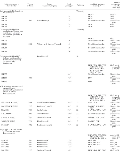
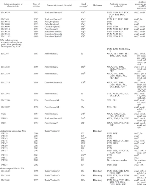
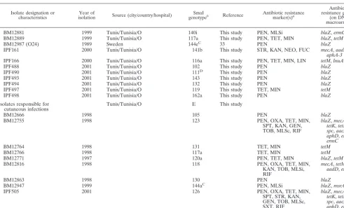
Bacterial isolates.The relevant characteristics of the 80S. aureusclinical isolates used to validate the DNA macroarray designed in this study are given in Table 1. The 44 staphylococcal, enterococcal, andEscherichia colistains used as substrates for PCR amplification of the genes chosen for the construction of the macroarray are reported in Table S1 in the supplemental material at http: //genopole.pasteur.fr/staph/.
DNA extraction.Total cellular DNAs were extracted and purified by use of a QIAamp DNA mini kit (Qiagen, Hilden, Germany). The method described by the supplier was modified by the inclusion of lysostaphin (Applied Microbiol-ogy), at a final concentration of 100 mg/liter, in the lysis step. RNAs were removed after 30 min of incubation at 37°C by the addition of 5 mg of RNase (DNase-free) (Roche, Meylan, France)/liter.
Comparative genome analysis, primer design, and PCR amplification.For the annotation and comparative analysis of the available genome sequences from the sevenS. aureusisolates cited above, the program CAAT-Box (12) was used. Genes whose nucleotide sequences exhibited⬍80% similarity were considered distinct. CAAT-Box uses the BLAST program, which presents the area of least similarity with the rest of the genome. The Primer3 program (http://www.broad .mit.edu/cgi-bin/primer/primer3-www.cg) identifies primer pairs in this specific area which are unlikely to produce nonspecific amplifications with regard to the seven sequencedS. aureusgenomes. The criteria used by CAAT-Box and Prim-er3 were as follows: match threshold, 21; maximum length of nonspecific PCR products, 3,000 bases; minimum PCR product length, 250 bases; optimum PCR product length, 400 to 500 bases; primer size, 18, 20, or 25 bases (minimum, optimum, and maximum sizes); primer melting temperature (Tm), 51, 55, or
60°C; % G⫹C, 25, 50, or 80%; maximum difference inTmfor a primer pair, 5°C.
Each of the 478 selected genes encoded at least 150 amino acids. Primers were designed to amplify a fragment of 400 to 500 bp specific for each gene. Each PCR was performed in a 100-l reaction volume containing 10 to 20 ng of DNA and a 1M concentration of each primer (Eurogentec, Liege, Belgium). The con-ditions used were an initial cycle of 5 min at 94°C, followed by 35 cycles of 1 min at 94°C, 1 min at 50°C, and 1 min at 72°C, with a final extension step of 7 min at 72°C. The concentration and size of each PCR product were verified by electro-phoresis using agarose gels.
Array construction.For array preparation, high-density nylon Performa mem-branes (Genetix, New Milton, United Kingdom) were soaked in TE solution (10 mM Tris [pH 7.6], 1 mM EDTA). Double spot blots of each PCR product were printed (50 ng of DNA in PCR buffer per spot) by a Qpix robot (Genetix). After spot deposition, DNAs were denatured and fixed on the membranes by incuba-tion for 15 min in 0.5 M NaOH–1.5 M NaCl. The membranes were then washed briefly in distilled water and stored wet at⫺20°C until use.
Hybridization.The cellular DNAs of theS. aureusstrains (50 ng) were labeled by use of a random priming DNA labeling kit (Roche Diagnostics GmbH, Penzberg, Germany) and 50Ci of 5⬘-[␣-33P]dCTP (Amersham, Piscataway, N.J.). Labeled probes were purified by use of a QIAquick nucleotide removal kit (Qiagen). The membranes were moistened in 2⫻SSC (0.3 M NaCl, 0.03 M sodium citrate) and prehybridized for 1 h in 10 ml of 5⫻SSPE (0.9 M NaCl, 6 mM NaH2PO4, 7.5 mM EDTA, pH 8), 4% sodium dodecyl sulfate, 1⫻Denhardt solution (0.02% Ficoll, 0.02% polyvinylpyrrolidone, 0.02% bovine serum albu-min), and 1 mg of denatured salmon sperm DNA. Hybridization was performed overnight at 65°C. Membranes were washed twice at room temperature and twice at 65°C in 0.5⫻SSPE–0.2% sodium dodecyl sulfate. Arrays were then sealed in polypropylene bags and exposed to a PhosphorImager screen for 24 h.
Verification of specificity of DNA macroarray.Of the 478 DNA fragments amplified, 106 were randomly chosen and sequenced. Sequencing of the PCR products was done with an ABI3700 capillary sequencer. For a test of correct spotting, the membranes loaded with the amplicons were hybridized with the cellular DNAs of theS. aureusstrains used as substrates in PCR amplifications. For 465 amplicons, the results were as expected, i.e., specific. Thirteen of the 478 genes selected were eliminated, either because two nonspecific DNA bands were amplified (1 gene) or because hybridization experiments revealed false-positive or -negative results (10 and 2 genes, respectively). The characteristics of the amplicons and the strains used as substrates, as well as the sequences of the
primers and their positions on the genome, are shown in Table S1 (http: //genopole.pasteur.fr/staph/).
Data analysis.For scanning, a Typhoon 9400 PhosphorImager (Molecular Dynamics) was used. Array Vision software (Imaging Research) was used for the quantification of the hybridization intensities and for normalization. For each spot, the hybridization intensity value was normalized by dividing it by the average of all significant intensity values on each membrane. For gene content analysis, a reference array was built by combining the average normalized data of two replicate hybridization experiments with the cellular DNAs of the strains used as substrates for PCR amplification. When a gene was known to be present either as a single copy or as multiple copies, the lowest significant intensity value corresponding to a single-copy gene was chosen. When a gene was known to be present in the tested strain used as a substrate, such as in the five strains whose genomes have been sequenced (N315 [19], Mu50 [19], COL [http://www.tigr.org /tdb/], MW2 [2], and NCTC8325 [http://www.genome.ou.edu/staph.html]), the ratio between the normalized signal intensity of the gene hybridized with the tested strain and that of the reference array was always higher than 0.3. Thus, the threshold for the presence of a gene or a variant related by at least 80% similarity was defined as 0.3. The data were then converted into a binary score as follows: atⱖ0.3, a gene was scored as present (score⫽1), and at⬍0.3, a gene was scored as absent (score⫽0).
The binary data were used to cluster the isolates hierarchically, using the program J-Express (9). The threshold adopted to distribute the isolates into clusters was that which enabled each of the outbreak-related isolates belonging to SmaI genotypes 100 or 101 (Table 1) to be grouped and distinguished from any of the other isolates.
Comparative analysis of the gene contents for different categories of isolates.
When categories ofnandmisolates are compared, the probability that a given gene is present by chance inn1isolates of the first category of isolates andn2 isolates of the second category is given by the following binomial formula:
p⫽
冉
nn1
冊冉
m n2
冊
qn1⫹n2共1⫺q兲n⫹m⫺n1⫺n2
whereqis estimated by maximum likelihood, using the equationq⫽(n1⫹n2)/(n
⫹m). A Bayesian approach based on the integration overqwith a uniform prior gives results similar to those presented in the sequel. pgis the normalized
probability, withgrepresenting the total number of genes investigated. The gene distribution was considered significant if the normalized probability, orpg, was
⬍0.10.
RESULTS
Choice of genes for construction of DNA macroarray.Based on a comparative analysis of the seven S. aureus genomes sequenced (N315 [19], Mu50 [19], COL [http://www.tigr.org /tdb/], MW2 [2], NCTC8325 [http://www.genome.ou.edu /staph.html], methicillin-susceptibleS.aureusstrain 476 [http: //www.sanger.ac.uk/Projects/S_aureus/], and EMRSA 16 strain 252 [http://www.sanger.ac.uk/Projects/S_aureus/]), we selected 397 genes for the macroarray. Among these strains, 305 of the genes were not shared by all of them and thus were candidate probes for typing. Although they were shared by the seven sequenced genomes, 92 additional genes were used. They in-cluded genes such asnuc(6) andsodM(34) for identification at the species level, genes encoding putative virulence proteins and factors involved in their regulation, and genes encoding proteins involved in antibiotic transport and resistance expres-sion.
Furthermore, 67 genes that were not detected in these seven
S. aureusgenomes were also spotted on the array because they encoded specific groups of proteins. (i) Genes encoding staph-ylococcal and enterococcal proteins mediating drug resistance were included. Thirteen antibiotic resistance genes were iden-tified in gram-positive species other thanS. aureus, as follows:
Staphylococcus hyicus, tetL; Staphylococcus cohnii, vatC and
fae-TABLE 1. Relevant characteristics of S.aureusclinical isolates
Isolate designation or
characteristics isolationYear of (city/country/hospital)Source genotypeSmaI b Reference Antibiotic resistancemarker(s)a,c
Antibiotic resistance gene(s)
(on DNA macroarrays)
Outbreak-related isolates from
the same ward This study
PEN⫹ blaZ⫹
IPF735 101 MLSi ermC
IPF736 101 MLSi ermC
IPF738 2000 Calais/France/A 101 No additional marker No additional
gene
IPF741 101 MLSi ermC
IPF743 101 MLSi ermC
Outbreak-related isolates producing exfoliative toxin A and responsible for scalded skin syndrome in neonates
This study
PEN⫹ blaZ
IPF308 100 No additional marker No additional
gene IPF310 2001 Villencuve St Georges/France/B 100 No additional marker No additional
gene
IPF311 100 No additional marker No additional
gene
IPF313 100 No additional marker No additional
gene Outbreak-related h-VISAb
isolates, indistinguishable from isolates previously detected at low frequencies
Paris/France/C 14
PEN, OXA, STR, TET, MIN, SPT, KAN, NEO, TOB, GEN, MLSc, PEF, RIF, FUC, h-VISA⫹
blaZ,mecA,tetM,
spc,aadD,aac A-aph D,ermA
IPF555 39aA No additional marker No additional
gene
IPF557 1999 39aA FOF No additional
gene
IPF562 39aA FOF No additional
gene MSRA isolates with decreased
susceptibility to glycopeptides, endemically spread in several European cities
PEN, OXA, STR, TET, MIN, SPT, KAN, NEO, TOB, GEN, MLSc, PEF, RIF⫹
blaZ,mecA,tetM,
spc,aadD,
aacA-aphD,
ermA
BM12612(CIP106757) 1998 Villiers St Denis/France/D 39aA 7 GISA, FOF No additional gene BM10828(CIP106759) 1993 Bordeaux/France/E 39aA 22 h-VISA,bSUL, FUC,
FOF No additionalgene
SPAIN E1 1989 Seville/Spain/- 39aA 23 h h-VISA,bSUL, FUC No additional
gene FINLAND E7 1990 Turku/Finland/- 39bA 23 No additional marker No additional
gene 97130(CIP106761) 1997 Toulouse/France/F 39cA 7 h-VISAb, FUC, FOF No additional
gene
96145(CIP106762) 1996 Blois/France/G 39dA 7 h-VISAb, FOF No additional
gene BM10829 1993 Bordeaux/France/E 39eA 22 h h-VISAb, SUL, FUC No additional
gene Phage-type 77 MSRA isolates
endemically spread in European cities
OXA, STR, TET, MIN, SPT, KAN, TOB, GEN, MLSc, PEF⫹
mecA,tetM,spc,
aacA-aphD,
ermA⫹
BM9290 1987 Paris/France/H 45aA PEN, RIF blaZ
BM9586 1987 Paris/France/C 45aA PEN, RIF, FOF blaZ,fos
BM12184 1987 Paris/France/C 45aA PEN, RIF, FOF blaZ,fos
BM12188 1987 Paris/France/C 45aA RIF No additional
gene
BM10761 1993 Toulouse/France/F 45bA PEN, NEO, RIF, FUC,
FOF, PRI, SGA, SXT
blaZ,aadD
[image:3.603.46.542.80.706.2]TABLE 1—Continued
Isolate designation or
characteristics isolationYear of Source (city/country/hospital) genotypeSmaI b Reference Antibiotic resistancemarker(s)a
Antibiotic resistance gene(s)
(on DNA macroarrays)
BM10759 1993 Toulouse/France/F 45cA PEN, NEO, RIF, FUC,
FOF, PRI, SGA, SXT
blaZ,aadD
BM9343 1987 Toulouse/France/F 45dA PEN, RIF, FUC, FOF blaZ,fos
BM10872 1992 Aalst/Belgium/I 45eA PEN blaZ
BM10888 1993 Aalst/Belgium/I 45eA PEN blaZ
BM10896 1994 Ghent/Belgium/J 45fA PEN, NEO blaZ,aadD
BM10914 1991 Paris/France/C 45fA PEN, NEO, RIF, FOF blaZ,aadD
BM10130 1989 Barcelona/Spain/K 45gA PEN, NEO, RIF blaZ,aadD
BM10138 1989 Barcelona/Spain/K 45gA PEN, NEO, RIF blaZ,aadD
BM12152 1989 Barcelona/Spain/K 45iA PEN, NEO, RIF blaZ,aadD
SGAr isolates whose streptogramin-resistant genes were previously investigated by PCR
PEN, KAN, NEO, SGA
⫹
BM3364 1981 Paris/France/C 13 OXA, TET, MIN, SPT,
TOB, GEN, MLSc blaZspc,mecA,aadE,,tetM,
aacA-aphD,
ermA,aphA-3,
vgaAv,vgaB,
vatB
BM12828 1999 Paris/France/C 16aB OXA, SPT, TOB,
MLSc, PRI, SXT, PEF
mecA,spc,aadD,
ermA,vatA,
vgbA
BM12830 1999 Paris/France/C 16aB OXA, SPT, TOB,
GEN, MLSc, PRI, PEF, RIF
mecA,spc,aadD,
aacA-aphD,
ermA,vatA,
vgbA
BM12714 1996 Grenoble/France/L 17bB OXA, SPT, TOB,
GEN, MLSc, PRI, SXT, PEF, FOF
blaZ,mecA,spc,
aadD, aacA-aphD,ermA,
vgaAv,vatB,
vgaB,dfrA
BM12942 1999 Paris/France/C 19 STR, MLSc, PRI, SUL,
SXT, FUC blaZA-3,aadE,ermC,aph,
vatA,vgbA
BM12286 1996 Paris/France/M 36a STR, PRI blaZ,aadE,aph
A-3,vatA,
vgbA
BM12827 1996 Paris/France/M 36a STR, PRI blaZ,aadE,
aphA-3,vatA,
vgbA
97233 1997 Paris/France/C 24hF OXA, TOB, MLSc,
PRI, SXT, PEF blaZaadD,mecA,vga Av,
IPF083 1998 Toulouse/France/F 24aF OXA, TOB, LIN, PEF blaZ,mecA,
aadD,vga Av
93184 1993 Paris/France/N 26F OXA, TOB, PRI, PEF,
RIF blaZaadD,mecA,vatA, ,
vgbA
Isolates from uninfected NCs Tunis/Tunisia/O This study
IPF139 2000 133 PEN, FOF blaZ,fos
IPF140 2001 139 PEN, blaZ
IPF143 2001 149aC PEN blaZ
IPF145 2001 155a PEN, MLSi, FUC, RIF blaZ,ermC
IPF147 2001 121b PEN, MLSi blaZ,ermC
IPF150 2001 137a PEN blaZ
IPF153 2001 128 PEN, blaZ
IPF157 2001 127 PEN, TET, MIN, STR,
KAN, NEO blaZaadE,tetK,aphA-3;tetM,
IPF159 2001 108D PEN blaZ
IPF511 2001 103 PEN blaZ
IPF520 2002 161 No resistance marker No resistance
gene
IPF524 2002 104 PEN, TET blaZ,tetK
Isolates responsible for BIs
BM12623 1998 Tunis/Tunisia/O 163 This study PEN, TET, STR, KAN
NEO, ERY blaZaphA-3,tetK,,aadEmsrA,
BM12633 1998 Tunis/Tunisia/O 136a This study PEN, STR, KAN NEO,
ERY blaZaphA-3,aadE,,msrA
BM12681 1997 Tunis/Tunisia/O 125a This study PEN, OXA, TET, MIN,
STR, SPT, KAN, GEN, TOB, RIF
blaZ, mecA,tetM,
spc, aacA-aphD,ermA
BM12685 1997 Tunis/Tunisia/O 151a This study PEN blaZ
BM12718 1998 Tunis/Tunisia/O 120a This study PEN, TET, MIN blaZ,tetM
calis,vanBandlsa; andEnterococcus gallinarum,vanC. These genes were chosen because of their possible transfer to S. aureus. (ii) Genes encoding factors known to be involved inS. aureus pathogenicity and structurally related proteins (e.g., toxins, adhesins, and enzymes involved in the biosynthesis of capsule or slime) were also included. (iii) Finally, genes en-coding proteins produced by mobile elements (transposons, insertion sequences, and plasmids) were spotted on the array. The negative control consisted of an amplicon corresponding to theStaphylococcus intermedius-specificnucIgene (6).
Thus, a total of 465 amplicons were spotted on the mem-branes. S. aureus strains N315, Mu50, COL, MW2, and NCTC8325 were used to amplify 385 intragenic fragments. The 80 other genes were previously amplified from 39 other strains (see Table S1 in the supplemental material [http://genopole .pasteur.fr/staph/]).
Distribution of the 465 genes among the 80S. aureusclinical isolates analyzed.The gene content of each of the 80 isolates is given in Table S2 in the supplemental material (http: //genopole.pasteur.fr/staph/). Of the 92 genes shared by the
seven sequenced genomes and used in the macroarray, 76, includingS. aureus nucandsodM, were detected in all isolates analyzed. Therefore, a total of 388 genes of this set were useful for typing.
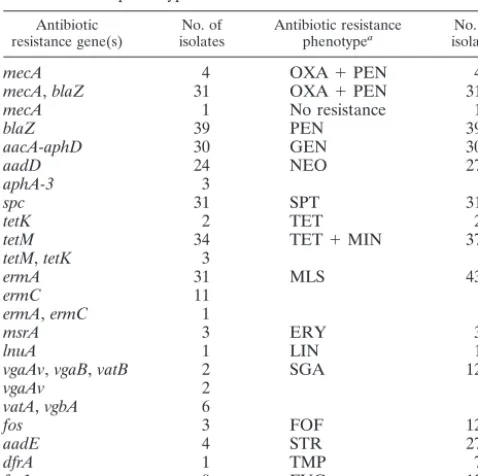
Antibiotic resistance genes and phenotypes.An analysis of the data reported in Table 1 enabled us to check whether the genes detected by hybridization were correlated with their phe-notypic expression in the isolates. As shown in Table 2, for 79 of the 80 isolates, each antibiotic resistance gene detected was associated with the corresponding phenotype. A singlemecA⫹
isolate was susceptible to-lactams.
The streptogramin resistance genes previously found by PCR with the isolates that were resistant to streptogramin A (15) (Table 1) were detectable by hybridization with the DNA macroarrays designed for this study. The intragenic amplicons from vgaA and vgaAv appear to be specific to each variant despite the 83.2% similarity relating them. This is due to the fact that the divergence is distributed along the entire se-quence of the gene variants, without⬎29 consecutive matching nucleotides between the amplicon and the variant gene.
Re-TABLE 1—Continued
Isolate designation or
characteristics isolationYear of Source (city/country/hospital) genotypeSmaI b Reference Antibiotic resistancemarker(s)a
Antibiotic resistance gene(s)
(on DNA macroarrays)
BM12881 1999 Tunis/Tunisia/O 140i This study PEN, MLSi blaZ,ermC
BM12889 1999 Tunis/Tunisia/O 117a This study PEN, TET, MIN blaZ,tetM
BM12987 (O24) 1989 Sweden 144eC 33 PEN blaZ
IPF161 2000 Tunis/Tunisia/O 141b This study STR, KAN, NEO, FUC mecA,aadE,
aphA-3
IPF166 2000 Tunis/Tunisia/O 116a This study PEN, TET, MIN, LIN tetM,lnuA
IPF488 2001 Tunis/Tunisia/O 102 This study PEN blaZ
IPF490 2001 Tunis/Tunisia/O 111D This study PEN blaZ
IPF493 2001 Tunis/Tunisia/O 143 This study PEN blaZ
IPF494 2001 Tunis/Tunisia/O 132 This study PEN blaZ
IPF497 2001 Tunis/Tunisia/O 119 This study TET, MIN tetM
IPF498 2001 Tunis/Tunisia/O 162a This study PEN blaZ
Isolates responsible for
cutaneous infections Tunis/Tunisia/O E This study
BM12666 1998 105 PEN blaZ
BM12755 1998 123 PEN, OXA, TET, MIN,
SPT, KAN, GEN, TOB, MLSc, RIF
blaZ,mecA,
tetK,tetM,
spc, aacA-aphD,ermA,
ermC
BM12764 1998 131 TET, MIN tetM
BM12766 1998 117a TET, MIN tetM
BM12771 1997 120a PEN, TET, MIN blaZ,tetM
BM12816 1998 118 PEN, OXA, TET, MIN,
KAN, TOB, MLSi, RIF
mecA,tetM,
aadD,ermC
BM12863 1998 130 PEN blaZ
BM12947 1999 144aC PEN, MLSi blaZ,msrA
IPF505 2001 126 PEN, OXA, TET, MIN,
SPT, STR, KAN, GEN, TOB, MLSc, SXT, RIF
blaZ,mecA,
tetK,tetM,
spc, aacA-aphD,ermA,
ermC aAbbreviations ERY, erythromycin; FUC, fucidic acid; FOF, fosfomycin; h-VISA, heterogeneous vancomycin-intermediateS. aureus; GEN, gentamicin; GISA,
glycopeptide intermediateS. aureus; KAN, kanamycin; LIN, lincomycin; MLSci, lincosamides-streptogramin B-inducible resistance; MLSc, macrolides-lincosamides-streptogramin B constitutive resistance; MIN, minocycline; NEO, neomycin; OXA, oxacillin; PEF, pefloxacin; PEN, penicillinase; PRI, pristinamycin; RIF, rifampin; SGA, streptogramin A; SPT, spectinomycin; STR, streptomycin; SUL, sulfonamides; SXT, trimethoprim-sulfamethoxazole; TET, tetracycline; TOB, tobramycin.
bStrains were clustered according to the following criteria proposed by Tenover et al. (32). (i) Strains were grouped in the same major genotype if their patterns
differed by no more than three bands (these strains were considered to be closely related and monoclonal). (ii) If patterns differed by between four and six bands, the strains were scored as being possibly related but were nevertheless classified into distinct genotypes to discriminate them from the closely related strains. (iii) If patterns differed by seven or more bands, strains were considered to be different. Major genotypes are designated by arabic numerals. Strains with indistinguishable patterns were classified within the same subtype. Subtypes are designated by arabic numerals with letter suffixes. Genotypes which include strains that are possibly related (less than seven bands with differences) are marked with a superscript letter.
[image:5.603.47.539.79.376.2]sistance to fosfomycin, streptomycin, trimethoprim, and fusidic acid, which can result from mutations in preexisting genes, are rarely associated with acquired genes (Table 2). In contrast, resistance to-lactams, aminocyclitols (except streptomycin), tetracycline, minocycline, macrolides, lincosamides, and strep-togramin B was correlated with the presence of at least one acquired gene (Table 2). Two of the 12 isolates that were resistant to streptogramin A (BM10761 and BM10759; Table 1) did not carry any of the investigated genes encoding resis-tance to this antibiotic.
TheS. aureus fosBgene, included in the arrays because of its similarity tofos, was found in 69 of the isolates, independent of their phenotypes of resistance to fosfomycin.
The combinations of genes carried by the transposons Tn554
(spe,ermA,tnpA, andtnpB), Tn5406(vgaAv,tnpA, andtnpB), and Tn4001 (aacA-aphD and IS256 tnp) were found in the isolates exhibiting the antibiotic resistance phenotypes medi-ated by these transposons. The genesblaZandtnp480, which are cocarried by Tn552, were associated with only 28 of the 70 isolates containing blaZ. As was stated previously (8), the genesaadE,sat4, andaphA-3, initially found in Tn5405, were always combined, and they were found in seven isolates in this study. This last combination was occasionally associated with
other Tn5405genes, i.e.,orfX(two isolates),orfXand IS1182 tnp (four isolates), ororfX, IS1182 tnp, and IS1181 tnp (one isolate).
Distribution of genes inmecAⴙisolates and isolates lacking
mecA.As shown in Table 1, 36 of the 80 tested isolates were
mecA⫹ and 44 lacked mecA. Several genes, including those
coding for antibiotic resistance and putative virulence factors, had a distribution which was significantly different (pg⬍0.1) for the two categories of isolates. The distribution of genes encoding putative toxins or adhesins is reported in Table 3. Interestingly, the enterotoxin-encoding genesseg,sei,sem,sen, andseo, codetected in the same pathogenicity island of theS. aureus N315 and Mu50 strains (19), were always associated with each other in our isolates and were significantly predom-inant in themecA-negative isolates (30 of 44 isolates) compare to themecA⫹isolates (1 of 36 isolates).
Distribution of genes in 16 BI isolates and 12 NC isolates.
Unrelated isolates were selected for a comparative analysis of BIs and nasal carriers (NCs) (Table 1). No significant differ-ences in the gene contents were observed between the BI and NC isolates when the 388 genes were taken into account for calculations of the probability that a given gene is present by chance. However, taking into account only 11 genes that were not shared by all isolates and that encode adhesins (sdrD,sdrC,
fnbA,fnbB, efb,map, cna,bbp, vwb, bap, andebpS), the two categories of isolates became significantly distinguishable (pg⫽ 0.059) by the presence of the sdrD gene, which codes for a putative SD (serine-aspartate) adhesin (18) and was detected in 15 of the 16 BI isolates compared to 7 of the 12 NC isolates.
Clustering of the 80S. aureusclinical isolates on the basis of their gene contents, as investigated with the DNA macroarray designed for this study.The hierarchical clustering of the iso-lates by neighbor joining is represented in the dendrogram shown in Fig. 1. First we checked whether the outbreak-related isolates (shown in gray boxes in the figure) were more closely linked to each other than to any of the other isolates.
[image:6.603.43.282.88.326.2]Within SmaI genotype 100 or 101 (Table 1), the isolates were more closely linked to each other. These isolates were included in this study because they were responsible for doc-umented acute outbreaks in the hospitals of Villeneuve St. Georges and Calais, France, respectively. Such isolates were not detected in the hospitals before the outbreaks. An analysis of their gene contents revealed the absence of two or seven widespread genes, respectively, which were detected in at least 84% of the other isolates. The four SmaI type 100 isolates lackedfnbBand MW2409, while the five SmaI type 101 isolates lacked set14, lukM, splcC, splD, vwb, emp, and SA0276. The absence of widespread genes confirmed the hypothesis of a close relationship between the isolates belonging to each of the two SmaI genotypes. As was found previously by PCR, the four
TABLE 2. Antibiotic resistance genes and their corresponding phenotypes in each of the 80 isolates
Antibiotic
resistance gene(s) isolatesNo. of Antibiotic resistancephenotypea isolatesNo. of
mecA 4 OXA⫹PEN 4
mecA,blaZ 31 OXA⫹PEN 31
mecA 1 No resistance 1
blaZ 39 PEN 39
aacA-aphD 30 GEN 30
aadD 24 NEO 27
aphA-3 3
spc 31 SPT 31
tetK 2 TET 2
tetM 34 TET⫹MIN 37
tetM,tetK 3
ermA 31 MLS 43
ermC 11
ermA,ermC 1
msrA 3 ERY 3
lnuA 1 LIN 1
vgaAv,vgaB,vatB 2 SGA 12
vgaAv 2
vatA,vgbA 6
fos 3 FOF 12
aadE 4 STR 27
dfrA 1 TMP 7
far1 0 FUC 12
aSee Table 1 for explanation of abbreviations. The phenotypes which are
conferred by acquired genes inS. aureusare reported.
TABLE 3. Comparative analysis of themecA⫹andmecA-negative isolates included in this study
Category isolatesNo. of No. ofgenotypesSmaI No. of clusters (basedon gene content)
No. of isolates harboring gene(s)a seg,sei,sem,
sen,seo entA cna bbp (SAV2595)sask
mecA⫹ 36 12 20 1 32 4 10 3
mecAmutant 44 33 32 30 9 20 38 21
aThep
[image:6.603.44.547.654.716.2]isolates from Villeneuve St. Georges, responsible for scalded skin syndrome in newborns (Table 1), carried the eta gene encoding the exfoliative toxin A. Moreover, the single SmaI 101 isolate that was distinguishable from the other four SmaI 101 isolates by its susceptibility to erythromycin lacked the
ermCgene that was present in the latter isolates (Table 1). The other outbreak-related isolates belonged to SmaI sub-type 39aA(IPF 555, IPF 557, and IPF 562) (14) or 45aA(BM
9586, BM 12184, nad BM 12188) (22) and were isolated in
hospital C (Paris) in 1999 and 1987, respectively (Table 1). Isolates belonging to SmaI genotype 39Awere phenotypically
recognizable because of their decreased susceptibility to gly-copeptides. Those belonging to SmaI genotype 45Aand phage
[image:7.603.111.477.123.572.2]that were isolated in several European countries and at time intervals of several years. These endemic isolates, which are considered putatively related according to their SmaI geno-types, were more linked to each other than to any of the 56 other isolates (Fig. 1). Note that some of them are clearly divergent in the dendrogram and that the mode of their link-age is not correlated to their SmaI genotype, but those con-sidered to be outbreak related are closely linked.
Clustering of the 80 clinical isolates after choice of thresh-old for hierarchical clustering dendrogram.For the distribu-tion of the isolates into clusters, it was necessary to choose a threshold for the dendrogram. For this purpose, the threshold adopted was that which enabled each of the outbreak-related isolates belonging to SmaI genotype 100 or 101 to be distin-guished from any of the other isolates. These isolates were taken into consideration because they were not detected be-fore the outbreaks, in contrast to the SmaI subtype 39aA or
45aA outbreak-related isolates. The choice of this threshold
enabled the discrimination of 52 clusters belonging to 45 SmaI genotypes among the 80 isolates (Fig. 1). In Table S2 in the supplemental material (http://genopole.pasteur.fr/staph/), the genes are listed according to the clusters to which they belong. With the selected threshold, a total of five clusters were found among the 10 SmaI type 39Aisolates and eight clusters
were found among the 14 SmaI type 45A isolates (Fig. 1).
Among these isolates, which are endemic in European cities, those collected in the same hospital or city were not necessarily the most closely linked. The three outbreak-related SmaI sub-type 39aAisolates collected in hospital C (Paris) in 1999 (IPF
555, IPF 557, and IPF 562) are linked in cluster 41, which includes another SmaI subtype 39aAisolate (BM 12612)
col-lected at Villiers St. Denis in 1998. Moreover, four of five isolates belonging to two SmaI subtypes, 45aAand 45dA, and
collected in three French hospitals in 1987 are within cluster 46 (BM 9290, BM 9343, BM 9586, and BM 12184). The fifth isolate, BM 12188, located in the separate but close cluster 47, was distinguishable by the lack of five drug resistance genes, namelyblaZ,qacA,qacC,CZ040, andCZ041, encoding -lac-tamase, resistance to antiseptics, organomercurial lyase, and mercuric reductase, respectively. Figure 2 shows the images resulting from scanning of the two DNA macroarrays hybrid-ized with the total cellular DNAs from the BM9290 and BM12188 isolates (Table 1).
Each of the isolates linked in clusters 6, 13, 37, and 40 belonged to the same SmaI genotypes. In contrast, the isolates linked in clusters 11, 14, and 33 belonged to unrelated SmaI genotypes, and cluster 31 contained two distinct but related SmaI genotypes. In addition, isolates with the same SmaI genotype, if it was 117 or 144, were separated. Note that the two isolates be-longing to SmaI genotype 144 had no epidemiological link since they were from distinct sources (Tunisia and Sweden) and were collected over a 10-year time interval. For this last case, the use of the DNA macroarray is more appropriate than the analysis of SmaI patterns for discrimination between the two isolates.
DISCUSSION
DNA macroarrays offer a rapid, robust, and easily standard-izable method for the simultaneous detection of several hun-dred genes of interest and may be used for analyses of
tran-scriptional expression in isolates grown under different in vitro and in vivo conditions. The 465 genes spotted on the DNA macroarray used in this study were chosen as probes in order to identifyS. aureusat the species level and to typeS. aureus
isolates. They included, in particular, genes encoding antibiotic resistance and putative virulence factors.
The detection of antibiotic resistance genes is particularly interesting when these genes mediate low antibiotic resistance levels that are not reproducibly detectable by antibiograms. This level of detection also contributes to the selection of isolates that carry genes that have not yet been described. By hybridization with 400- to 500-bp amplicons, mutations in pre-existing genes associated with antibiotic resistance cannot be visualized and would necessitate hybridization with oligonucle-otides. For 79 of the 80 clinical isolates tested, the resistance phenotype conferred by each of the detected resistance genes was expressed, whereas onemecA⫹isolate was susceptible to
-lactams. This high correlation demonstrated an extensive and satisfactory choice of antibiotic resistance genes spotted on the membranes. For the two related streptogramin A-resis-tant isolates, the lack of any known staphylococcal or entero-coccal gene conferring resistance to this antibiotic is probably due to the presence of a gene(s) that has not yet been de-scribed.
The assessment of the presence of all knownS. aureusgenes encoding putative virulence factors may contribute to the de-termination of the pathogenic potential correlated with partic-ular types of infection and to the identification of emerging pathotypes. In this study, we checked whether some genes were more prevalent in isolates responsible for BIs than in isolates from uninfected NCs. For this purpose, only unrelated isolates from our collection were included. This constraint explains why the numbers of isolates analyzed were 16 BI isolates and 12 NC isolates. Despite the fact that BIs were contracted by children outside the hospital, several patients were infected by
S. aureusisolates that were considered monoclonal on the basis of their SmaI patterns. Although a few genes, includingsdrD, encoding a putative SD adhesin, appeared predominant in one of the two categories of isolates, the differences were not sig-nificant when the 388 genes used for typing were taken into account for the calculation of the probability that a given gene is present by chance. Thus, a larger number of unrelated iso-lates from various sources merits further analysis. However, when only the 11 genes encoding putative adhesins were taken into account, the higher prevalence ofsdrDin BI isolates than in NC isolates became significant. Some SD proteins were shown to bind fibrinogen (ClfA [21], ClfB [25], and SdrG [16]) or bone sialoprotein (Bbp) (33), but the ability of SdrD to bind a matrix protein(s) has not been investigated. The impact of
sdrDinactivation merits evaluation in an animal model of BIs. The significantly distinct distribution of some genes encod-ing enterotoxins or adhesins among the mecA⫹ and mecA
-negative isolates in this study (Table 3) may not be the case among isolates from various sources. Indeed, most of the 80 isolates tested were collected in France and Tunisia, and the
mecA⫹isolates belonged to a limited number of SmaI
geno-types. Nevertheless, the low frequency of cna detection in
mecA⫹isolates has been reported already by Booth et al. (5).
analysis of a large number of genes was expected to yield more discrimination between the isolates than the typing methods based on sequencing of a limited number of genes or on the analysis of SmaI patterns, which depends on the number and locations of SmaI sites in the genome. This was confirmed by this study, for the mecA⫹ isolates were endemic to several
European cities and were collected at large time intervals
(SmaI genotypes 39A and 45A). Among the latter isolates,
those considered to be outbreak related in the same hospital were found in the same or in a close cluster(s): cluster 41 or 46-47. In such a context, the typing method proposed in this study provides more discrimination of the isolates responsible for acute outbreaks than the determination of SmaI patterns. For the other isolates, if we excluded the three pairs which
FIG. 2. Images resulting from scanning of the DNA macroarrays hybridized with the total cellular DNAs from two isolates. (A) Isolate BM9290 (cluster 46). (B) Isolate BM12188 (cluster 47). Even though they belonged to the same SmaI subtype (45aA), the two isolates were found in two
were linked in the same cluster despite belonging to unrelated SmaI types, our results revealed a correlation between the modes of isolate clustering based on the two typing methods, i.e., the analysis of gene contents and the SmaI patterns. In-deed, the isolates belonging to the same or related SmaI types appeared to be more linked to each other than to those be-longing to unrelated SmaI types.
In conclusion, the typing method proposed here performed better than that based on the analysis of SmaI patterns, in particular for distinguishing outbreak-related isolates from those that are endemic to a particular area. It also has the advantages of being faster and providing additional informa-tion concerning the gene contents of interest. This macroarray should be updated when additional genes are described and also needs to be validated for the analysis of the transcription of genes in order to evaluate the levels of gene expression which may be correlated with particular types of infections. The method described here can also be performed with glass slides and fluorescent labeling in order to be more amenable to automation for routine analyses.
ACKNOWLEDGMENTS
For part of this work, S. Trad received a grant from Fondation pour la Recherche Me´dical (FRM). The isolate MW2 was obtained through the Network on Antimicrobial Resistance in Staphylococcus aureus (NARSA) Program supported under NIAID, NIH, contract no. N01-AI-95359.
We thank the biologists and NARSA who provided several of the strains used in this study and Iain Old for reviewing the manuscript.
REFERENCES
1. Anonymous.2002. First U.S. case of vancomycin-resistantStaphylococcus aureusinfection reported; patient has chronic renal failure. Dialysis Trans-plant.2002:602–603.
2. Baba, T., F. Takeuchi, M. Kuroda, H. Yuzawa, K. Aoki, A. Oguchi, Y. Nagai, N. Iwama, K. Asano, T. Naimi, H. Kuroda, L. Cui, K. Yamamoto, and K. Hiramatsu.2002. Genome and virulence determinants of high virulence community-acquired MRSA. Lancet359:1819–1827.
3. Becker, K., A. W. Friedrich, G. Lubritz, M. Weilert, G. Peters, and C. Von Eiff.2003. Prevalence of genes encoding pyrogenic toxin superantigens and exfoliative toxins among strains ofStaphylococcus aureusisolated from blood and nasal specimens. J. Clin. Microbiol.41:1434–1439.
4. Berger-Ba¨chi, B.1997. Resistance not mediated by-lactamase (methicillin-resistance), p. 158–174.InK. B. Crossley and G. L. Archer (ed.), The staphylococci in human disease. Churchill Livingstone, New York, N.Y. 5. Booth, M. C., L. M. Pence, P. Mahasreshti, M. C. Callegan, and M. S.
Gilmore.2001. Clonal associations amongStaphylococcus aureusisolates from various sites of infection. Infect. Immun.69:345–352.
6. Chesneau, O., J. Allignet, and N. El Solh.1994. Three thermonuclease gene probes designed for rapid identification ofS. aureus,S. hyicus, andS. inter-medius, p. 83–85.InR. Mo¨llby, J.-I. Flock, C. E. Nord, and B. Christensen (ed.), Staphylococci and staphylococcal infections. Gustav Fischer Verlag, Stuttgart, Germany.
7. Chesneau, O., A. Morvan, and N. E. Solh.2000. Retrospective screening for heterogeneous vancomycin resistance in diverse Staphylococcus aureus clones disseminated in French hospitals. J. Antimicrob. Chemother.45:887– 890.
8. Derbise, A., S. Aubert, and N. El Solh.1997. Mapping the regions carrying the three contiguous antibiotic resistance genesaadE,sat4, andaphA-3in the genomes of staphylococci. Antimicrob. Agents Chemother.41:1024– 1032.
9. Dysvik, B., and I. Jonassen.2001. J-Express: exploring gene expression data using Java. Bioinformatics17:369–370.
10. Enright, M. C., N. P. Day, C. E. Davies, S. J. Peacock, and B. G. Spratt.2000. Multilocus sequence typing for characterization of methicillin-resistant and methicillin-susceptible clones ofStaphylococcus aureus. J. Clin. Microbiol.
38:977–986.
11. Fitzgerald, J. R., D. E. Sturdevant, S. M. Mackie, S. R. Gill, and J. M. Musser.2001. Evolutionary genomics of Staphylococcus aureus: insights into the origin of methicillin-resistant strains and the toxic shock syndrome epi-demic. Proc. Natl. Acad. Sci. USA98:8821–8826.
12. Frangeul, L., P. Glaser, C. Rusniok, C. Buchrieser, E. Duchaud, P. Dehoux, and F. Kunst.2004. CAAT-box, contigs-assembly and annotation tool-box for genome sequencing projects. Bioinformatics20:790–797.
13. Grundmann, H., S. Hori, M. C. Enright, C. Webster, A. Tami, E. J. Feil, and T. Pitt.2002. Determining the genetic structure of the natural population of
Staphylococcus aureus: a comparison of multilocus sequence typing with pulsed-field gel electrophoresis, randomly amplified polymorphic DNA anal-ysis, and phage typing. J. Clin. Microbiol.40:4544–4546.
14. Guerin, F., A. Buu-Hoi, J. Mainardi, G. Kac, N. Colardelle, S. Vaupre, L. Gutmann, and I. Podglajen.2000. Outbreak of methicillin-resistant Staphy-lococcus aureuswith reduced susceptibility to glycopeptides in a Parisian hospital. J. Clin. Microbiol.38:2985–2988.
15. Haroche, J., A. Morvan, M. Davi, J. Allignet, F. Bimet, and N. El Solh.2003. Clonal diversity among streptogramin A-resistantStaphylococcus aureus iso-lates collected in French hospitals. J. Clin. Microbiol.41:586–591. 16. Hartford, O., L. O’Brien, K. Schofield, J. Wells, and T. Foster.2001. The Fbe
(SdrG) protein of Staphylococcus epidermidis HB promotes bacterial ad-herence to fibrinogen. Microbiology147:2545–2552.
17. Hiramatsu, K.2001. Vancomycin-resistant Staphylococcus aureus: a new model of antibiotic resistance. Lancet Infect. Dis.1:147–155.
18. Josefsson, E., D. O’Connell, T. Foster, I. Durussel, and J. A. Cox.1998. The binding of calcium to the B-repeat segment of SdrD, a cell surface protein of Staphylococcus aureus. J. Biol. Chem.273:31145–31152.
19. Kuroda, M., T. Ohta, I. Uchiyama, T. Baba, H. Yuzawa, I. Kobayashi, L. Cui, A. Oguchi, K. Aoki, Y. Nagai, J. Lian, T. Ito, M. Kanamori, H. Matsumaru, A. Maruyama, H. Murakami, A. Hosoyama, Y. Mizutani-Ui, N. K. Taka-hashi, T. Sawano, R. Inoue, C. Kaito, K. Sekimizu, H. Hirakawa, S. Kuhara, S. Goto, J. Yabuzaki, M. Kanehisa, A. Yamashita, K. Oshima, K. Furuya, C. Yoshino, T. Shiba, M. Hattori, N. Ogasawara, H. Hayashi, and K. Hira-matsu.2001. Whole genome sequencing of methicillin-resistant Staphylo-coccus aureus. Lancet357:1225–1240.
20. McCormick, J. K., J. M. Yarwood, and P. M. Schlievert.2001. Toxic shock syndrome and bacterial superantigens: an update. Annu. Rev. Microbiol.
55:77–104.
21. McDevitt, D., P. Francois, P. Vaudaux, and T. Foster.1994. Molecular characterization of the clumping factor (fibrinogen receptor) of Staphylo-coccus aureus. Mol. Microbiol.11:237–248.
22. Morvan, A., S. Aubert, C. Godard, and N. El Solh.1997. Contribution of a typing method based on IS256-probing ofSmaI-digested cellular DNA to discrimination of European phage-type 77 methicillin-resistant Staphylococ-cus aureusstrains. J. Clin. Microbiol.35:1415–1423.
23. Murchan, S., M. Kaufmann, A. Deplano, R. de Ryck, M. Struelens, C. E. Zinn, V. Fussing, S. Salmenlinna, J. Vuopio-Varkila, N. El Solh, C. Cuny, W. Witte, P. Tassios, N. Legakis, W. van Leeuwen, A. van Belkum, A. Vindel, I. Laconcha, J. Garaizar, S. Haeggman, B. Olsson-Liljequist, U. Ransjo, G. Coombes, and B. Cookson.2003. Harmonization of pulsed-field gel electro-phoresis protocols for epidemiological typing of strains of methicillin-resis-tantStaphylococcus aureus: a single approach developed by consensus in 10 European laboratories and its application for tracing the spread of related strains. J. Clin. Microbiol.41:1574–1585.
24. Musser, J. M., and R. K. Selander.1990. Genetic analysis of natural popu-lations ofStaphylococcus aureus, p. 59–67.InR. P. Novick (ed.), Molecular biology of the staphylococci. VCH Publishers, New York, N.Y.
25. Ni Eidhin, D., S. Perkins, P. Francois, P. Vaudaux, M. Hook, and T. Foster.
1998. Clumping factor B (ClfB), a new surface-located fibrinogen-binding adhesin of Staphylococcus aureus. Mol. Microbiol.30:245–257.
26. Novick, R. P.2003. Autoinduction and signal transduction in the regulation of staphylococcal virulence. Mol. Microbiol.48:1429–1449.
27. Oliveira, D. C., A. Tomasz, and H. de Lencastre.2001. The evolution of pandemic clones of methicillin-resistant Staphylococcus aureus: identifica-tion of two ancestral genetic backgrounds and the associated mec elements. Microb. Drug Resist.7:349–361.
28. Paulsen, I. T., N. Firth, and R. A. Skurray.1997. Resistance to antimicrobial agents other than-lactams, p. 175–212.InK. B. Crossley and G. L. Archer (ed.), The staphylococci in human disease. Churchill Livingstone, New York, N.Y.
29. Peacock, S. J., C. E. Moore, A. Justice, M. Kantzanou, L. Story, K. Mackie, G. O’Neill, and N. P. Day.2002. Virulent combinations of adhesin and toxin genes in natural populations ofStaphylococcus aureus. Infect. Immun.70:
4987–4996.
30. Projan, S. J., and R. P. Novick.1997. The molecular basis of pathogenicity, p. 55–81.InK. B. Crossley and G. L. Archer (ed.), The staphylococci in human disease. Churchill Livingstone, New York, N.Y.
31. Sabat, A., J. Krzyszton-Russjan, W. Strzalka, R. Filipek, K. Kosowska, W. Hryniewicz, J. Travis, and J. Potempa.2003. New method for typing Staph-ylococcus aureusstrains: multiple-locus variable-number tandem repeat anal-ysis of polymorphism and genetic relationships of clinical isolates. J. Clin. Microbiol.41:1801–1804.
33. Tung, H., B. Guss, U. Hellman, L. Persson, K. Rubin, and C. Ryden.2000. A bone sialoprotein-binding protein from Staphylococcus aureus: a member of the staphylococcal Sdr family. Biochem. J.345:611–619.
34. Valderas, M. W., J. W. Gatson, N. Wreyford, and M. E. Hart.2002. The superoxide dismutase gene sodMis unique to Staphylococcus aureus: absence ofsodMin coagulase-negative staphylococci. J. Bacteriol.184:
2465–2472.
35. van Leeuwen, W., C. Jay, S. Snijders, N. Durin, B. Lacroix, H. A. Verbrugh, M. C. Enright, A. Troesch, and A. van Belkum.2003. Multilocus sequence typing ofStaphylococcus aureuswith DNA array technology. J. Clin. Micro-biol.41:3323–3326.