expansion and function of adoptively
transferred antitumor CD8
+
T cells
Claudia Wrzesinski, … , Steven A. Rosenberg, Nicholas P.
Restifo
J Clin Invest.
2007;
117(2)
:492-501.
https://doi.org/10.1172/JCI30414
.
Depleting host immune elements with nonmyeloablative regimens prior to the adoptive
transfer of tumor-specific CD8
+T cells significantly enhances tumor treatment. In the current
study, superior antitumor efficacy was achieved by further increasing the intensity of
lymphodepletion to a level that required HSC transplantation. Surprisingly, the HSC
transplant and not the increased lymphodepletion caused a robust expansion of adoptively
transferred tumor-specific CD8
+T cells. The HSC-driven cell expansion of effector, but not
of naive, CD8
+T cells was independent of in vivo restimulation by MHC class I–expressing
APCs. Simultaneously, HSCs also facilitated the reconstitution of the host lymphoid
compartment, including inhibitory elements, not merely via the production of progeny cells
but by enhancing the expansion of cells that had survived lymphodepletion. Profound
lymphodepletion, by myeloablation or by genetic means, focused the nonspecific HSC
boost preferentially toward the transferred tumor-specific T cells, leading to successful tumor
treatment. These findings indicate that CD8
+T cell–mediated tumor responses can be
efficiently driven by HSCs in the myeloablative setting and have substantial implications for
the design of new antitumor immunotherapies.
Research Article
Oncology
Find the latest version:
Research article
492 The Journal of Clinical Investigation http://www.jci.org Volume 117 Number 2 February 2007
Hematopoietic stem cells promote the
expansion and function of adoptively
transferred antitumor CD8
+
T cells
Claudia Wrzesinski, Chrystal M. Paulos, Luca Gattinoni, Douglas C. Palmer, Andrew Kaiser, Zhiya Yu, Steven A. Rosenberg, and Nicholas P. Restifo
National Cancer Institute, NIH, Bethesda, Maryland, USA.
Depleting host immune elements with nonmyeloablative regimens prior to the adoptive transfer of
tumor-specific CD8
+T cells significantly enhances tumor treatment. In the current study, superior antitumor efficacy
was achieved by further increasing the intensity of lymphodepletion to a level that required HSC
transplan-tation. Surprisingly, the HSC transplant and not the increased lymphodepletion caused a robust expansion
of adoptively transferred tumor-specific CD8
+T cells. The HSC-driven cell expansion of effector, but not of
naive, CD8
+T cells was independent of
in vivo restimulation by MHC class I–expressing APCs. Simultaneously,
HSCs also facilitated the reconstitution of the host lymphoid compartment, including inhibitory elements,
not merely via the production of progeny cells but by enhancing the expansion of cells that had survived
lym-phodepletion. Profound lymphodepletion, by myeloablation or by genetic means, focused the nonspecific
HSC boost preferentially toward the transferred tumor-specific T cells, leading to successful tumor treatment.
These findings indicate that CD8
+T cell–mediated tumor responses can be efficiently driven by HSCs in the
myeloablative setting and have substantial implications for the design of new antitumor immunotherapies.
Introduction
The adoptive cell transfer (ACT) of ex vivo activated autologous tumor-reactive T cells is currently one of the most promising approaches for the treatment of patients with advanced mela-noma (1–3). Immunization with therapeutic cancer vaccines can substantially increase the number of circulating tumor-reactive CD8+ CTLs and trigger tumor reduction in some cases, but
nei-ther the numbers nor the functions of these cells correlate with objective clinical responses (4). These findings may indicate that the T cells induced by vaccination are functionally tolerized. The growth of tumor-reactive T cells ex vivo, unencumbered by the mechanisms that enforce functional tolerance in vivo, enables these self/tumor antigen–specific cells to become fully activated (5, 6). The ex vivo preparation of tumor-specific T cells for adop-tive transfer also makes it possible to deplete host lymphocytes prior to the administration of antitumor T cells. Lymphodeple-tion prior to the transfer of tumor-reactive CD8+ T cells has been
shown to enhance tumor destruction in mice (7–10). When these findings were translated to human trials, 51% of the patients experienced an objective response (11). In the setting of adoptive transfer for the treatment of solid tumors, the current lymphode-pleting regimens are termed “nonmyeloablative” because they transiently deplete lymphocytes and do not lethally damage the host bone marrow and thus do not require HSC transfer (9, 12).
A lymphopenic environment can be induced by depleting endogenous immune cells via total body irradiation (TBI) or che-motherapy. The numbers of endogenous lymphocytes are tightly regulated by the host, and destruction of lymphocytes triggers the
homeostatic correction of the size of the lymphocyte compart-ment (13, 14). CD8+ T cells that are expanding into a
lymphope-nic environment acquire a memory-like phenotype and express high levels of CD44, Ly6C, and CD122 (15, 16). Lymphocyte activation after immunodepletion is also seen in the setting of adoptively transferred T cells, where this activation is due, in part, to the increased availability of homeostatic cytokines that result from the removal of endogenous host cells acting as competi-tors for these cytokines (9). The specific homeostatic cytokines that play major roles include IL-7 and IL-15, cytokines that uti-lize the common γ chain receptor (γC) (14). Decreases in levels of
host inhibitory elements, such as CD4+CD25+Foxp3+ Tregs, also
facilitate the activation of adoptively transferred T cells (17, 18). Evidence for the increased activation of adoptively transferred T cells into immunodepleted hosts includes results of ex vivo analy-ses of IFN-γ release and tumor cytolysis and in vivo measurement of tumor destruction (9, 15, 16). The signal strength required to trigger proliferation and activation is considerably reduced, and low-affinity antigens such as self antigens and tumor antigens become sufficient triggering signals (16, 19, 20).
Although ACT is a promising approach for the treatment of advanced melanoma patients, there is substantial opportunity for improvement (1, 21). Since nonmyeloablative lymphodeple-tion prior to ACT has been shown to improve tumor treatment outcome in mice and humans, we sought to explore whether we could significantly augment tumor destruction by increasing the intensity of lymphodepletion beyond the level currently used in ACT clinical trials. We employed the pmel-1 TCR-Tg mouse model wherein CD8+ T cells recognize the mouse gp100 tumor antigen,
as this antigen is often a target in human clinical trials (6). As a tumor model, we treated established B16 tumors, which express the gp100 melanoma antigen. Prior to the adoptive transfer of pmel-1 CD8+ T cells, the intensity of the lymphodepletion used
to precondition the host was increased to a level that required an
Nonstandard abbreviations used: ACT, adoptive cell transfer; β2m, β2
-microglobu-lin; MSC, myeloid suppressor cell; NT, no treatment; rh2, recombinant human IL-2; TBI, total body irradiation; TIL, tumor-infiltrating lymphocyte.
Conflict of interest: The authors have declared that no conflict of interest exists. Citation for this article:J. Clin. Invest.117:492–501 (2007). doi:10.1172/JCI30414.
HSC (lin–/c-kit+) transplant to rescue the mice from lethality. We
found that increasing the intensity of lymphodepletion can result in enhanced tumor destruction, a result that has major implica-tions for the design of current ACT-based immunotherapies. Transplantation of HSCs, as part of the myeloablative regimen, significantly enhanced the expansion and the antitumor impact of adoptively transferred self/tumor antigen–reactive T cells. Based upon this central observation, we investigated the requirements of HSC-driven lymphocyte expansion.
Results
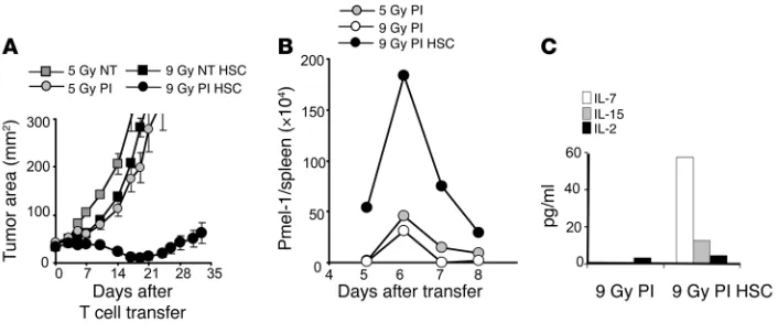
Myeloablative TBI with HSC transplant significantly enhances adoptive T cell therapy. We have previously reported that a nonmyeloabla-tive conditioning regimen delivered prior to the adopnonmyeloabla-tive transfer of antitumor T cells and IL-2 results in a 51% objective response in patients with metastatic melanoma (12). We hypothesized that the efficiency of adoptively transferred CD8+ T cells would
be enhanced by increasing the intensity of immunodepletion. To maximize lymphopenia, B16 tumor–bearing mice received a mye-loablative dose of TBI (9 Gy) accompanied by a transplantation of 105 lin–/c-kit+ syngeneic HSCs. Adoptive transfer of preactivated
transgenic pmel-1 CD8+ T cells mediated tumor regression that
was strikingly better after a myeloablative preparative regimen (9 Gy) than after a preparative regimen that spared the bone mar-row (5 Gy) (P = 0.0036, 5 Gy versus9 Gy; Figure 1A).
Proliferation of transferred CD8+ T cells into a myeloablated host is driven
by the HSC transplant. To evaluate the mechanism accounting for increased tumor treatment in mice receiving ACT after TBI and HSC transplant, the persistence of transferred pmel-1 CD8+ T cells in vivo
was assessed. The numbers of pmel-1 CD8+ T cells recovered from
the spleens of mice receiving the 9 Gy plus HSCs regimen were greater than those found in mice receiving 5 Gy (Figure 1B). We hypothesized that increasing the intensity of lymphode-pletion would enhance the expan-sion of the adoptively transferred T cells by the more complete elimina-tion of regulatory host elements and cytokine sinks. However, the mere reduction in the levels of regulatory host elements and cytokine sinks was not sufficient to increase T cell proliferation (Figure 1B). Instead, it appeared that the HSC transplant, rather than the increased ablation per se, was driving the expansion of the adoptively transferred pmel-1 CD8+ T cells (Figure 1B). We also
observed that elevated serum levels of IL-7 and IL-15, key regulators of homeostatic CD8+ T cells expansion,
were only measurable when HSCs were administered in the myeloabla-tive setting. Increased cytokine levels were transient, peaking 5 days after the HSC transplant in myeloablated animals (Figure 1C). Given our find-ing that the augmented prolifera-tion of adoptively transferred pmel-1 CD8+ T cells was driven not by a more intense lymphodepletion but
by the activities of the HSCs after the 9-Gy regimen, we sought to further analyze the requirements for this proliferation.
HSC-driven T cell proliferation and tumor response are not affected by the activation state of transferred T cells. In our previous work, the antitumor effectiveness of tumor-specific pmel-1 CD8+ T cells was
dependent on stimulation with cognate antigen in vivo, regardless of whether the pmel-1 CD8+ T cells were naive or activated prior
to their adoptive transfer (6, 22, 23). While lymphodepletion with a nonmyeloablating (5-Gy) regimen prior to ACT improved the efficacy of tumor treatment, antigen stimulation in vivo by a vac-cine remained an absolute requirement for tumor treatment (9). However, vaccine was not necessary in order to induce significant tumor treatment when we transferred pmel-1 CD8+ T cells into
mice receiving a more intensive preparative regimen (9 Gy TBI) that were reconstituted with an HSC transplant (Supplemental Figure 1; supplemental material available online with this article; doi:10.1172/JCI30414DS1). It appeared that adoptively trans-ferred pmel-1 CD8+ T cells that had been preactivated in vitro
became functionally activated in the absence of antigen-specific vaccination in myeloablated animals. Because the experiments represented in Figure 1 employed CD8+ pmel-1 CD8+ T cells that
had been stimulated with antigen prior to their adoptive transfer, it remained unclear whether preactivation in vitro was required for HSC-driven expansion. Thus, we evaluated the proliferation and the antitumor efficacy of pmel-1 CD8+ T cells that were naive or
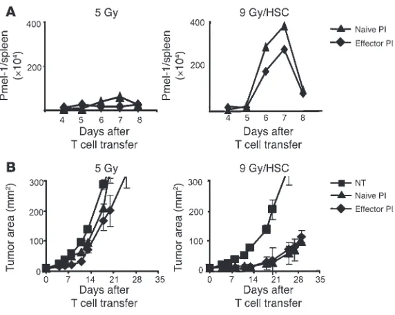
preactivated in vitro. In nonmyeloablated (5 Gy) control mice, little proliferation of naive or effector pmel-1 CD8+ T cells was observed
(Figure 2A, left panel). In contrast, significant proliferation was seen in myeloablated hosts when pmel-1 CD8+ T cells were
trans-Figure 1
Myeloablative TBI (9 Gy) with HSC transplant significantly enhances ACT and drives T cell expan-sion. (A) Increasing a nonmyeloablative regimen to a myeloablative one augmented ACT-mediated
tumor treatment. C57BL/6 tumor-bearing mice were irradiated with 5 Gy or 9 Gy and received an HSC transplant (HSC); were left untreated as a control (NT); or received transfer of 1 × 10⁶ effector pmel-1
CD8+ T cells and rhIL-2 (PI). Tumor treatment efficacy was strongly improved in myeloablated mice
(P = 0.0036; 5 Gy PI versus 9 Gy PI HSC). Results for tumor area are the mean of measurements from 6 mice per group (±SEM). Data are representative of 3 independent experiments. (B) HSC transplant
drives the proliferation of transferred pmel-1 CD8+ T cells in a myeloablated host. Cultured
gene-marked (Thy1.1+) pmel-1 CD8+ T cells (1 × 10⁶) were transferred with rhIL-2 into a nonmyeloablated
or myeloablated host with or without an HSC transplant. At indicated days, the absolute numbers of adoptively transferred CD8+ T cells in the spleen of tumor-bearing mice were analyzed. Data shown
represent 3 mice pooled per group. The experiment was performed 3 times, with similar results. (C)
[image:3.585.46.399.82.233.2]research article
494 The Journal of Clinical Investigation http://www.jci.org Volume 117 Number 2 February 2007
ferred independently of the in vitro preactivation (Figure 2A, right panel). In the nonmyeloablated (5 Gy) host, neither in vitro stim-ulated nor naive pmel-1 CD8+ T cells induced significant tumor
treatment (Figure 2B, left panel). However, both naive and effector pmel-1 CD8+ T cells mediated tumor treatment with comparable
effectiveness in the myeloablative setting (9 Gy/HSC: P = 0.0003, no treatment [NT] versus treatment with naive pmel CD8+ T cells
and recombinant human IL-2 [rhIL-2] [PI]; 9 Gy/HSC: P = 0.0002, NT versus effector PI;9 Gy/HSC: P = 0.76, naiveversus effector PI; Figure 2B). This indicated that pmel-1 CD8+ T cells did not
require in vitro preactivation to reach their full antitumor capacity in the myeloablated setting. It is important to note that complete cures were not always observed. Even in the treatment groups in the 9 Gy/HSC setting, the tumor eventually grew. In addition to the intensity of lymphodepletion, the number of cells as well as their differentiation stage significantly impact the tumor treat-ment capacity of adoptively transferred T cells (22).
HSC-driven pmel-1 CD8+ T cell expansion requires in vitro or in vivo TCR
engagement. The antitumor activity of pmel-1 CD8+ T cells in
non-myeloablated mice was absolutely dependent on vaccine-induced
antigen presentation through MHC class I–express-ing APCs (22). However, as shown above (Figure 1), in myeloablated mice, ACT mediated tumor destruction in the absence of in vivo vaccination, even when naive pmel-1 CD8+ T cells were transferred. Because the
anti-tumor reactivity was vaccine independent, we evaluated whether the expression of host MHC class I by endog-enous host cells, HSCs, or their progeny APCs was required. We tested this question in mice that lack β2
-microglobulin (β2m) and are virtually devoid of
func-tional MHC class I (24). Transgenic in vitro activated pmel-1 CD8+ T cells were transferred into 9
Gy–irradi-ated β2m-deficient hosts that had been reconstituted
with HSCs derived from WT or β2m-deficient donors.
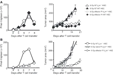
The expansion of in vitro activated antitumor T cells was robust in the presence or absence of β2m (Figure 3A,
left panel). Most importantly, significant tumor treat-ment was observed in HSC-transplanted, myeloablated hosts in the absence of β2m (Figure 3A, right panel;
9 Gy in β2m–/– mice: P = 0.001, NTversus effector PI).
This tumor treatment efficacy was comparable to that seen in myeloablated WT mice that received an HSC transplant (Figure 3A, right panel;9 Gy effector PI:
P = 0.9, WT versus β2m–/–).
We next tested the in vivo activities of naive CD8+
T cells compared with antigen-experienced effector CD8+ T cells in myeloablated mice lacking β2m
expres-sion transplanted with β2m–/– HSCs (Figure 3B). In
sharp contrast to the functional activities of preacti-vated effector cells adoptively transferred into hosts lacking β2m (Figure 3A), adoptively transferred naive
pmel-1 CD8+ T cells did not proliferate (Figure 3B, left
panel) and did not mediate a measurable therapeutic impact in the tumor-bearing setting (Figure 3B, right panel; P = 0.96, NT versus naive PI). Thus, while acti-vated cells can proliferate and destroy tumor in the absence of host β2m expression, naive cell proliferation
and function were dependent on the expression of β2m
by the host. This result showed that the HSC-driven proliferation and antitumor function required MHC class I–dependent TCR triggering of the transferred pmel-1 CD8+
T cells and that TCR triggering of pmel-1 CD8+ T cells could be
accomplished either in vivo or in vitro.
HSCs drive pmel-1 CD8+ T cell expansion but not tumor response in
nonmyeloablated mice. HSC-driven proliferation of pmel-1 CD8+ T
cells was observed after 9 Gy TBI, in which case these HSCs were required for host survival after myeloablation (Figure 1B), but it was unclear whether HSCs would drive the expansion of CD8+
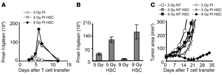
T cells when they were given after a nonmyeloablative prepara-tive regimen, where they were not required to rescue mice from lethality. HSCs were transplanted after 5 Gy TBI, and levels of transferred pmel-1 transgenic T cells in the spleen were analyzed. HSCs increased the proliferation of pmel-1 CD8+ T cells in the
nonmyeloablative (5-Gy) setting (Figure 4A). This proliferation was equivalent to that seen in mice receiving an HSC transplant after 9 Gy TBI. The kinetics of HSC-driven pmel-1 CD8+ T cell
expansions were similar to those seen in the 9-Gy TBI setting. These results were seen reproducibly upon analysis of individual mice (Figure 4B; P = 0.02, 5 Gy versus 5 Gy/HSC; P = 0.0009, 9 Gy versus 9 Gy/HSC; P = 0.24, 5 Gy/HSC versus 9 Gy/HSC).
Surpris-Figure 2
HSCs drive the proliferation and antitumor activity of naive and effector pmel-1 CD8+ T cells in mice treated with a myeloablative preparative regimen and an
HSC transplant. (A) The expansion of naive and effector pmel-1 CD8+ T cells is
comparable in myeloablated hosts with HSC transplant. Gene-marked (Thy1.1+)
naive or effector pmel-1 CD8+ T cells (1 × 10⁶) were adoptively transferred with
rhIL-2 into a nonmyeloablated host (left panel) or a myeloablated host receiving an HSC transplant (right panel). The absolute numbers of pmel-1 CD8+ T cells in
the spleens of treated animals were enumerated on the days indicated. Spleens of 3 mice per group were pooled at each time point. This experiment was performed 3 times, with similar results. (B) Naive and effector pmel-1 CD8+ T cells elicit a
similar tumor treatment in myeloablated hosts with HSC transplant. Naive or effec-tor pmel-1 CD8+ T cells (1 × 10⁶) were transferred with rhIL-2 in nonmyeloablated
(left panel) or myeloablated hosts with HSC transplant (right panel). Both naive and effector pmel-1 CD8+ T cells mediated significant tumor treatment (9 Gy/HSC:
[image:4.585.44.330.80.308.2]ingly, the HSC-driven expansion observed in the nonmyeloablative setting did not result in enhanced antitumor activity (Figure 4C;
P = 0.22, 5 Gy versus 5 Gy/HSC). Thus, the HSC-induced spike in antitumor T cell number was not associated with measurable tumor destruction. Although myeloablation was not essential for HSC-driven T cell proliferation, it was important for the transla-tion into effective tumor treatment. We previously reported that Tregs and cellular sinks for cytokines could inhibit the antitumor activities of adoptively transferred CD8+ T cells (9, 17). We
hypoth-esized that HSCs might not only cause the expansion of adoptively transferred T cells, but might also provide support for host cells that survive 5 Gy TBI, including cells capable of restraining the antitumor immune response.
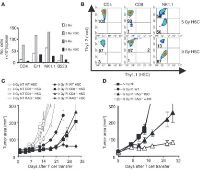
HSCs drive host cells surviving TBI that hinder the effectiveness of anti-tumor T cells. To determine the impact of HSCs on host immune cells that survived lymphodepletion, we analyzed the number of a variety of cellular subsets in the spleen after nonmyeloablative (5 Gy) and myeloablative (9 Gy) irradiation. Myeloablative con-ditioning regimens reduced the number of cells present 5 days after irradiation compared with the nonmyeloablative condition (Figure 5A). We observed that an HSC transplant increased the
numbers of CD4+ T cells, Gr1+
cells, NK cells, and B cells in both the 5-Gy and 9-Gy settings. Thus, HSCs sup-ported the expansion of not only adoptively transferred CD8+ T cells but a variety of
endogenous host cells as well (Figure 5A). Because HSCs can differentiate into each of the above-mentioned cell types (25), we sought to deter-mine whether these different immune cells were deriva-tives from the HSCs or were host cells that had survived TBI. Studies using congenic markers indicated that virtu-ally all lymphocytes obtained 7 days after the transplant were derived from host cells that survived TBI (Figure 5B). Thus, HSCs drove the prolif-eration of adoptively trans-ferred transgenic CD8+ T
cells and increased the num-bers of splenic lymphocytes not via progeny cells but by promoting the expansion of endogenous host cells. These expanded endogenous lymphocyte compartments included inhibitory elements such as CD4+CD25+Foxp3+
Tregs, as well as endogenous CD8+ T cells and NK cells.
CD4+CD25+Foxp3+ Tregs
have been shown to negatively modulate the functionality of adoptively transferred tumor-reactive CD8+ T cells, resulting in
diminished treatment efficacy (17, 18, 26, 27). Endogenous CD8+
T and NK cells can act as cellular sinks for homeostatic cytokines and compete for these cytokines with the adoptively transferred tumor-reactive CD8+ T cells, thereby reducing the efficacy of tumor
treatment mediated by the transferred tumor-reactive CD8+ T cells
(9, 28–30). Thus, HSC transplantation increased the levels of sur-viving endogenous host cells that might have abrogated pmel-1 CD8+ T cell antitumor efficacy.
To directly test whether these surviving host cells were capable of negatively influencing pmel-1 CD8+ T cell–mediated tumor
destruction, we measured the treatment efficacy of adoptively transferred pmel-1 CD8+ T cells in WT hosts and hosts that were
genetically deficient in lymphocyte subsets. Biological differences in the tumor treatment efficacy emerged when suboptimal num-bers of adoptively transferred T cells were transferred (Figure 5C). Treatment was improved in myeloablated mice that were knock-outs for CD4 (P = 0.017, 9 Gy PI WT versus 9 Gy CD4–/–), CD8
(P = 0.004, 9 Gy PI WT versus 9 Gy CD8–/–), as well as RAG (P = 0.0017,
9 Gy PI WT versus9 Gy RAG–/–) (Figure 5C). These findings indicated
[image:5.585.44.417.81.331.2]that surviving host cells, even after 9 Gy TBI, were able to reduce the
Figure 3
HSCs drive effector, but not naive, pmel-1 CD8+ T cell expansion and tumor treatment in the absence of host
β2m. (A) HSC-driven effector pmel-1 CD8+ T cell expansion and tumor treatment is MHC class I independent.
Effector pmel-1 CD8+ T cells (1 × 10⁶) were transferred together with rhIL-2 into 9-Gy irradiated β2m–/– or WT
mice that had received the matched HSC transplant and had 10-day established B16 tumors. T cell expansion (left panels) and tumor treatment (right panels) were evaluated at the indicated days. Control groups were left untreated. Effector pmel-1 CD8+ T cells showed similar proliferation and tumor treatment in β2m–/– and
WT mice (9 Gy effector PI: P = 0.9, WT versus β2m–/–; 9 Gy in β2m–/–: P = 0.001, NTversus effector PI). (B) In
β2m–/– mice, only effector, but not naive, pmel-1 CD8+ showed HSC-driven T cell expansion and tumor
treat-ment. Effector or naive pmel-1 CD8+ T cells were transferred into myeloablated tumor-bearing β2m–/– mice
receiving a β2m–/– HCS transplant. Naive pmel-1 CD8+ T cells showed neither proliferation nor significant
tumor treatment (P = 0.96, NT versus naive PI). Proliferation curves represent the numbers of gene-marked pmel-1 CD8+ T cells found in the spleens of 3 mice pooled per group per time point. Results for tumor area
research article
496 The Journal of Clinical Investigation http://www.jci.org Volume 117 Number 2 February 2007
effectiveness of pmel-1 CD8+ T cell tumor treatment. Endogenous
CD4+ T cells, which contain Tregs, and endogenous CD8+ T cells
were able to negatively impact the pmel-1 CD8+ T cell tumor
treat-ment capacity. An inhibitory role for NK1.1+ cells was not detected
in the myeloablative setting (data not shown) but was revealed by their depletion in nonmyeloablated RAG–/– hosts (P = 0.01;
5 Gy PI RAG–/– ISO versus 5 Gy PI RAG–/–α-NK; Figure 5D). Thus,
endogenous host cells strongly influenced the tumor treatment capacity of transferred CD8+ T cells even when they were present in
small numbers as a result of TBI-mediated immunodepletion.
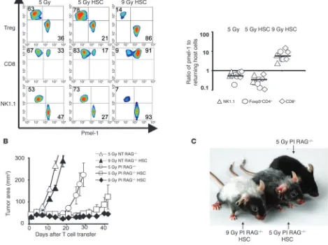
The antitumor T cell/host cell ratio determines tumor treatment out-come. We evaluated the impact of the nonmyeloablative (5-Gy) and myeloablative (9-Gy) preparative regimens on the ratios of effector pmel-1 CD8+ T cells to remaining inhibitory elements (Foxp3+CD4+
cells, CD8+ cells, and NK1.1+ cells). Three congenic markers were
used to identify the ablated host cells (Thy1.2+) in mice that
received TBI followed by HSC transplantation (Thy1.1+) with the
adoptive transfer of preactivated pmel-1 CD8+ T cells (Ly5.1+)
(Figure 6, A and B, left and right panels). We found that compared with mice receiving a nonmyeloablative regimen without HSC transplant, mice receiving myeloablative TBI with HSC transplant had significantly higher ratios of transferred pmel-1 effector CD8+
T cells to host regulatory elements including Foxp3+CD4+ Tregs
(P = 0.0006, 5 Gy versus 9 Gy/HSC), CD8+ T cells (P = 0.003, 5 Gy
versus 9 Gy/HSCs), and NK1.1+ cells (P = 0.0006, 5 Gy versus 9 Gy/
HSC) (Figure 6B). When HSCs were transferred into the nonmye-loablated and myenonmye-loablated animals, the ratios of antitumor T cells to host immunoregulatory cells still remained greater after 9 Gy TBI. Although the total number of pmel-1 CD8+ T cells was comparable
in myeloablated and nonmyeloablated animals with an HSC trans-plant, the ratios were significantly in favor of the transferred pmel-1 CD8+ T cells in the myeloablated animals, measured for pmel-1
CD8+ T cells versus Foxp3+CD4+ Tregs (P = 0.0005, 5 Gy/HSC
ver-sus 9 Gy/HSC), CD8+ T cells
(P = 0.002, 5 Gy/HSC versus 9 Gy/HSC), and NK1.1+ cells
(P = 0.0005, 5 Gy/HSC versus 9 Gy/HSC) (Figure 6B). These findings were consistent with the observation that HSCs enhanced the adoptively trans-ferred T cells as well as endo-genous host cells (Figure 5A). Thus, although the absolute numbers of transferred pmel-1 CD8+ T cells were
compa-rable in the 5-Gy and 9-Gy settings (Figure 4, A and B), it appeared that the numbers of host immunoregulatory cells limited the therapy in the nonmyeloablated setting.
HSC transplant augmented tumor treatment capacity of pmel-1 CD8+ T cells upon the
reduction of host inhibitory ele-ments. We evaluated whether the increased number of transferred T cells observed after an HSC transplant could result in an enhanced tumor treatment if there are no or few inhib-itory elements to be driven by the HSC transplant. In this scenario, the HSC transplant increases the number of transferred T cells but not of inhibitory elements, since they are genetically removed. Pmel-1 CD8+ T cells were transferred with an HSC transplant into
RAG–/– hosts, which are genetically lymphodepleted (Figure 6B).
Giving an HSC transplant in RAG–/– mice significantly augmented
the efficacy of tumor-reactive pmel-1 CD8+ T cells in a
nonmy-eloablated (5-Gy irradiated) host (P = 0.038, 5 Gy PI RAG–/– versus
5 Gy PI RAG–/– HSC; Figure 6B) but not in control WT mice in
the same experiment (data not shown). These results confirmed the hypothesis that enhanced treatment can be achieved by focus-ing HSC support on transferred pmel-1 CD8+ T cells in an
envi-ronment where regulatory elements are genetically reduced. The antitumor capacity of the transferred pmel-1 CD8+ T cells
corre-lated with the amount of autoimmune vitiligo resulting from the destruction of normal melanocytes that express gp100, the target antigen of pmel-1 CD8+ T cells (Figure 6C). Thus, HSCs drove the
expansion of transferred tumor-reactive T cells in both 5-Gy and 9-Gy regimens but 9 Gy TBI mediated a more complete depletion of endogenous host cells, focusing the HSC-mediated lymphocyte expansion on the adoptively transferred antitumor cells.
Discussion
Endogenous host cells can impact the functionality of adop-tively transferred tumor-reactive T cells and can negaadop-tively influence their tumor treatment capacity (9, 17, 18). A regimen in which endogenous host cells are removed by nonmyeloabla-tive, lymphodepletion conditioning has been used successfully in mice and in patients to enhance the antitumor efficacy of adoptively transferred T cells (9, 12). Increasing the lymphode-pletion to achieve a myeloablative regimen requiring an HSC transplant significantly augmented tumor treatment. The
aug-Figure 4
HSCs drive pmel-1 CD8+ T cell expansion in nonmyeloablated and myeloablated mice, but tumor treatment
is only achieved in myeloablated mice. (A and B) HSC transplantation–driven transgenic T cell expansion
is not dependent on the intensity of immunodepletion. Mice received a preparative regimen of either 5 Gy or 9 Gy TBI, which was followed by effector pmel-1 CD8+ T cells (1 × 10⁶) and rhIL-2 with or without HSC
transplantation. (A) Absolute numbers of adoptively transferred congenic marked pmel-1 CD8+ T cells in the
spleen were enumerated on the days indicated. Results shown were derived from pooled splenocytes of 3 mice per group per time point (A) or from 3 individual mice (±SEM) assessed on day 6 (B) (P = 0.0009, 9 Gy versus 9 Gy/HSC; P = 0.02, 5 Gy versus 5 Gy/HSC; P = 0.24, 5 Gy/HSC versus 9 Gy/HSC). (C) HSC-driven
expansion of pmel-1 CD8+ T cells is therapeutically effective in myeloablated, but not in nonmyeloablated, WT
mice. Animals bearing tumors established for 10 days were either left as controls (NT) or received, prior to the transfer of 1 × 10⁶ effector pmel-1 CD8+ T cells and rhIL-2, either 5 Gy TBI, 5 Gy TBI and an HSC transplant,
[image:6.585.44.419.80.208.2]mented tumor treatment efficacy was associated with increased antitumor T cell expansion. The reason for the increased T cell proliferation was not merely the lymphopenic environment in myeloablated animals but the HSC transplant, which induced the expansion of transferred tumor-reactive pmel-1 CD8+ T
cells. This was also the case in the nonmyeloablative setting, but tumor treatment was only seen when the HSCs were trans-ferred into myeloablated animals. The expansion of antitumor T cells with tumor destruction can also be triggered by antigen-specific vaccination (6). While vaccination preferentially drives the expansion of antitumor T cells, HSCs drive tumor-specific T cells and endogenous host immunoregulatory cells with equal vigor. Even in very small numbers, these host cells inhibit the antitumor activity of pmel-1 CD8+ T cells. Thus,
myeloabla-tion significantly reduces the number of surviving host cells, drastically shifting the ratio of tumor-reactive pmel-1 CD8+ T
cells to endogenous host cells in favor of the pmel-1 CD8+ T
cells, allowing improved tumor treatment. The ratio of effector to suppressor cells has been recently proposed to be a predic-tor of tumor treatment outcome in mice (31) and survival in patients with ovarian cancer (32, 33). Besides CD4+CD25+Foxp3+
Tregs, CD11b+Gr1+ myeloid suppressor cells (MSCs) have been
described to suppress the functionality of tumor-specific T lymphocytes (34–36). By increasing the intensity of ablation, the level of MSCs in the tumor might be decreased, leading to improved tumor treatment. In addition to lowering levels of suppressive elements, intensive lymphodepletion also reduces the numbers of endogenous host cells such as CD8+ and NK
[image:7.585.96.490.80.413.2]cells, which consume homeostatic cytokines, thereby leading to elevated levels of IL-7 and IL-15. Additionally we showed that an HSC transplant in 9-Gy irradiated animals also increased the level of theses cytokines in the serum. IL-7 and IL-15 are main inducers of homeostatic T cell proliferation and activation and can impact the transferred T cells. Thus, the interaction of the
Figure 5
HSCs drive expansion of host cells surviving TBI, hindering the effectiveness of antitumor T cells. (A) HSC transplants increase the numbers of
splenic CD4+ T cells and Gr-1+, NK, and B cells found in irradiated mice. Absolute numbers of splenic CD4+, Gr1+, NK1.1+, or B220+ cells were
determined 5 days after mice received 5 Gy or 9 Gy TBI with or without HSC transplant. Mice received 1 × 10⁶ pmel-1 CD8+ T cells and rhIL-2. (B)
HSC-driven recovery is derived from host cells, not HSC progeny. Thy1.1+ HSCs were transplanted into Thy1.2+ hosts that had received either 5
Gy or 9 Gy TBI and rhIL-2. Data shown are from 3 pooled spleens per group at day 7. (C and D) Returning host cells can abrogate pmel-1 CD8+
T cell tumor treatment. (C) Tumor-bearing myeloablated WT, CD4–/–, CD8–/–, or RAG–/– mice with a syngeneic HSC transplant were left untreated
as control or received suboptimal amounts (5 × 10⁵) of effector pmel-1 CD8+ T cells with rhIL-2. P = 0.017, 9 Gy PI WT versus 9 Gy CD4–/–;
P = 0.004, 9 Gy PI WT versus 9 Gy CD8–/–; P = 0.0017, 9 Gy PI WT versus 9 Gy RAG–/–. (D) Tumor-bearing nonmyeloablated WT or RAG–/– mice
were left untreated as control or received 1 × 10⁶ effector pmel-1 CD8+ T cells and rhIL-2. NK1.1 cell–depleting (α-NK) or isotope control (ISO)
antibodies were given every 5 days, starting at day 0 and ending on day 20 (P = 0.01, 5 Gy PI RAG–/– ISO versus 5 Gy PI RAG–/–α-NK). Results
research article
498 The Journal of Clinical Investigation http://www.jci.org Volume 117 Number 2 February 2007
HSC transplant with the adoptively transferred T cells is likely to impact the quality of the T cells, but the exact changes in the quality of the transferred T cells remain to be elucidated.
TBI may facilitate tumor destruction by a variety of factors in addition to immune depletion. Irradiation, especially inten-sive radiation, can directly damage tumor cells and supporting tumor stroma. However, we have previously shown that local irradiation of the B16 tumor up to 10 Gy does not significantly impact on the tumor growth and does not improve the activity of adoptively transferred TCR-restricted T cells (9). Furthermore,
chemotherapeutic agents as well as irradiation are described to modulate the immunogenicity of tumor cells in mouse and human. Irradiation reportedly upregulates the expression of surface molecules such as MHC class I, Fas, and ICAM-1 (37), which can enhance tumor recognition by adoptively transferred T cells. In addition, irradiation can modify the peptide reper-toire, increase peptide production and antigen presentation, and enhance MHC class I expression by tumor cell lines (38). This might have particular relevance to tumor recognition by endogenous host cells with an open repertoire. Thus,
increas-Figure 6
The antitumor effectiveness of HSC-driven CD8+ T cell proliferation is dependent on the reduction of host inhibitory elements. (A and B) Ratios
of adoptively transferred effector pmel-1 CD8+ T cells (CD8+Ly5.1+) relative to returning host Treg cells (Foxp3+CD4+), host CD8+Th1.1+ cells,
and host NK1.1+ cells are shifted toward pmel-1 CD8+ T cells in myeloablated mice. Thy1.2+ host mice received a preparative regimen of 5 Gy
or 9 Gy with an HSC transplantation from Thy1.1+ mice, which was followed by the adoptive transfer of 1 × 10⁶ effector (Ly5.1+) pmel-1 CD8+ T
cells and rhIL-2. Control mice received 5 Gy TBI in the absence of an HSC transplant. Splenocytes obtained 1 week after HSC transplant were simultaneously analyzed for adoptively transferred gene-marked pmel-1 CD8+ and reconstituting host cells (left panel). Flow cytometry data
from pooled splenocytes from 3 mice were used to calculate the ratios of adoptively transferred pmel-1 CD8+ T cells, and cumulative results
from 3 independent experiments (3 mice per experiment) are shown in the right panel. The ratios of transferred pmel-1 T cells to host immune cell subsets were significantly higher after 9 Gy TBI with an HSC transplant than after 5 Gy irradiation with or without an HSC transplant. The levels of significance are as follows: for of NK cells (P = 0.0006, 5 Gy versus 9 Gy/HSC, P = 0.0005, 5 Gy/HSC versus 9 Gy/HSC), Foxp3+CD4+
Tregs (P = 0.0006, 5 Gy versus 9 Gy/HSC, P = 0.0005, 5 Gy/HSC versus 9 Gy/HSC) and CD8+ T cells (P = 0.003, 5 Gy versus 9 Gy/HSC,
P = 0.002, 5 Gy/HSC versus 9 Gy/HSC). (B) HSC-driven pmel-1 CD8+ T cell proliferation can augment tumor treatment in RAG–/– mice. RAG–/–
mice bearing 10-day tumors were irradiated with 5 Gy or 9 Gy with or without an HSC transplant and were treated with 1 × 10⁶ effector pmel-1
CD8+ T cells with rhIL-2. Control mice were left untreated. Data are representative of 3 independent experiments, each with 5 mice per group.
[image:8.585.58.527.100.450.2]ing the intensity of the TBI or chemotherapy-based preparative regimen may enhance the efficacy of immunotherapies based on the adoptive transfer of antitumor T cells through a variety of mechanisms; importantly, the use of more intense myeloablative regimens must be balanced against their toxicities (39).
Previous work has shown that nonmyeloablative lymphodeple-tion significantly increased the efficacy of ACT given in combina-tion with IL-2 (9); however, vaccinacombina-tion with an altered-self ligand– based vaccine was required. Thus, the lymphopenia induced by the regimens previously employed did not activate adoptively trans-ferred antitumor CD8+ T cells to a level required to mediate the
treatment of established tumor (6, 9, 17). Surprisingly, vaccina-tion was not required in the setting of a myeloablative condivaccina-tion- condition-ing regimen prior to ACT-based immunotherapy (Supplemental Figure 1). Why did the requirements for T cell activation change so dramatically when the preparative regimen was increased from 5 Gy TBI to 9 Gy TBI? The most likely scenario was that a more intensive lymphodepletion resulted in a more complete depletion of cellular cytokine sinks and CD4+CD25+Foxp3+ Tregs. Indeed,
we found that although cells of hematopoietic origin were deplet-ed by a nonmyeloablative TBI regimen (5 Gy), the administration of a myeloablative dose of TBI depleted host endogenous myeloid and lymphoid cells an additional 10-fold.
Vaccine independence may be a critical feature in the translation of these findings to ACT-based therapies in humans, as vaccines for adoptively transferred, polyclonal T cells are not always avail-able. In humans, tumor-reactive T cells are usually derived from tumor-infiltrating lymphocytes (TILs) that are expanded ex vivo using IL-2. TILs are further selected by their ability to specifically secrete IFN-γ when cultured with autologous or allogeneic MHC-matched tumor cell lines. Before tumor-reactive TILs are trans-ferred into the patient, they are expanded using CD3-specific antibodies, IL-2, and irradiated allogeneic peripheral-blood mono-nuclear feeder cells (1). Approximately 50% of the tumor-reactive TILs have specificity against epitopes from unknown cancer anti-gens. In the absence of suitable vaccines, tumor-reactive CD8+ T
cells can be activated and expanded in vivo upon transfer into an HSC-transplanted, myeloablated host.
In the field of antiviral immunotherapy, valuable experience has been gained in the transfer of antigen-specific T cells into highly lymphodepleted patients with an autologous bone marrow trans-plant. Patients who are lymphopenic after organ or bone marrow transplant are at high risk for developing CMV- or EBV-mediated disease (40, 41). Immunotherapeutic strategies aim to restore the virus-specific cellular immunity by transferring allogeneic or autologous virus-reactive CTLs (42, 43), which restored the cellu-lar immune deficiency in highly immune-suppressed patients with limited toxicity and efficiently treat CMV- and EBV-associated disease (40, 44–46). In myeloma patients treated with high-dose chemotherapy and autologous stem cell transplantation, a bet-ter clinical outcome was associated with more rapid lymphocyte recovery after the transplant (47). In order to augment the recovery and function of autologous T cells in myeloma patients, June and colleagues stimulated antigen-primed autologous T cells ex vivo and combined vaccine therapy with adoptive T cell transfer into lymphodepleted patients. They were able to raise clinically relevant immunity in adults within 1 month after transplantation (48).
HSCs are known to provide new progeny cells (25), and HSC transplantation has been used in tumor therapy to rescue bone marrow function after high-dose chemotherapy (49). Depending
on the cell type, the recovery time of HSC-derived progeny can take several days to several months, but little is known about how HSCs influence the homeostatic proliferation of endogenous host cells or transferred T cells. In this study, syngeneic HSCs not only pro-vided new progeny, but also supported the proliferation and sur-vival of adoptively transferred self/tumor-reactive CD8+ T cells that
can mediate tumor destruction. HSC-driven T cell expansion was not limited to myeloablated animals but also could be observed in nonmyeloablated animals that received HSC transplant. By studying the requirements for the HSC-driven T cell expansion, we found that both naive and antigen-experienced T cells under-went HSC-driven expansion similar to homeostatic proliferation (13, 15, 16). TCR engagement of naive T cells was required because HSC-driven T cell expansion of naive T cells was seen in WT but not in β2m–/– animals. In contrast, for antigen-experienced CD8+
T cells, HSC-driven expansion did not require MHC class I expres-sion on host cells. Key regulators of peripheral T cell homeostasis are homeostatic cytokines, especially IL-7 (13, 14, 50–52) and IL-15 (13, 53, 54). While IL-15 mainly regulates memory T cell homeo-stasis, IL-7 is required for the homeostatic expansion of naive and memory T cells. We found elevated serum levels of both IL-7 and IL-15 after HSC transplantation in the myeloablative setting. The elevation was only transient — the peak occurred on day 5 after the HSC transplant — and corresponded with the observed expansion of transferred T cells as well as surviving endogenous host cells, perhaps indicating that these cells were consuming the homeo-static cytokines. The exact origin and induction of these cytokines after the HSC transplant requires further investigation.
Hematopoiesis has been closely investigated in the field of HSC transplantation (55) and could provide insight into the mecha-nism of HSC support of adoptively transferred T cells. Bone marrow–derived lin– cells prolong the survival of myeloablated
animals via soluble factors (56). HSC support is not limited to T cells but is open to a variety of different cell types, such as NK cells, B cells, and monocytes. To influence several different cell types, HSCs could provide multiple factors or a single pluripotent factor that can impact a variety of cell types. One well-studied fac-tor is IGF-1, which has been reported to enhance hematopoiesis, thymopoiesis, as well as T cell and B cell function (57). The IGF-1 receptor is found on thymocytes, T cells, B cells, and NK cells. IGF-1 can be released by HSCs and bone marrow stromal cells (58) and has been shown to increase the function of peripheral T cells, B cells, NK cells, and macrophages. IGF-1 may also have an antiapoptotic effect on peripheral T cells (59, 60), which per-haps explains the survival of endogenous lymphocytes that have received a damaging insult from TBI or chemotherapy. Other growth factors and cytokines have been reported to provide radio-protection when administered either before or shortly after the radiation exposure (56). For example, the administration of stem cell factor (SCF), Flt-3 ligand, TPO, IL-3, and SDF-1 together sig-nificantly prolonged the survival of myeloablated mice (61, 62). Radioprotection by these factors is probably mediated through an antiapoptotic effect on hematopoietic progenitor cells. Recently it has been shown that bone marrow lin– cells can release soluble
research article
500 The Journal of Clinical Investigation http://www.jci.org Volume 117 Number 2 February 2007
transferred T cells capable of destroying large tumor burdens in the absence of cancer vaccines. The finding that myeloablative lymphodepletion enabled HSCs to drive the expansion and func-tion of adoptively transferred CD8+ T cells in the absence of
com-peting host cells can have substantial implications for the design of new antitumor immunotherapies.
Methods
Mice and tumor lines. All mice used in these experiments were bred and
housed at NIH facilities. Female pmel-1 TCR-Tg mice were crossed with C57BL/6-Thy1.1+–Tg or C57BL/6-Ly5.1+–Tg mice (The Jackson
Laborato-ry) to derive pmel-1–Thy1.1+ or pmel-1–Ly5.1+ double-Tg mice
(C57BL/6-pmel-1–Thy1.1+ mice are now available at http://www.jax.org). Female
C57BL/6, RAG–/–, CD4–/–, CD8–/– (The Jackson Laboratory), and β2m–/–
mice (Taconic) were used as recipients in ACT experiments. Experiments were conducted with the approval of the National Cancer Institute Animal Use and Care Committee. B16-F10 (H-2b), a spontaneous, transplantable
gp100+ murine melanoma, was maintained in culture medium (CM).
In vitro activation of pmel-1 CD8+ T cells. Pmel-1 splenocytes were isolated as
described previously (23) and cultured in the presence of 1 μM hgp10025–33
and CM containing 30 IU/ml of rhIL-2 (Chiron). Cells were used for adop-tive transfer 6–7 days after the start of the culture.
Adoptive transfers. Mice 6–12 weeks of age (n = 5–6 for all groups) were
injected subcutaneously with 2 × 10⁵ to 5 × 10⁵ B16-F10 melanoma cells and
treated 10–14 days later with intravenous adoptive transfer of in vitro–acti-vated pmel-1 CD8+ T splenocytes. Lymphopenia was induced by
nonmy-eloablative (5 Gy) or mynonmy-eloablative (9 Gy) TBI of tumor-bearing mice on the day of HSC transfer, which was 1 day prior to pmel-1 CD8+ T cell transfer.
HSCs were extracted from the bone marrow by lineage depletion with strep-tavidin-coated magnetic beads (Dynabeads M-280 Streptavidin; Dynal Bio-tech) against biotin-labeled antibodies (γδ T cell receptor, αβ T cell receptor, CD4, CD8a, NK1.1, Gr-1, B220, Ter-119, CD2, CD11b) (BD Biosciences) fol-lowed by a c-kit enrichment with CD117 MicroBeads (Miltenyi Biotec) and cultured for 18 hours in 10% DMEM with 50 ng recombinant mouse IL-3 (rmIL-3), 500 ng rmIL-6, and 500 ng rmSCF (PeproTech); 1 × 10⁵ cells were
administered the following day directly after the irradiation. NK cells were depleted by intraperitoneal administration of 100 μg/mouse of NA/LE anti-NK1.1 antibody (BD Biosciences) every 5 days for a total of 5 doses. NA/LE
anti-Ig2aK (100 μg/mouse) was used as control antibody (BD Biosciences).
Mice were vaccinated with 2 × 10⁷ PFU of a previously described (23)
recom-binant fowlpox virus expressing human gp100 (rFPhgp100; Therion Bio-logics). rhIL-2 (108 μg/dose; Chiron) was administered by intraperitoneal injection twice daily for a total of 6 doses. Tumors were measured using calipers, and the products of the perpendicular diameters were recorded. All experiments were performed in a blinded, randomized fashion and per-formed independently at least twice, with similar results.
Enumeration of cells in vivo and assessment of cytokine serum levels. At indicated time points, spleens were resected, and adoptively transferred CD8+Thy1.1+
or CD8+Ly5.1+ pmel-1 CD8+ T cells were enumerated as previously
described (23). Samples were analyzed by flow cytometry for B220, Gr1, CD8a, CD4, NK1.1, and Foxp3 expression (BD Biosciences). Cell numbers were calculated by multiplying the total number of live cells by the percent-age of B220+, Gr1+, CD8a+, CD4+ T, Foxp3+, CD4+, or NK1.1+ cells. Serum
was collected from the tail vein at day 5 after HSC transplantation and assayed by murine LINCOplex cytokine assay (Linco Research).
Statistics. Tumor graphs were compared using ANOVA. P values less than
0.05 were considered significant.
Acknowledgments
This research was supported by the Intramural Research Pro-gram of the NIH, National Cancer Institute, Center for Cancer Research. The authors would like to thank A. Mixon and S. Farid of the Flow Cytometry Unit (Surgery Branch of the NCI) for flow cytometry analyses; D. Jones for excellent technical support; and L. Cassard, B. Heemskerk, and D.J. Powell Jr. for critical review of the manuscript.
Received for publication September 21, 2006, and accepted in revised form November 21, 2006.
Address correspondence to: Claudia Wrzesinski or Nicholas P. Restifo, National Cancer Institute, National Institutes of Health, Bethesda, Maryland 20892, USA. Phone: (301) 451-6948; Fax: (301) 496-0011; E-mail: [email protected] (C. Wrzesinski). Phone: (301) 496-4904; Fax: (301) 496-0011; E-mail: restifo@ nih.gov (N.P. Restifo).
1. Gattinoni, L., Powell, D.J., Jr., Rosenberg, S.A., and Restifo, N.P. 2006. Adoptive immunotherapy for cancer: building on success. Nat. Rev. Immunol. 6:383–393.
2. Foster, A.E., and Rooney, C.M. 2006. Improving T cell therapy for cancer. Expert Opin. Biol. Ther. 6:215–229.
3. Yee, C., et al. 2002. Adoptive T cell therapy using antigen-specific CD8+ T cell clones for the treat-ment of patients with metastatic melanoma: in vivo persistence, migration, and antitumor effect of transferred T cells. Proc. Natl. Acad. Sci. U. S. A. 99:16168–16173.
4. Rosenberg, S.A., Yang, J.C., and Restifo, N.P. 2004. Cancer immunotherapy: moving beyond current vaccines. Nat. Med.10:909–915.
5. Klebanoff, C.A., Gattinoni, L., and Restifo, N.P. 2006. CD8+ T-cell memory in tumor immunology and immunotherapy. Immunol. Rev.211:214–224. 6. Overwijk, W.W., et al. 2003. Tumor regression and
autoimmunity after reversal of a functionally toler-ant state of self-reactive CD8+ T cells. J. Exp. Med. 198:569–580.
7. Cheever, M.A., Greenberg, P.D., and Fefer, A. 1980. Specificity of adoptive chemoimmunother-apy of established syngeneic tumors. J. Immunol. 125:711–714.
8. North, R.J. 1982. Cyclophosphamide-facilitated adoptive immunotherapy of an established tumor depends on elimination of tumor-induced suppres-sor T cells. J. Exp. Med.155:1063–1074.
9. Gattinoni, L., et al. 2005. Removal of homeostatic cytokine sinks by lymphodepletion enhances the efficacy of adoptively transferred tumor-specific CD8+ T cells. J. Exp. Med.202:907–912.
10. Maine, G.N., and Mule, J.J. 2002. Making room for T cells. J. Clin. Invest.110:157–159. doi:10.1172/ JCI200216166.
11. Dudley, M.E., et al. 2005. Adoptive cell transfer therapy following non-myeloablative but lym-phodepleting chemotherapy for the treatment of patients with refractory metastatic melanoma. J. Clin. Oncol. 23:2346–2357.
12. Dudley, M.E., et al. 2002. Cancer regression and autoimmunity in patients after clonal repopu-lation with antitumor lymphocytes. Science. 298:850–854.
13. Goldrath, A.W., et al. 2002. Cytokine requirements for acute and basal homeostatic proliferation of naive and memory CD8+ T cells. J. Exp. Med. 195:1515–1522.
14. Schluns, K.S., and Lefrancois, L. 2003. Cytokine control of memory T-cell development and survival. Nat. Rev. Immunol. 3:269–279.
15. Cho, B.K., Rao, V.P., Ge, Q., Eisen, H.N., and Chen, J. 2000. Homeostasis-stimulated proliferation drives naive T cells to differentiate directly into memory T cells. J. Exp. Med. 192:549–556. 16. Kieper, W.C., and Jameson, S.C. 1999. Homeostatic
expansion and phenotypic conversion of naive T cells in response to self peptide/MHC ligands. Proc. Natl. Acad. Sci. U. S. A. 96:13306–13311. 17. Antony, P.A., et al. 2005. CD8+ T cell immunity
against a tumor/self-antigen is augmented by CD4+ T helper cells and hindered by natu-rally occurring T regulatory cells. J. Immunol. 174:2591–2601.
18. Turk, M.J., et al. 2004. Concomitant tumor immunity to a poorly immunogenic melanoma is prevented by regulatory T cells. J. Exp. Med. 200:771–782.
19. Viret, C., Wong, F.S., and Janeway, C.A., Jr. 1999. Designing and maintaining the mature TCR rep-ertoire: the continuum of self-peptide: self-MHC complex recognition. Immunity. 10:559–568. 20. Goldrath, A.W., and Bevan, M.J. 1999. Low-affinity
ligands for the TCR drive proliferation of mature CD8+ T cells in lymphopenic hosts. Immunity. 11:183–190.
lymphocytes. Science. 314:126–129.
22. Gattinoni, L., et al. 2005. Acquisition of full effector function in vitro paradoxically impairs the in vivo antitumor efficacy of adoptively transferred CD8+ T cells. J. Clin. Invest. 115:1616–1626. doi:10.1172/ JCI24480.
23. Palmer, D.C., et al. 2004. Vaccine-stimulated, adop-tively transferred CD8+ T cells traffic indiscrimi-nately and ubiquitously while mediating specific tumor destruction. J. Immunol. 173:7209–7216. 24. Zijlstra, M., et al. 1990. Beta 2-microglobulin
defi-cient mice lack CD4-8+ cytolytic T cells. Nature. 344:742–746.
25. Kondo, M., et al. 2003. Biology of hematopoietic stem cells and progenitors: implications for clinical application. Annu. Rev. Immunol. 21:759–806. 26. Shen, S., et al. 2005. Control of homeostatic
proliferation by regulatory T cells. J. Clin. Invest. 115:3517–3526. doi:10.1172/JCI25463.
27. Antony, P.A., et al. 2006. Interleukin-2-dependent mechanisms of tolerance and immunity in vivo. J. Immunol. 176:5255–5266.
28. Prlic, M., Blazar, B.R., Farrar, M.A., and Jameson, S.C. 2003. In vivo survival and homeostatic proliferation of natural killer cells. J. Exp. Med. 197:967–976. 29. Klebanoff, C.A., et al. 2004. IL-15 enhances the in
vivo antitumor activity of tumor-reactive CD8+ T cells. Proc. Natl. Acad. Sci. U. S. A. 101:1969–1974. 30. Koka, R., et al. 2003. Interleukin
(IL)-15Ralpha-deficient natural killer cells survive in normal but not IL-15Ralpha-deficient mice. J. Exp. Med. 197:977–984.
31. Quezada, S.A., Peggs, K.S., Curran, M.A., and Alli-son, J.P. 2006. CTLA4 blockade and GM-CSF com-bination immunotherapy alters the intratumor balance of effector and regulatory T cells. J. Clin. Invest. 116:1935–1945. doi:10.1172/JCI27745. 32. Sato, E., et al. 2005. Intraepithelial CD8+
tumor-infiltrating lymphocytes and a high CD8+/regula-tory T cell ratio are associated with favorable prog-nosis in ovarian cancer. Proc. Natl. Acad. Sci. U. S. A. 102:18538–18543.
33. Zhang, L., et al. 2003. Intratumoral T cells, recur-rence, and survival in epithelial ovarian cancer. N. Engl. J. Med. 348:203–213.
34. Bronte, V., et al. 1998. Apoptotic death of CD8+ T lymphocytes after immunization: induction of a suppressive population of Mac-1+/Gr-1+ cells. J. Immunol. 161:5313–5320.
35. Gallina, G., et al. 2006. Tumors induce a subset of inflammatory monocytes with immunosup-pressive activity on CD8+ T cells. J. Clin. Invest.
116:2777–2790. doi:10.1172/JCI28828.
36. Frey, A.B. 2006. Myeloid suppressor cells regulate the adaptive immune response to cancer. J. Clin. Invest. 116:2587–2590. doi:10.1172/JCI29906. 37. Garnett, C.T., et al. 2004. Sublethal irradiation of
human tumor cells modulates phenotype result-ing in enhanced killresult-ing by cytotoxic T lymphocytes. Cancer Res.64:7985–7994.
38. Reits, E.A., et al. 2006. Radiation modulates the peptide repertoire, enhances MHC class I expres-sion, and induces successful antitumor immuno-therapy. J. Exp. Med. 203:1259–1271.
39. Muranski, P., et al. 2006.Increased intensity lym-phodepletion and adoptive immunotherapy — how far can we go? Nat. Clin. Pract. Oncol. 3:668–681. 40. Walter, E.A., et al. 1995. Reconstitution of cellular
immunity against cytomegalovirus in recipients of allogeneic bone marrow by transfer of T-cell clones from the donor. N. Engl. J. Med. 333:1038–1044. 41. Papadopoulos, E.B., et al. 1994. Infusions of donor
leukocytes to treat Epstein-Barr virus-associated lym-phoproliferative disorders after allogeneic bone mar-row transplantation. N. Engl. J. Med. 330:1185–1191. 42. Haque, T., et al. 1998. Reconstitution of EBV-spe-cific T cell immunity in solid organ transplant recipients. J. Immunol. 160:6204–6209.
43. Haque, T., et al. 2002. Treatment of Epstein-Barr-virus-positive post-transplantation lymphoprolif-erative disease with partly HLA-matched allogeneic cytotoxic T cells. Lancet. 360:436–442.
44. Rooney, C.M., et al. 1998. Infusion of cytotoxic T cells for the prevention and treatment of Epstein-Barr virus-induced lymphoma in allogeneic trans-plant recipients. Blood. 92:1549–1555.
45. Riddell, S.R., et al. 1992. Restoration of viral immu-nity in immunodeficient humans by the adoptive transfer of T cell clones. Science. 257:238–241. 46. Heslop, H.E., et al. 1996. Long-term restoration of
immunity against Epstein-Barr virus infection by adoptive transfer of gene-modified virus-specific T lymphocytes. Nat. Med. 2:551–555.
47. Porrata, L.F., et al. 2001. Early lymphocyte recov-ery predicts superior survival after autologous hematopoietic stem cell transplantation in mul-tiple myeloma or non-Hodgkin lymphoma. Blood. 98:579–585.
48. Rapoport, A.P., et al. 2005. Restoration of immu-nity in lymphopenic individuals with cancer by vaccination and adoptive T-cell transfer. Nat. Med. 11:1230–1237.
49. Barlogie, B., et al. 2004. Treatment of multiple myeloma. Blood. 103:20–32.
50. Tan, J.T., et al. 2001. IL-7 is critical for homeostatic proliferation and survival of naive T cells. Proc. Natl.
Acad. Sci. U. S. A. 98:8732–8737.
51. Schluns, K.S., Kieper, W.C., Jameson, S.C., and Lefrancois, L. 2000. Interleukin-7 mediates the homeostasis of naive and memory CD8 T cells in vivo. Nat. Immunol. 1:426–432.
52. Fry, T.J., and Mackall, C.L. 2005. The many faces of IL-7: from lymphopoiesis to peripheral T cell main-tenance. J. Immunol. 174:6571–6576.
53. Tan, J.T., et al. 2002. Interleukin (IL)-15 and IL-7 jointly regulate homeostatic proliferation of mem-ory phenotype CD8+ cells but are not required for memory phenotype CD4+ cells. J. Exp. Med. 195:1523–1532.
54. Alves, N.L., Hooibrink, B., Arosa, F.A., and van Lier, R.A. 2003. IL-15 induces antigen-independent expansion and differentiation of human naive CD8+ T cells in vitro. Blood. 102:2541–2546. 55. van den Brink, M.R., Alpdogan, O., and Boyd, R.L.
2004. Strategies to enhance T-cell reconstitution in immunocompromised patients. Nat. Rev. Immunol. 4:856–867.
56. Zhao, Y., Zhan, Y., Burke, K.A., and Anderson, W.F. 2005. Soluble factor(s) from bone marrow cells can rescue lethally irradiated mice by protecting endog-enous hematopoietic stem cells. Exp. Hematol. 33:428–434.
57. Clark, R., Strasser, J., McCabe, S., Robbins, K., and Jardieu, P. 1993. Insulin-like growth factor-1 stimu-lation of lymphopoiesis. J. Clin. Invest. 92:540–548. 58. Stuart, C.A., Meehan, R.T., Neale, L.S., Cintron,
N.M., and Furlanetto, R.W. 1991. Insulin-like growth factor-I binds selectively to human periph-eral blood monocytes and B-lymphocytes. J. Clin. Endocrinol. Metab. 72:1117–1122.
59. Welniak, L.A., Sun, R., and Murphy, W.J. 2002. The role of growth hormone in T-cell development and reconstitution. J. Leukoc. Biol. 71:381–387. 60. Dobashi, H., Sato, M., Tanaka, T., Tokuda, M., and
Ishida, T. 2001. Growth hormone restores gluco-corticoid-induced T cell suppression. FASEBJ. 15:1861–1863.
61. Drouet, M., et al. 2004. Single administration of stem cell factor, FLT-3 ligand, megakaryocyte growth and development factor, and interleukin-3 in combination soon after irradiation prevents nonhuman primates from myelosuppression: long-term follow-up of hematopoiesis. Blood. 103:878–885.