0095-1137/96/$04.0010
Copyrightq1996, American Society for Microbiology
Three Species of Borrelia burgdorferi Sensu Lato (B. burgdorferi
Sensu Stricto, B. afzelii, and B. garinii) Identified from
Cerebrospinal Fluid Isolates by Pulsed-Field Gel
Electrophoresis and PCR
ULRICH BUSCH,* CECILIA HIZO-TEUFEL, REINHARD BOEHMER,
VOLKER FINGERLE, HANS NITSCHKO, BETTINA WILSKE,
AND
VERA PREAC-MURSIC
Max von Pettenkofer-Institut fu
¨r Hygiene und Medizinische Mikrobiologie,
Ludwig-Maximilians Universita
¨t, D-80336 Munich, Germany
Received 16 October 1995/Returned for modification 12 December 1995/Accepted 5 February 1996
A total of 36 European Borrelia burgdorferi sensu lato cerebrospinal fluid isolates (mainly from southern
Germany) were analyzed by pulsed-field gel electrophoresis (PFGE) for large restriction fragment pattern
(LRFP) and linear plasmid profiles. Analyzing this large panel of isolates, we detected all three species of
B. burgdorferi sensu lato pathogenic for humans in cerebrospinal fluid from patients with Lyme
neuroborre-liosis by PFGE typing after MluI digestion: 21 B. garinii (58%), 10 B. afzelii (28%), and 4 B. burgdorferi sensu
stricto (11%) strains as well as 1 isolate with bands characteristic of both B. afzelii and B. garinii. Species
classification by PFGE typing was confirmed by 16S rRNA-specific PCR. Eighteen isolates (11 B. garinii, 6
B. afzelii, and 1 B. burgdorferi sensu stricto isolate) were further characterized by LRFP with four different
restriction enzymes (ApaI, KspI, SmaI, and XhoI). All B. afzelii isolates showed identical patterns for each
restriction enzyme group. Considerable heterogeneity was demonstrated within the B. garinii group.
Subse-quent analysis of plasmid profiles revealed only marginal differences for B. afzelii strains but different patterns
for B. garinii isolates. In one B. afzelii strain we found a linear plasmid of about 110 kbp not described before.
LRFP analysis by PFGE is a suitable tool for the molecular characterization of B. burgdorferi sensu lato strains
and allows determination not only of the species but also of the subtypes within B. garinii.
The spirochete Borrelia burgdorferi sensu lato is the causative
agent of Lyme borreliosis and is transmitted to humans
pri-marily by ticks of the genus Ixodes (13). Initially identified as
one species (27a), B. burgdorferi sensu lato has recently been
delineated into three pathogenic species: B. burgdorferi sensu
stricto, B. garinii, and B. afzelii (6, 15).
Lyme borreliosis exhibits a broad array of clinical
manifesta-tions, e.g., skin disorders like erythema migrans and
acroder-matitis chronica atrophicans, carditis, arthritis, and
neurologi-cal symptoms (e.g., lymphocytic meningoradiculitis [Bannwarth’s
syndrome] and meningoencephalitis) (38, 39, 49, 52). Lyme
neuroborreliosis (LNB) may occur with or without antecedent
erythema migrans or other symptoms (26, 29, 38, 50). In
Ger-many, Bannwarth’s syndrome is the most common
presenta-tion of LNB, whereas subacute basilar meningitis, with or
with-out unilateral or bilateral facial palsy, is most common in the
United States (37, 38). B. burgdorferi sensu lato was first
cul-tured from cerebrospinal fluid (CSF) in the United States by
Steere et al. (50) and in Europe by Preac-Mursic et al. (42).
B. burgdorferi sensu lato has been only rarely isolated from
patients with neuroborreliosis (28, 43–45, 50). Thus, culture
isolation has not become clinical practice. However, B.
burg-dorferi sensu lato has also been isolated from the CSF of
seronegative patients (41, 43, 44). Presentations of Lyme
dis-ease vary in different geographic regions; e.g., mild LNB is
more common in the United States, whereas severe
neurolog-ical and late skin manifestations like acrodermatitis chronica
atrophicans occur more often in Europe (1, 40, 42, 49, 53).
These observations suggest that clinical outcome might depend
on infection with strains of different species and pathogenic
potentials.
In order to evaluate this aspect, we characterized a large
panel of 36 isolates from the CSF of patients with
neurobor-reliosis. We were mainly interested in seeing which species
were involved in this disorder. To characterize these isolates,
pulsed-field gel electrophoresis (PFGE) typing was used for
designation of the isolates and to place them into the
respec-tive species as described by Belfaiza et al. (8). For 18 strains,
we compared the species differentiation obtained by PFGE
with those obtained by an established PCR based upon 16S
rRNA sequence described by Marconi and Garon (33). We
found predominantly B. garinii species and, less frequently,
B. afzelii and B. burgdorferi sensu stricto species.
MATERIALS AND METHODS
Bacterial strains.B. burgdorferi strains (see Table 1) were isolated between
1984 and 1993 in the Max von Pettenkofer-Institut from the CSF of patients (mainly from southern Germany) with symptoms and diagnoses of LNB (e.g., lymphocytic meningoradiculitis [Bannwarth’s syndrome] and lymphocytic men-ingitis). All CSF samples were clear upon receipt in the laboratory. For the present study the strains were grown in modified Kelly medium for 4 to 5 days at 338C (44) to a cell density of 108
cells per ml. Only strains subcultured less than 11 times were used. Cells were harvested by centrifugation and were washed three times in TN buffer (10 mM Tris-OH/HCl [pH 7.6], 1 M NaCl).
Restriction analysis of the whole genome and plasmid separation.Borrelial
strains were embedded in agarose blocks, lysed with lysozyme, and digested with proteinase K as described previously (14). For plasmid profile analysis, agarose sheets were used without further treatment. For species differentiation, the DNAs of all strains were digested with MluI. Additionally, to compare hetero- or homogeneity within one species, the DNAs of 18 strains were digested with ApaI, * Corresponding author. Mailing address: Max von
Pettenkofer-Institut fu¨r Hygiene und Medizinische Mikrobiologie, Ludwig-Maxi-milians Universita¨t, Pettenkoferstr. 9a, D-80336 Munich, Germany. Phone: 089-51605225. Fax: 089-5380584.
1072
on May 15, 2020 by guest
http://jcm.asm.org/
KspI, SmaI, and XhoI restriction enzymes under the conditions recommended by
the supplier (Boehringer Mannheim, Mannheim, Germany). PFGE was done with a CHEF (contour-clamped homogeneous electric field electrophoresis) DR (dynamic regulated) III apparatus (Bio-Rad, Munich, Germany). The DNA fragments obtained after ApaI digestion were separated for 30 h with pulse times of 1 to 20 s (SmaI and XhoI, 1 to 30 s; KspI and MluI, 1 to 40 s). The plasmid analysis was done with pulse times of 0.9 to 2.5 s for 30 h. Lambda concatemers with a monomer size of 48.5 kbp and Marker II (Boehringer Mannheim) were used as length markers.
16S rRNA-specific PCR.DNA isolation was done as described by Luft et al.
(32). As a target for PCR amplification, the gene coding for 16S rRNA was selected. We applied the B. burgdorferi sensu lato-specific primers and species-specific primers for B. burgdorferi sensu stricto, B. afzelii, and B. garinii as pub-lished by Marconi and Garon (33). PCR was performed under the conditions described by Marconi and colleagues (33, 34) with the Hybaid OmniGene ther-mocycler (MWG-Biotech, Ebersberg, Germany), with 1 min of denaturation at 948C, 1 min of annealing at 478C, and 1.5 min of extension at 728C for 25 cycles. The amplified DNA was separated by electrophoresis with a 2% agarose gel containing ethidium bromide to stain the bands.
OspA serotyping.Strains were analyzed for their OspA serotypes as described
previously (56). Whole-cell lysates of borreliae (7.5 mg of protein per lane) were separated in sodium dodecyl sulfate (SDS)–15% polyacrylamide gels and were transferred to nitrocellulose by the semidry technique. After blocking, the blots were reacted with a panel of eight OspA-specific monoclonal antibodies. Binding of antibodies was detected with horseradish peroxidase-conjugated rabbit anti-mouse immunoglobulins diluted 1:1,000 (Dakopatts, Copenhagen, Denmark).
RESULTS
Genome analysis by PFGE typing (LRFP pattern after MluI
digestion).
The CSF isolates were grouped into the different
species according to their typical large restriction fragment
patterns (LRFPs) described by Belfaiza et al. (8), with one
minor exception that in our experimental setup the specific
band identifying B. burgdorferi was 145 kbp instead of the
previously described band of 135 kbp.
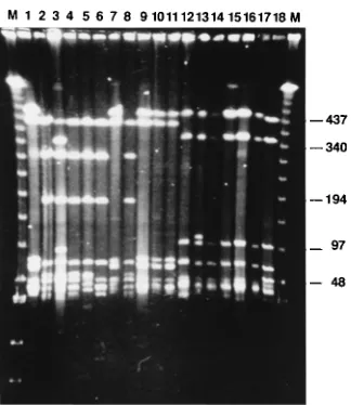
LRFP after MluI digestion showed two characteristic bands
of 220 and 80 kbp for the 21 B. garinii strains (PBi, PBr, PHei,
PMek, PHe, PRef, PWa, PFe, PMue, PFlk, and PSh [Fig. 1,
lanes 1 to 11, respectively] and PLi, PKi, PSeS, PScf, PBaEII,
PFin, POhm, PSoR, PFei, and PLa [data not shown]). Three
characteristic bands of 460, 320, and 90 kbp were observed for
the 10 B. afzelii strains (PLap, PMel, PAlt, PStb, PSpe, and
PBoj [Fig. 1, lanes 12 to 17, respectively] and PBo, PHo, PKr,
and PHa [data not shown]). One band of 145 kbp was typical
for the B. burgdorferi sensu stricto strains PKa1 (Fig. 1, lane 18)
and PFra, PHas, and PStm (data not shown). Remarkably, one
strain (strain PFCa) showed the three bands characteristic for
B. afzelii and the two specific bands characteristic for B. garinii.
This patient seems to have had a mixed infection (Table 1).
The SmaI LRFP revealed the specific bands for B. afzelii and
B. garinii for strain PFCa.
LRFP after ApaI, KspI, SmaI, and XhoI digestions.
We
ran-domly selected 11 B. garinii and 6 B. afzelii strains and analyzed
them with four different restriction enzymes to examine the
strains for hetero- or homogeneity within the species. Also,
one isolate of B. burgdorferi sensu stricto was examined. To
obtain a small number of fragments, restriction enzymes which
recognize GC-rich DNA sequences were chosen. Only bands
larger than 70 kbp were considered for analysis because
smaller bands could be plasmid bands.
B. garinii strains were heterogeneous. Strains PBi, PWa,
[image:2.612.66.298.70.210.2]PMue, PFlk, and PSh showed an identical pattern after
diges-tion with SmaI (fragments of 260, 135, 125, 105, and 90 kbp;
Fig. 2, lanes 1, 7, 9, 10, and 11), KspI (band sizes of 475 and 425
kbp; see Fig. 4, lanes 1, 7, 9, 10, and 11), ApaI (band sizes of
115, 105, 85, and 70 kbp; data not shown), and XhoI (band sizes
of 170, 160, 95, 90, 85, 80, and 70 kbp; Fig. 3, lanes 1, 7, 9, 10,
and 11). These strains were grouped together in group LRFP
1; they all shared OspA serotype 4. Three other B. garinii
strains, strains PBr, PMek, and PFe, revealed five bands with
sizes of 260, 240, 110, 80, and 75 kbp after SmaI digestion (Fig.
2, lanes 2, 4, and 8), three bands with sizes of 415, 290, and 175
kbp after KspI digestion (Fig. 4, lanes 2, 4, 8), four bands of
160, 150, 90, and 80 kbp after XhoI digestion (Fig. 3, lanes 2, 4,
8), and six bands of 120, 105, 90, 85, 80, and 75 kbp after ApaI
digestion (data not shown). These three strains were grouped
together in LRFP 2 and shared OspA serotype 3.
Strains PHei, PHe, and PRef showed different patterns after
digestion with the four different restriction enzymes. These
strains belonged to OspA serotypes 5, 6, and 7, respectively,
and were grouped together in LRFP 3 (Fig. 2 to 4, lanes 3, 5,
and 6).
B. afzelii PLap, PMel, PAlt, PStb, PSpe, and PBoj were
homogeneous after digestion with the four different restriction
enzymes. The strains showed identical patterns after SmaI
digestion (six bands of 145, 135, 125, 115, 85, and 80 kbp; Fig.
2, lanes 12 to 17), after KspI digestion (475, 385, and 110 kbp;
Fig. 4, lanes 12 to 17), after XhoI digestion (170, 125, 110, 80,
and 70 kbp; Fig. 3, lanes 12 to 17), and after ApaI digestion
(160, 155, and 75 kbp bands; data not shown). One strain,
strain PMel, showed an additional band after digestion with
SmaI (110 kbp), KspI (120 kbp), and ApaI (110 kbp). This band
was identified as a plasmid band (Fig. 5).
Plasmid analysis.
The 11 B. garinii strains revealed six to
eight linear plasmid bands of between 15 and 70 kbp. Five of
the B. garinii strains (strains PBi, PWa, PMue, PFlk, and PSh;
group LRFP 1) showed a unique plasmid pattern: three
plas-mids larger than 50 kbp and three to four plasplas-mids of between
20 and 30 kbp. Three of the B. garinii strains (strains PBr,
PMek, and PFe; group LRFP 2) showed very similar plasmid
patterns, with a characteristic four- to five-band pattern of
between 30 and 40 kbp (data not shown). The remaining three
strains showed different patterns. The six B. afzelii strains
showed similar plasmid patterns, with six to eight bands. All
strains exhibited four plasmids of approximately 65, 40, 30, and
27 kbp. Strain PMel showed a unique plasmid of 110 kbp; this
plasmid has not been described before for B. afzelii (Fig. 5, lane
13). B. burgdorferi sensu stricto PKa1 showed four plasmids of
approximately 60, 43, 40, and 30 kbp.
16S rRNA-specific PCR.
To confirm the validity of our
spe-cies determination by PFGE typing, a spespe-cies-specific PCR of
16S rRNA was performed for 18 strains. With the B.
burgdor-feri sensu lato primers, a specific 350-bp amplicon was
gener-ated from all isolates (expected band size, 357 bp) (Fig. 6a).
FIG. 1. LRFPs after MluI digestion. Lanes 1 to 11, B. garinii PBi, PBr, PHei,PMek, PHe, PRef, PWa, PFe, PMue, PFlk, and PSh, respectively, identified by two specific band of 220 and 80 kbp; lanes 12 to 17, B. afzelii PLap, PMel, PAlt, PStb, PSpe, and PBoj, respectively, characterized by three bands of 460, 320, and 90 kbp; lane 18, a B. burgdorferi sensu stricto strain (strain PKa1) characterized by one specific band of 145 kbp; lanes M, marker (lambda concatemer ladder; monomer size, 48.5 kbp). Numbers on the right are in kilobase pairs.
on May 15, 2020 by guest
http://jcm.asm.org/
Eleven strains revealed an amplicon of approximately 570 bp
(expected band size, 574 bp) only with the B. garinii-specific
primers (Fig. 6b, lanes 1 to 11). These strains were identified as
B. garinii. Six strains were identified as B. afzelii by an amplicon
of approximately 590 bp (expected band size, 591 bp) only with
a specific primer pair for B. afzelii (Fig. 6c, lanes 12 to 17).
With the specific primers for B. burgdorferi sensu stricto, only
one strain showed an amplicon of approximately 570 bp
(ex-pected band size, 574 bp) and was identified as B. burgdorferi
sensu stricto (Fig. 6d, lane 18). The PCR findings
corre-sponded completely to the results obtained by PFGE typing.
DISCUSSION
We set out to examine 36 isolates from the CSF of patients
with neuroborreliosis by using PFGE typing. This is the largest
number of CSF isolates analyzed until now. Among these CSF
isolates, we found 21 B. garinii isolates (58%), 10 B. afzelii
isolates (28%), 4 B. burgdorferi sensu stricto strains (11%), and
one isolate with a mixed pattern. To confirm these findings, we
examined 18 randomly selected strains by 16S rRNA-specific
PCR (33). Both methods revealed identical results. This is the
first report on the differentiation of isolates from the CSF of
patients with neuroborreliosis by both methods.
The degree of heterogeneity or homogeneity within the
spe-cies of B. garinii (11 strains) and B. afzelii (6 strains) was
ad-ditionally examined by LRFP after digestion of genomic DNA
with four different restriction enzymes (ApaI, KspI, SmaI, and
XhoI). This method has been successfully used to analyze
[image:3.612.60.556.84.430.2]iso-lates from ticks for heterogeneity or homogeneity within the
species without subsequent hybridization with specific probes
(14). Our six B. afzelii isolates from CSF showed identical
patterns after digestion with each of four different restriction
[image:3.612.320.554.568.705.2]FIG. 2. LRFPs after SmaI digestion. The lanes are the same as those de-scribed in the legend to Fig. 1.
TABLE 1. CSF strains used in the study
No. Strain Yr of isolation Place of residence Species OspA serotypea
1 PBib 1984 Southern Germany B. garinii 4
2 PBr 1984 Southern Germany B. garinii 3
3 PHei 1987 Northern Germany B. garinii 5
4 PMek 1992 Southern Germany B. garinii 3
5 PHe 1993 Southern Germany B. garinii 6
6 PRef 1988 Southern Germany B. garinii 7
7 PWab 1990 Southern Germany B. garinii 4
8 PFeb 1992 Southern Germany B. garinii 3
9 PMue 1992 Southern Germany B. garinii 4
10 PFlk 1990 Southern Germany B. garinii 4
11 PSh 1987 Southern Germany B. garinii 4
12 PLap 1988 Southern Germany B. afzelii 0
13 PMel 1990 Southern Germany B. afzelii X
14 PAltb 1988 Southern Germany B. afzelii 2
15 PStbb 1987 Southern Germany B. afzelii 0
16 PSpeb 1986 Southern Germany B. afzelii 2
17 PBojb 1989 Poland B. afzelii 0
18 PKa1b 1983 Southern Germany B. burgdorferi sensu stricto 1
19 PLi 1988 Southern Germany B. garinii 5
20 PKi 1992 Southern Germany B. garinii X
21 PSeS 1990 Southern Germany B. garinii 6
22 PScf 1992 Southern Germany B. garinii 4
23 PBaEII 1990 Southern Germany B. garinii 4
24 PFin 1991 Southern Germany B. garinii 4
25 POhm 1991 Southern Germany B. garinii 6
26 PSoR 1989 Southern Germany B. garinii 6
27 PFeib 1986 Southern Germany B. garinii 4
28 PLa 1988 Northern Germany B. garinii 3
29 PBo 1987 Southern Germany B. afzelii 0
30 PHo 1992 Southern Germany B. afzelii 0
31 PKr 1992 Southern Germany B. afzelii 2
32 PHa 1992 Northern Germany B. afzelii 2
33 PFra 1991 Southern Germany B. burgdorferi sensu stricto 1
34 PHasb 1992 Southern Germany B. burgdorferi sensu stricto 1
35 PStmb 1992 Northern Germany B. burgdorferi sensu stricto 1
36 PFCa 1992 Southern Germany B. afzelii/B. garinii 214
aSerotypes according to Wilske et al. (56). b
Isolates from patients with Bannwarth’s syndrome.
on May 15, 2020 by guest
http://jcm.asm.org/
enzymes, thus reflecting homogeneity within the isolates of the
B. afzelii group investigated. Among 20 B. afzelii isolates from
different origins, Belfaiza et al. (8) found indistinguishable
patterns after MluI digestion; 15 of 20 isolates had identical
SmaI patterns, but 5 of 20 isolates revealed different SmaI
patterns because of one missing or additional DNA fragment.
Characterization of strains from skin and CSF by Boehmer et
al. (11) revealed marginal differences within the B. afzelii
spe-cies after hybridization with different probes. By contrast,
Chetcuti et al. (17) observed major variations in the B. afzelii
group when they investigated strains isolated from ticks and
skin biopsy specimens. Our B. afzelii isolates from skin also
showed a heterogeneous pattern (unpublished data). These
differences are possibly explained by the different biological
sources (CSF, skin, and ticks) of the B. afzelii strains
investi-gated. While other investigators found at least marginal
differ-ences, our investigation with only B. afzelii strains from CSF
showed homogeneity within this group. Homogeneity within
B. afzelii strains isolated from CSF but heterogeneity within
B. afzelii strains from different sources suggests that subgroups
may exist within the B. afzelii species. These subgroups may
exhibit different pathogenic potentials and/or different
affini-ties to various tissues. Members of these subgroups may
pref-erentially penetrate into the CSF, while others may remain
localized in the skin. More strains must be examined to
vali-date these findings.
These closely related strains of B. afzelii were further
char-acterized by plasmid analysis since this technique has been
successfully used for strain differentiation (7, 48, 59). The
plas-mid pattern analysis revealed no major differences, another
strong indication for homogeneity within the B. afzelii isolates
from CSF. Xu and Johnson (59) also found only minor
differ-ences within the B. afzelii strains by analyzing the plasmid
patterns of strains from different sources. However, one
inter-esting finding was the existence of a 110-kbp plasmid
(approx-imate size) in one of our B. afzelii isolates (strain PMel).
Plasmids of this size have, to our knowledge, not been
de-scribed for the three species of B. burgdorferi sensu lato
patho-genic for humans but have been described for B. japonica,
group 21038, and relapsing fever borreliae (10, 16, 47).
In contrast to B. afzelii, B. garinii isolates from CSF show
het-erogeneity in their LRFPs, representing at least three groups
(LRFPs 1, 2, and 3). Comparison of the LRFPs with OspA
serotypes (56) revealed that LRFP 1 correlates with OspA
serotype 3 and that LRFP 2 correlates with OspA serotype 4.
The group LRFP 3 includes only a single isolate of OspA
serotypes 5, 6, and 7. This heterogeneity could also be detected
by plasmid analysis. When comparing the three different
meth-ods, there was a clear correlation between the plasmid pattern,
LRFPs 1 and 2, as well as OspA serotype 3 and 4, respectively.
However, a larger number of strains needs to be analyzed to
confirm these results. Our findings of heterogeneity within the
B. garinii species ties in with the observations made by using
other methods (5, 11, 12, 18, 23, 30, 46, 55, 56, 60).
[image:4.612.361.512.69.291.2]It has been speculated that the different clinical
manifesta-tions of Lyme borreliosis might be specifically correlated with
FIG. 3. LRFPs after XhoI digestion. The lanes are the same as thosede-scribed in the legend to Fig. 1.
[image:4.612.99.263.70.268.2]FIG. 4. LRFPs after KspI digestion. The lanes are the same as those de-scribed in the legend to Fig. 1.
FIG. 5. Plasmid profiles of B. afzelii PLap (lane 12), PMel (lane 13), PAlt (lane 14), PStb (lane 15), PSpe (lane 16), and PBoj (lane 17) and the B.
burg-dorferi sensu stricto strain (lane 18); lane M, marker (lambda DNA digested with HindIII).
on May 15, 2020 by guest
http://jcm.asm.org/
[image:4.612.99.261.521.708.2]certain subtypes of B. burgdorferi sensu lato (2, 3, 12, 51, 56,
58). Many investigators have observed B. afzelii to be prevalent
among skin isolates from Europe (6, 15, 51, 54, 56, 58). In
contrast, it has been discussed and is controversial whether a
similar association of certain subtypes of B. burgdorferi sensu
lato with isolates from CSF can be established. Since it is
difficult to isolate borreliae from the CSF, only a few isolates
have been investigated. Whereas only B. garinii has been
iso-lated from the CSF of five patients from Scandinavia (30),
analysis of 11 CSF isolates from patients in Germany
demon-strated the presence of all three species (56). Van Dam et al.
(51) found by rRNA gene restriction analysis of 10 isolates
from patients with extracutaneous symptoms 9 B. garinii and 1
B. burgdorferi sensu stricto strains. Those investigators
specu-lated that B. garinii might be associated with extracutaneous
symptoms and that B. afzelii might be associated with
cutane-ous manifestations of Lyme disease. Using PCR, Eiffert et al.
(22) found five different ospA sequences in isolates from the
CSF of 12 pediatric patients with LNB and thus confirmed the
heterogeneity of CSF isolates for OspA serotypes and
se-quences as well as the prevalence of B. garinii-associated OspA
types described by Wilske et al. (56) for 18 strains from
Eu-rope. Demaerschalck et al. (21) demonstrated ospA DNA from
all three species in one CSF sample and B. burgdorferi sensu
stricto and B. garinii ospA DNA in another CSF sample.
Our results demonstrate considerable heterogeneity among
CSF isolates from Europe. All three species have been isolated
from patients presenting with classical Bannwarth’s syndrome.
Thus, this type of neurological disease is not associated with a
certain species. One patient was even shown to have a mixed
infection of B. afzelii and B. garinii by PFGE typing and OspA
serotyping (55a). This observation was verified by the SmaI
LRFP, which revealed the bands specific for B. afzelii and B.
garinii. This is the first description of isolates from human CSF
consisting of two different strains and confirms infection with
more than one strain, as suggested by PCR (21, 36), in humans.
Halperin (25) discussed the fact that a milder form of
neu-roborreliosis is more frequent in the United States than in
Europe. It appears that the clinical outcome depends not only
on infection with strains of different species. Certain types of
outer surface proteins (OspA, OspC, and OspD) (9, 35, 57) are
associated with certain species (27, 34, 50a, 51a, 55b, 56) and
may play a role in pathogenesis (20, 24). In addition,
platelet-binding activity (19), expression of lectin activity that promotes
bacterial attachment to glycosaminoglycans (31), binding to
glycosphingolipids (4), and other as yet unknown factors may
be involved in the pathogenesis of the disease.
In conclusion, we found a considerable degree of
heteroge-neity among 36 isolates from CSF. This could explain the
contradictory results obtained previously by analysis of small
numbers of isolates from CSF. This heterogeneity has
impor-tant implications for the microbiological diagnosis
(serodiag-nosis and PCR) as well as the development of a borrelia
vac-cine.
ACKNOWLEDGMENTS
We thank C. Schweizer, U. Wilhelm, I. Pradel, and M. Bergerhausen for excellent technical help and G. Ruckdeschel for generous support.
REFERENCES
1. Ackermann, R., B. Rehse-Kupper, E. Gollmer, and R. Schmitt. 1988. Chronic neurologic manifestation of erythema migrans borreliosis. Ann. N. Y. Acad. Sci. 539:16–23.
2. Anthonissen, F. M., M. De Kesel, P. P. Hoet, and G. H. Bigaignon. 1994. Evidence for the involvement of different genospecies of Borrelia in the clinical outcome of Lyme disease in Belgium. Res. Microbiol. 145:327–331. 3. Assous, M. V., D. Postic, G. Paul, P. Nevot, and G. Baranton. 1993. Western blot analysis of sera from Lyme borreliosis patients according to the genomic species of the Borrelia strains used as antigens. Eur. J. Clin. Microbiol. Infect. Dis. 12:261–268.
4. Backenson, P. B., J. L. Coleman, and J. L. Benach. 1995. Borrelia burgdorferi shows specificity of binding to glycosphingolipids. Infect. Immun. 63:2811–2817. 5. Balmelli, T., and J. C. Piffaretti. 1995. Association between different clinical
manifestations of Lyme disease and different species of Borrelia burgdorferi sensu lato. Res. Microbiol. 146:329–340.
6. Baranton, G., D. Postic, I. Saint Girons, P. Boerlin, J. C. Piffaretti, M.
Assous, and P. A. D. Grimont.1992. Delineation of Borrelia burgdorferi sensu
[image:5.612.73.540.70.288.2]stricto, Borrelia garinii sp. nov., and group VS461 associated with Lyme borreliosis. Int. J. Syst. Bacteriol. 42:378–383.
FIG. 6. PCR amplification with 16S rRNA-specific primer. Lanes 1 to 11, B. garinii strains (PBi, PBr, PHei, PMek, PHe, PRef, PWa, PFe, PMue, PFlk, and PSh, respectively); lanes 12 to 17, B. afzelii PLap, PMel, PAlt, PStb, PSpe, and PBoj, respectively; lane 18, B. burgdorferi sensu stricto strain (strain PKa1); lane M, marker DNA (pBR322 DNA digested with MspI). (a) PCR amplification with B. burgdorferi sensu lato-specific primer. (b) PCR amplification with B. garinii-specific primer. (c) PCR amplification with B. afzelii-specific primer. (d) PCR amplification with B. burgdorferi sensu stricto-specific primer.
on May 15, 2020 by guest
http://jcm.asm.org/
7. Barbour, A. G. 1988. Plasmid analysis of Borrelia burgdorferi, the Lyme disease agent. J. Clin. Microbiol. 26:475–478.
8. Belfaiza, J., D. Postic, E. Bellenger, G. Baranton, and I. Saint Girons. 1993. Genomic fingerprinting of Borrelia burgdorferi sensu lato by pulsed-field gel electrophoresis. J. Clin. Microbiol. 31:2873–2877.
9. Bergstro¨m, S., V. G. Bundoc, and A. G. Barbour.1989. Molecular analysis of linear plasmid-encoded major surface proteins, OspA and OspB, of the Lyme disease spirochete Borrelia burgdorferi. Mol. Microbiol. 3:479–486. 10. Bergstro¨m, S., C. F. Garon, A. G. Barbour, and J. MacDougall.1992.
Extra-chromosomal elements of spirochetes. Res. Microbiol. 143:623–628. 11. Boehmer, R. H., G. Will, U. Busch, C. Schweizer, S. Jauris-Heipke, D.
Ro¨ssler, B. Wilske, and V. Preac-Mursic.1995. Genetic variability of
Bor-relia burgdorferi sensu lato strains demonstrated by restriction fragment
length polymorphism and probe hybridisation. Med. Microbiol. Lett. 4:180– 188.
12. Boerlin, P., O. Peter, A. G. Bretz, D. Postic, G. Baranton, and J. C. Piffaretti. 1992. Population genetic analysis of Borrelia burgdorferi isolates by multilocus enzyme electrophoresis. Infect. Immun. 60:1677–1683.
13. Burgdorfer, W., A. G. Barbour, S. F. Hayes, J. L. Benach, E. Grunwald, and
J. P. Davis.1982. Lyme disease—a tick-borne spirochetosis? Science 216:
1317–1319.
14. Busch, U., C. Hizo-Teufel, R. Boehmer, B. Wilske, and V. Preac-Mursic. 1995. Molecular characterization of Borrelia burgdorferi sensu lato strains by pulsed-field gel electrophoresis. Electrophoresis 16:744–747.
15. Canica, M. M., F. Nato, L. Du Merle, J. C. Mazie, G. Baranton, and D.
Postic.1993. Monoclonal antibodies for identification of Borrelia afzelii sp.
nov. associated with late cutaneous manifestations of Lyme borreliosis. Scand. J. Infect. Dis. 25:441–448.
16. Casjens, S., M. Delange, H. L. Ley III, P. Rosa, and W. M. Huang. 1995. Linear chromosomes of Lyme disease agent spirochetes: genetic diversity and conservation of gene order. J. Bacteriol. 177:2769–2780.
17. Chetcuti, M., M. Blot, and J. Meyer. 1994. Genomic variations among
Bor-relia afzelii strains. Med. Microbiol. Lett. 3:423–430.
18. Cinco, M., R. De Giovannini, P. Fattorini, F. Florian, and G. Graziosi. 1993. Classification of Italian isolates of Borrelia burgdorferi into three genomic groups. Microbiologica 16:323–332.
19. Coburn, J., S. W. Barthold, and J. M. Leong. 1994. Diverse Lyme disease spirochetes bind integrinaIIbb3on human platelets. Infect. Immun. 62:5559– 5567.
20. Comstock, L. E., E. Fikrig, R. J. Shoberg, R. A. Flavell, and D. D. Thomas. 1993. A monoclonal antibody to OspA inhibits association of Borrelia
burg-dorferi with human endothelial cells. Infect. Immun. 61:423–431.
21. Demaerschalck, I., A. B. Messaoud, M. De Kesel, B. Hoyois, Y. Lobet, P.
Hoet, G. Bigaignon, A. Bollen, and E. Godfroid.1995. Simultaneous
pres-ence of different Borrelia burgdorferi genospecies in biological fluids of Lyme disease patients. J. Clin. Microbiol. 33:602–608.
22. Eiffert, H., A. Ohlenbusch, H. J. Christen, R. Thomssen, A. Spielman, and
F. R. Matuschka.1995. Nondifferentiation between Lyme disease
spiro-chetes from vector ticks and human cerebrospinal fluids. J. Infect. Dis. 171: 476–479.
23. Filipuzzi-Jenny, E., M. Blot, N. Schmid-Berger, J. Meister-Turner, and J.
Meyer.1993. Genetic diversity among Borrelia burgdorferi isolates: more than
three genospecies? Res. Microbiol. 144:295–304.
24. Fuchs, H., R. Wallich, M. M. Simon, and M. D. Kramer. 1994. The outer surface protein A of the spirochete Borrelia burgdorferi is a plasmin(ogen) receptor. Proc. Natl. Acad. Sci. USA 91:12594–12598.
25. Halperin, J. J. 1991. North American Lyme neuroborreliosis. Scand. J. Infect. Dis. 77:74–80.
26. Hansen, K., and A. M. Lebech. 1992. The clinical and epidemiological profile of Lyme neuroborreliosis in Denmark 1985–1990. Brain 115:399–423. 27. Jauris-Heipke, S., G. Liegl, V. Preac-Mursic, D. Roessler, E. Schwab, E.
Soutschek, G. Will, and B. Wilske.1995. Molecular analysis of genes
encod-ing outer surface protein C (OspC) of Borrelia burgdorferi sensu lato: rela-tionship to ospA genotype and evidence of lateral gene exchange of ospC. J. Clin. Microbiol. 33:1860–1866.
27a.Johnson, R. C., G. P. Schmid, F. W. Hyde, A. G. Steigerwalt, and D. J.
Brenner.1984. Borrelia burgdorferi sp. nov.: etiologic agent of Lyme disease.
Int. J. Syst. Bacteriol. 34:496–497.
28. Karlsson, M., K. Hovind-Hougen, B. Svenungsson, and G. Stiernstedt. 1990. Cultivation and characterization of spirochetes from cerebrospinal fluid of patients with Lyme borreliosis. J. Clin. Microbiol. 28:473–479.
29. Kristoferitsch, W. 1989. Neuropathien bei Lyme-Borreliose. Ph.D. thesis. Springer Wien, New York.
30. Lebech, A. M., K. Hansen, B. Wilske, and M. Theisen. 1994. Taxonomic classification of 29 Borrelia burgdorferi strains isolated from patients with Lyme borreliosis: a comparison of five different phenotypic and genotypic typing schemes. Med. Microbiol. Immunol. 183:325–341.
31. Leong, J. M., P. E. Morrissey, E. Ortega-Barria, M. E. A. Pereira, and J.
Coburn. 1995. Hemagglutination and proteoglycan binding by the Lyme
disease spirochete, Borrelia burgdorferi. Infect. Immun. 63:874–883. 32. Luft, B. J., C. R. Steinman, H. C. Neimark, B. Muralidhar, T. Rush, M.
Finkel, M. Kunkel, and R. J. Dattwyler.1992. Invasion of the central nervous
system by Borrelia burgdorferi in acute disseminated infection. JAMA 267: 1364–1367.
33. Marconi, R. T., and C. F. Garon. 1992. Development of polymerase chain reaction primer sets for diagnosis of Lyme disease and for species-specific identification of Lyme disease isolates by 16S rRNA signature nucleotide analysis. J. Clin. Microbiol. 30:2830–2834.
34. Marconi, R. T., D. S. Samuels, R. K. Landry, and C. F. Garon. 1994. Analysis of the distribution and molecular heterogeneity of the ospD gene among the Lyme disease spirochetes: evidence for lateral gene exchange. J. Bacteriol.
176:4572–4582.
35. Norris, S. J., C. J. Carter, J. K. Howell, and A. G. Barbour. 1992. Low-passage-associated proteins of Borrelia burgdorferi B31: characterization and molecular cloning of OspD, a surface-exposed, plasmid-encoded lipoprotein. Infect. Immun. 60:4662–4672.
36. Oksi, J., M. Marjama¨ki, K. Koski, J. Nikoskelainen, and M. K. Viljanen.
1995. Bilateral facial palsy and meningitis caused by borrelia double infec-tion. Lancet 345:1583–1584. (Letter.)
37. Pachner, A. R. 1995. Early disseminated Lyme disease: Lyme meningitis. Am. J. Med. 98:30S–43S.
38. Pfister, H. W., K. M. Einha¨upl, B. Wilske, and V. Preac-Mursic. 1986. Bannwarth’s syndrome and the enlarged neurological spectrum of arthro-pod-borne borreliosis. Zentralbl. Bakteriol. Parasitenkd. Infektionskr. Hyg. Abt. 1 Orig. Reihe A 263:343–347.
39. Pfister, H. W., W. Kristoferitsch, and C. Meier. 1993. Early neurological involvement (Bannwarth’s syndrome), p. 152–167. In K. Weber and W. Burgdorfer (ed.), Aspects of Lyme borreliosis. Springer, Berlin.
40. Pfister, H. W., B. Wilske, and K. Weber. 1994. Lyme borreliosis: basic science and clinical aspects. Lancet 343:1013–1016.
41. Preac-Mursic, V., H. W. Pfister, H. Spiegel, R. Burk, B. Wilske, S. Reinhardt,
and R. Boehmer.1993. First isolation of Borrelia burgdorferi from an iris
biopsy. J. Clin. Neurol. Ophthalmol. 13:155–161.
42. Preac-Mursic, V., G. Schierz, H. W. Pfister, K. Einha¨upl, B. Wilske, and K.
Weber.1984. Isolierung einer Spirocha¨te aus Liquor cerebrospinalis bei
Meningoradiculitis-Bannwarth. Mu¨nch. Med. Wochenschr. 126:275–276. 43. Preac-Mursic, V., K. Weber, H. W. Pfister, B. Wilske, B. Gross, A. Baumann,
and J. Prokop.1989. Survival of Borrelia burgdorferi in antibiotically treated
patients with Lyme borreliosis. Infection 17:355–359.
44. Preac-Mursic, V., B. Wilske, and G. Schierz. 1986. European Borrelia
burg-dorferi isolated from humans and ticks: culture conditions and antibiotic
susceptibility. Zentralbl. Bakteriol. Parasitenkd. Infektionskr. Hyg. Abt. 1 Orig. Reihe A 263:112–118.
45. Preac-Mursic, V., B. Wilske, G. Schierz, H. W. Pfister, and K. Einha¨upl.
1984. Repeated isolation of spirochetes from the cerebrospinal fluid of a patient with meningoradiculitis Bannwarth Eur. J. Clin. Microbiol. 3:564– 565. (Letter.)
46. Ro¨ssler, D., H. Eiffert, S. Jauris-Heipke, G. Lehnert, V. Preac-Mursic, J.
Teepe, T. Schlott, E. Soutschek, and B. Wilske.1995. Molecular and
immu-nological characterization of the p83/100 protein of various Borrelia
burgdor-feri sensu lato strains. Med. Microbiol. Immunol. 184:23–32.
47. Saint-Girons, I., S. J. Norris, U. Go¨bel, J. Meyer, E. M. Walker, and R.
Zuerner.1992. Genome structure of spirochetes. Res. Microbiol. 143:615–
621.
48. Sta¨lhammar-Carlemalm, M., E. Jenny, L. Gern, A. Aeschlimann, and J.
Meyer.1990. Plasmid analysis and restriction fragment length
polymor-phisms of chromosomal DNA allow a distinction between Borrelia
burgdor-feri strains. Zentralbl. Bakteriol. Parasitenkd. Infektionskr. Hyg. Abt. 1 Orig.
274:28–39.
49. Steere, A. C. 1989. Medical progress—Lyme disease. N. Engl. J. Med. 321: 586–596.
50. Steere, A. C., R. L. Grodzicki, A. N. Kornblatt, J. E. Craft, A. G. Barbour, W.
Burgdorfer, G. P. Schmid, E. Johnson, and S. E. Malawista.1983. The
spirochetal etiology of Lyme disease. N. Engl. J. Med. 308:733–740. 50a.Theisen, M., M. Borre, M. J. Mathiesen, B. Mikkelsen, A. M. Lebech, and K.
Hansen.1995. Evolution of the Borrelia burgdorferi outer surface protein
OspC. J. Bacteriol. 177:3036–3044.
51. Van Dam, A. P., H. Kuiper, K. Vos, A. Widjojokusumo, B. M. De Jongh, L.
Spanjaard, A. C. P. Ramselaar, M. D. Kramer, and J. Dankert.1993.
Different genospecies of Borrelia burgdorferi are associated with distinct clinical manifestations of Lyme borreliosis. Clin. Infect. Dis. 17:708–717. 51a.Wallich, R., C. Helmes, U. E. Schaible, Y. Lobet, S. E. Moter, M. D. Kramer,
and M. M. Simon.1992. Evaluation of genetic divergence among Borrelia
burgdorferi isolates by use of OspA, fla, HSP60, and HSP70 gene probes.
Infect. Immun. 60:4856–4866.
52. Weber, K., and W. Burgdorfer (ed.). 1993. Aspects of lyme borreliosis. Springer, Berlin.
53. Weber, K., G. Schierz, B. Wilske, and V. Preac-Mursic. 1984. European erythema-migrans disease and related disorders. Yale J. Biol. Med. 57:463– 471.
54. Wienecke, R., N. Zo¨chling, U. Neubert, E. M. Schlu¨pen, M. Meurer, and
M. Volkenandt.1994. Molecular subtyping of Borrelia burgdorferi in erythema
migrans and acrodermatitis chronica atrophicans. J. Invest. Dermatol. 103:19– 22.
on May 15, 2020 by guest
http://jcm.asm.org/
55. Will, G., S. Jauris-Heipke, E. Schwab, U. Busch, D. Ro¨ssler, E. Soutschek, B.
Wilske, and V. Preac-Mursic.1995. Sequence analysis of ospA genes shows
homogeneity within Borrelia burgdorferi sensu stricto and Borrelia afzelii strains but reveals major subgroups within the Borrelia garinii species. Med. Microbiol. Immunol. 184:73–80.
55a.Wilske, B., U. Busch, H. Eiffert, V. Fingerle, H.-W. Pfister, D. Ro¨ssler, and
V. Preac-Mursic.1996. Diversity of OspA and OspC among cerebrospinal
fluid isolates of Borrelia burgdorferi sensu lato from patients with neurobor-reliosis in Germany. Med. Microbiol. Immunol. 184:195–201.
55b.Wilske, B., S. Jauris-Heipke, R. Lobentanzer, I. Pradel, V. Preac-Mursic, D.
Roessler, E. Soutschek, and R. C. Johnson.1995. Phenotypic analysis of the
outer surface protein C (OspC) of Borrelia burgdorferi sensu lato by mono-clonal antibodies: relationship to genospecies and OspA serotype. J. Clin. Microbiol. 33:103–109.
56. Wilske, B., V. Preac-Mursic, U. B. Go¨bel, B. Graf, S. Jauris-Heipke, E.
Soutschek, E. Schwab, and G. Zumstein.1993. An OspA serotyping system
for Borrelia burgdorferi based on reactivity with monoclonal antibodies and OspA sequence analysis. J. Clin. Microbiol. 31:340–350.
57. Wilske, B., V. Preac-Mursic, S. Jauris-Heipke, A. Hofmann, I. Pradel, E.
Soutschek, E. Schwab, G. Will, and G. Wanner.1993. Immunological and
molecular polymorphisms of OspC, an immunodominant major outer sur-face protein of Borrelia burgdorferi. Infect. Immun. 61:2182–2191. 58. Wilske, B., V. Preac-Mursic, G. Schierz, R. Ku¨hbeck, A. G. Barbour, and M.
Kramer.1988. Antigenic variability of Borrelia burgdorferi. Ann. N. Y. Acad.
Sci. 539:126–143.
59. Xu, Y., and R. C. Johnson. 1995. Analysis and comparison of plasmid profiles of Borrelia burgdorferi sensu lato strains. J. Clin. Microbiol. 33:2679–2685. 60. Zingg, B. C., R. N. Brown, R. S. Lane, and R. B. LeFebvre. 1993. Genetic
diversity among Borrelia burgdorferi isolates from wood rats and kangaroo rats in California. J. Clin. Microbiol. 31:3109–3114.