Amendment history:
Corrigendum
(February 2014)
Intramuscular viral delivery of paraplegin
rescues peripheral axonopathy in a model of
hereditary spastic paraplegia
Marinella Pirozzi, … , Alberto Auricchio, Elena I. Rugarli
J Clin Invest.
2006;
116(1)
:202-208.
https://doi.org/10.1172/JCI26210
.
Degeneration of peripheral motor axons is a common feature of several debilitating
diseases including complicated forms of hereditary spastic paraplegia. One such form is
caused by loss of the mitochondrial energy-dependent protease paraplegin.
Paraplegin-deficient mice display a progressive degeneration in several axonal tracts, characterized by
the accumulation of morphological abnormal mitochondria. We show that adenoassociated
virus–mediated (AAV-mediated) intramuscular delivery of paraplegin halted the progression
of neuropathological changes and rescued mitochondrial morphology in the peripheral
nerves of paraplegin-deficient mice. One single injection before onset of symptoms
improved the motor performance of paraplegin-deficient mice for up to 10 months, indicating
that the peripheral neuropathy contributes to the clinical phenotype. This study provides a
proof of principle that gene transfer may be an effective therapeutic option for patients with
paraplegin deficiency and demonstrates that AAV vectors can be successfully employed for
retrograde delivery of an intracellular protein to spinal motor neurons, opening new
perspectives for several hereditary axonal neuropathies of the peripheral nerves.
Research Article
Neuroscience
Find the latest version:
Intramuscular viral delivery of paraplegin
rescues peripheral axonopathy in a model
of hereditary spastic paraplegia
Marinella Pirozzi,1 Angelo Quattrini,2 Gennaro Andolfi,1 Giorgia Dina,2
Maria Chiara Malaguti,2 Alberto Auricchio,1 and Elena I. Rugarli1
1Telethon Institute of Genetics and Medicine (TIGEM), Naples, Italy. 2Department of Neurology,
University Vita-Salute, San Raffaele Scientific Institute, Milan, Italy.
Degeneration of peripheral motor axons is a common feature of several debilitating diseases including
compli-cated forms of hereditary spastic paraplegia. One such form is caused by loss of the mitochondrial
energy-depen-dent protease paraplegin. Paraplegin-deficient mice display a progressive degeneration in several axonal tracts,
characterized by the accumulation of morphological abnormal mitochondria. We show that adenoassociated
virus–mediated (AAV-mediated) intramuscular delivery of paraplegin halted the progression of
neuropatho-logical changes and rescued mitochondrial morphology in the peripheral nerves of paraplegin-deficient mice.
One single injection before onset of symptoms improved the motor performance of paraplegin-deficient mice
for up to 10 months, indicating that the peripheral neuropathy contributes to the clinical phenotype. This study
provides a proof of principle that gene transfer may be an effective therapeutic option for patients with
paraple-gin deficiency and demonstrates that AAV vectors can be successfully employed for retrograde delivery of an
intracellular protein to spinal motor neurons, opening new perspectives for several hereditary axonal
neuropa-thies of the peripheral nerves.
Introduction
Hereditary spastic paraplegia (HSP) is a neurodegenerative disor-der characterized by progressive loss of motor axons of the corti-cospinal tracts and of sensory axons of the fasciculus gracilis by a dying-back mechanism (1–4). HSP patients complain of relentless weakness, spasticity, and impairment of the vibratory sense in the lower limbs and are ultimately confined to a wheelchair. Currently, this debilitating condition has no cure.
HSP affects 1 in every 10,000 individuals and is genetically het-erogeneous, with 28 loci identified so far (5). Approximately 30% of HSP cases are inherited as autosomal-recessive traits. One of these recessive forms is due to loss-of-function mutations in the SPG7 gene, encoding paraplegin (6). Although the original family described with SPG7 mutations displayed a relatively pure phe-notype, most cases are complicated by additional neurological symptoms such as cortical and cerebellar atrophy, optic atrophy, amyotrophia, and peripheral neuropathy (7).
Paraplegin is a subunit of an energy-dependent protease in the inner mitochondrial membrane (the m-AAA protease) (8). Studies in yeast have shown that the m-AAA protease is involved in protein qual-ity control activities in the inner mitochondrial membrane, such as ensuring the removal of nonassembled or misfolded membrane pro-teins and controlling ribosome assembly in mitochondria and thereby the formation of respiratory complexes and ATP synthase (9, 10).
We previously developed a mouse model for paraplegin deficiency by knocking out the first 2 exons of the mouse Spg7 gene (11). Para-plegin-deficient mice are characterized by a late-onset distal
axo-nopathy of spinal, optic, and peripheral axons, thus mimicking the human disease. Axonal degeneration is preceded by axonal swelling due to accumulation of organelles and neurofilaments, a sign of impaired axonal transport. Interestingly, paraplegin-deficient mice begin to show impaired motor performance on the rotarod appara-tus at 4 months of age, 3 months before the first signs of pathology in the spinal cord are detected. The onset of the clinical phenotype correlates with the appearance of mitochondrial morphological abnormalities in synaptic terminals and in distal regions of spinal axons, suggesting that dysfunction of a subset of mitochondria is at the basis of the pathogenic process. This result is puzzling, since paraplegin is widely expressed in mitochondria from all tissues.
HSP due to paraplegin deficiency has several ideal features for exploiting a gene replacement therapeutic strategy: the onset is late, the progression of symptoms is relatively slow, and the molecular basis of the disease is caused by complete loss of function. The large window of time between the onset of motor symptoms and the actual loss of axons in the mouse model suggests that therapeutic options could be successfully exploited for several years after the disease is diagnosed. However, an open question is whether it is possible to successfully revert the accumulation of abnormal mitochondria in synaptic terminals of affected axons.
One challenge in designing therapies for neurological conditions based on gene delivery is the specific targeting of the affected neu-rons. Recently, a few studies have unraveled the ability of certain viral vectors to be retrogradely transported by axons, opening the path for novel routes of administration. Notably, both adenoas-sociated virus (AAV) vectors and lentiviral vectors based on equine infectious anemia virus have been shown to transduce spinal motor neurons after intramuscular injection (12–15).
In this study, we asked whether restoring paraplegin expres-sion in mitochondria is efficient in preventing, halting, or revers-ing the mitochondrial abnormalities and the clinical and
neuro-Nonstandard abbreviations used: AAV, adenoassociated virus; ALS, amyotrophic lateral sclerosis; GC, genome copies; HSP, hereditary spastic paraplegia.
Conflict of interest: The authors have declared that no conflict of interest exists.
research article
The Journal of Clinical Investigation http://www.jci.org Volume 116 Number 1 January 2006 203 pathological phenotype of Spg7–/– mice. As a proof of principle, we
resorted to Spg7 gene replacement in the peripheral nerves by use of recombinant AAV vectors delivered intramuscularly. We found that this approach was successful in halting the progression of neuropathology in the peripheral nerves of paraplegin-deficient mice. Furthermore, mitochondrial morphological abnormalities were partially rescued. Surprisingly, Spg7 gene replacement in the peripheral nerves before onset of symptoms ameliorated the rotarod performance of Spg7–/– mice, suggesting that peripheral nerve involvement plays an important role in the manifestation of motor symptoms in the mouse model.
Results
Peripheral neuropathy in Spg7–/– mice. We previously described a pro-gressive axonopathy in the sciatic nerve of Spg7–/– mice (11). More in-depth analyses allowed us to detect a few axoplasmic altera-tions as early as 10 months of age in the distal nerve, while frank signs of degeneration appeared at 18 months (Figure 1, A–D). Axonal changes and degenerating axons were also observed at 18 months in the purely sensory saphenous nerve (data not shown), indicating that Spg7–/– mice are affected by a motor-sen-sory peripheral neuropathy. We asked whether ultrastructural alterations of mitochondria precede these changes, as previ-ously observed in the spinal cord (11). We found ultrastructural changes in sciatic nerve mitochondria of Spg7–/– mice as early as 6 months of age, compared with age-matched controls (Figure 1, E and F). The neuromuscular junctions appeared normal until 18 months of age, but were almost completely denervated at 24 months (Figure 1, G and H).
Retrograde targeting of spinal motor neurons by intramuscular delivery of AAV vectors. We tested the ability of AAV vectors derived from dif-ferent serotypes (AAV2/1, AAV2/2, AAV2/5, AAV2/7, and AAV2/8, where the first number defines the genome and the second the capsid of origin) encoding for β-gal to efficiently transduce spinal motor neurons after a single unilateral intramuscular injection in the gastrocnemius muscles of adult mice. Four weeks after injec-tion, the muscle and the spinal cord were analyzed for expression of the reporter gene. We found that both AAV2/1 and AAV2/2 transduced spinal motor neurons after intramuscular delivery (Figure 2). Transduction was restricted to the motor neurons corresponding to the injected side. Quantification revealed that up to 47 and 82 motor neurons in the lumbar spinal cord were positive for LacZ staining following intramuscular injection of LacZ-expressing vectors AAV2/1 and AAV2/2 (AAV2/1-LacZ and AAV2/2-LacZ), respectively. These numbers are consistent with the labeling of a significant percentage of motor neurons innervating the gastrocnemius (16, 17). Notably, no known correlation exists between the efficiency of a given serotype of AAV to transduce the skeletal muscle and the retrograde transport to motor neurons.
To confirm that the transduction of spinal motor neurons derives from retrogradely transported viral vectors, we admin-istered AAV2/1-LacZ intravenously. Although this procedure allowed efficient delivery of β-gal to the skeletal muscles (18), we did not observe transduction of spinal motor neurons (data not shown). These data confirm that the AAV vectors do not cross the adult blood-brain barrier and suggest that they must come in direct contact with the presynaptic terminals of projecting neu-rons to be internalized and retrogradely transported.
[image:3.585.84.244.79.396.2]Intramuscular delivery of AAV2/2-Spg7 restores paraplegin expression in spinal cord mitochondria. Since paraplegin is totally deficient in Spg7–/– mice (11), we constructed AAV2/1 and AAV2/2 vectors expressing mouse Spg7 cDNA (AAV2/1-Spg7 and AAV2/2-Spg7) and tested their ability to reconstitute paraplegin expression in the skeletal muscle and the spinal cord of Spg7–/– mice following injection in the quadriceps and gastrocnemius muscles. Six months after injection, we isolated mitochondria from the skeletal muscle and spinal cord and assessed paraplegin expression by Western blot analysis. A pro-tein corresponding to correctly processed paraplegin was present in skeletal muscle mitochondria in the case of both AAV2/1-Spg7 and AAV2/2-Spg7. In contrast, paraplegin was detected in spinal cord mitochondria only after AAV2/2 delivery, not AAV2/1 (Figure 3), probably due to the lower efficiency of the latter serotype in trans-ducing the spinal motor neurons after intramuscular injection.
Figure 1
Peripheral neuropathy in Spg7–/– mice. (A–D) Semithin sections of the
distal sciatic nerve of a 6-month-old WT mouse (A) and Spg7–/– mice at 6
months (B), 10 months (C), and 18 months (D) of age. Axonal changes, characterized by the accumulation of dense material (arrows), are appreciated in Spg7–/– mice at 10 months and increase in severity
at 18 months. (E and F) Electron micrographs of the sciatic nerve of 6-month-old WT (E) and Spg7–/– (F) mice. A few axons already contain
abnormal mitochondria in Spg7–/– mice (asterisks). (G and H)
Confo-cal images of neuromuscular junctions from teased muscular fibers of 24-month-old WT (G) and Spg7–/– (H) mice double-labeled with
AAV-Spg7 delivered to the skeletal muscle halts the progression of neu-ropathology in Spg7–/– mice. To explore the efficacy of Spg7 cDNA replacement in blocking the progression of the neuropathologi-cal cascade in Spg7–/– mice, we injected AAV2/2-Spg7 (n = 12) or AAV2/1-Spg7 (n = 8) unilaterally into both the quadriceps and the gastrocnemius of 10-month-old Spg7–/– mice. The contralateral side was injected with an AAV-LacZ vector to provide an internal control. We selected 10-month-old animals because at this age the motor phenotype was established and the first neuropathologi-cal changes in the peripheral nerves were limited but detectable (Figure 1C). In HSP patients this would correspond to an early symptomatic stage of the disease. The injected mice were kept for 6 months and then sacrificed for neuropathological analysis.
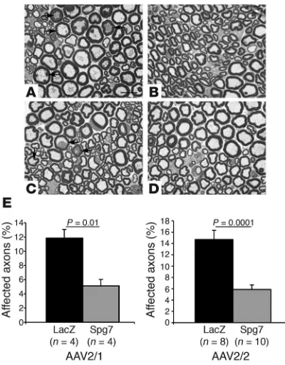
The effect of Spg7 replacement was first assessed in semithin sec-tions of the distal part of the sciatic nerve. As expected, the sciatic nerve on the side injected with AAV-LacZ vector displayed several axons showing signs of axonopathy, such as the presence of inclu-sions and deposits, and a few degenerating axons (Figure 4, A and C). This phenotype was greatly reduced in the sciatic nerve correspond-ing to the side where the muscles had been injected with either AAV2/1-Spg7 or AAV2/2-Spg7 (Figure 4, B and D). To obtain a quantitative measure of the rescue of the phenotype, we performed a morphometric analysis and counted the percentage of axons showing unequivocal signs of axonopathy in random sections. Both AAV2/1-Spg7– and AAV2/2-Spg7–treated animals showed a statistically significant decrease in the number of affected axons in their sciatic nerves (Figure 4E). Consistent with the amelioration of the peripheral neuropathy, the skeletal muscle showed decreased signs of denervation in the treated side (Supplemental Figure 1; available online with this article; doi:10.1172/JCI26210DS1).
Paraplegin replacement rescues mitochondrial morphological abnormali-ties. In Spg7–/– mice, morphological abnormal mitochondria accu-mulated in axons of the sciatic nerve as early as 6 months of age (Figure 1F) and involved a consistent percentage of axons at 10 months, the age at which we injected the vectors. As the reason for the mitochondrial abnormalities is still poorly understood, an important question is whether they are reversible and can be res-cued by restoring paraplegin function in mitochondria. To this
end, we performed ultrastructural studies in the group of animals injected with AAV2/2 vectors and found that the number of axons containing abnormal mitochondria and accu-mulation of neurofilaments and other aggre-gates was significantly reduced in the sciatic nerve from the side treated with AAV2/2-Spg7 compared with the AAV-LacZ–injected side (Figure 5). Strikingly, quantification showed that the percentage of axons with abnormal mitochondria in the side treated with the AAV2/2-Spg7 vector was even significantly lower than that at the time the viral vector was administered, as assessed in untreated Spg7–/– mice at 10 months (Figure 5H). This result strongly suggests that paraplegin delivery not only stops the progression of the mitochon-drial phenotype, but also partially rescues the abnormal morphology of the organelles.
Replacement of paraplegin in the peripheral nerve is sufficient to improve motor performance. The neuropathological rescue following gene replacement in the peripheral nerve might influence the motor phenotype of Spg7–/– mice. To address this issue, we turned to a different experimental paradigm, treating Spg7–/– mice bilaterally at 3 months of age before any identifiable motor deficit and fol-lowing their motor performance for several months through the rotarod test. The rotarod test is a useful means to assess the motor deficit of paraplegin-deficient mice from adulthood until about 15 months of age (11). After this age, the compliance of the mice low-ers, regardless of genotype, and the test renders less reliable data.
[image:4.585.44.357.82.212.2]We treated a cohort of 13 Spg7–/– mice at 3 months of age bilat-erally with AAV2/2-Spg7. As a control, another group of Spg7–/– mice (n = 13) were treated bilaterally with AAV-LacZ. At this age, the Spg7–/– mice do not display any identifiable clinical or path-ological phenotype. The motor performance of the 2 groups of mice was followed for 10 months through monthly rotarod tests and compared with that of Spg7+/+ animals of the same age (n = 9). Spg7–/– mice treated with the control AAV-LacZ vector started to show a decrease in their performance compared with WT animals
Figure 2
Retrograde transduction of spinal motor neurons by AAV vectors. Expression of β-gal activity in the skeletal muscle (A) and spinal cord (B) 4 weeks after injection of different AAV-LacZ vector serotypes in the gastrocnemius muscle. (A) AAV2/1 shows remarkable transduction of skeletal muscle, followed by AAV2/7 and AAV2/5. Conversely, the muscle transduction level for AAV2/2 and AAV2/8 is low. (B) Longitudinal sections of lumbar spinal cord to visualize targeting of motor neurons by retrograde transport of AAV (left side injected with AAV-LacZ). β-Gal activity restricted to motor neurons is visible only with AAV2/2 and AAV2/1. Magnification, ×10.
Figure 3
Paraplegin expression in skeletal muscle and spinal cord mitochondria after AAV-mediated intramuscular delivery of Spg7 cDNA. Western blot analysis of paraplegin on mitochondrial extracts from the gas-trocnemius and the spinal cord of Spg7–/– mice injected
[image:4.585.317.525.541.619.2]research article
The Journal of Clinical Investigation http://www.jci.org Volume 116 Number 1 January 2006 205 at about 4 months (Figure 6A), as we previously described (11).
Notably, we observed a statistically significant improvement of the rotarod performance in the group of Spg7–/– mice treated with the vector encoding paraplegin compared with those treated with the control AAV-LacZ vector (Figure 6A). This improvement was observed starting 2 months after treatment and was maintained for the entire period of observation.
To ascertain that the improvement in motor performance on the rotarod was due to a decrease in the number of affected axons in the treated animals, we analyzed their sciatic nerves and per-formed a morphometric analysis. We found that the percentage of axons showing neuropathological changes in the sciatic nerves of animals injected with AAV-Spg7 was significantly less than that observed in the mice treated with AAV-LacZ (Figure 6B).
Since the sciatic nerve is a mixed motor and sensory nerve, we wanted to determine whether part of the therapeutic effect is due to the rescue of sensory neurons. Paraplegin-deficient mice are
[image:5.585.63.263.81.337.2]indeed affected by a progressive retrograde axonopathy of sen-sory fibers in peripheral nerves and in spinal central pathways (ref. 11 and data not shown). A subset of these fibers, arising in the muscle spindles and conveying the sense of joint position, may have been targeted after intramuscular delivery of the viral vector. Some of these afferent fibers enter the spinal cord from the dorsal roots and ascend the spinal cord directly in the fas-ciculus gracilis. To this end, we performed semithin sections and ultrastructural studies of the fasciculus gracilis at cervical level in the same 2 groups of animals that were subjected to the rotarod analysis. These animals were injected bilaterally at 3 months and analyzed at 13 months. We did not find any statistically signifi-cant improvement in the cohort of mice treated with AAV-Spg7 versus AAV-LacZ (data not shown).
Figure 4
Rescue of the neuropathological phenotype in the peripheral nerve. (A–D) Semithin sections of the distal sciatic nerves in 16-month-old
Spg7–/– mice. Six months before the neuropathological studies, mice
were treated with a single intramuscular injection of AAV-LacZ on the left side (A and C) and either AAV2/1-Spg7 (B) or AAV2/2-Spg7 (D) on the right side. Note the decreased number of affected axons (arrows) in the nerves from the side treated with the Spg7 vectors. (E) Morpho-metric quantification of the percentage of affected axons for animals treated with AAV2/1 and AAV2/2 vectors. The P value of Student’s t
[image:5.585.332.507.381.743.2]test is shown. Scale bar: 15 µm (A–D).
Figure 5
Rescue of the ultrastructural mitochondrial abnormalities. (A–G) Elec-tron micrographs of the sciatic nerves in 16-month-old Spg7–/– mice.
Taken together, our data allow us to conclude that retrograde deliv-ery of paraplegin to spinal motor neurons through AAV vectors has an impact on the motor phenotype in paraplegin-deficient mice.
Discussion
Gene transfer to the peripheral nervous system has great therapeu-tic potential for the treatment of many neurological diseases, such as spinal muscular atrophy, amyotrophic lateral sclerosis (ALS), hereditary axonal motor neuropathies, and HSP. These diseases are characterized by degeneration of cell bodies and axons of spinal motor neurons of varying severity. Beside palliative treatments, no efficacious therapy is available yet. We used a HSP mouse model, due to the lack of the mitochondrial protein paraplegin, to exploit the efficacy of AAV-mediated gene delivery in the skeletal muscle to rescue degeneration of motor peripheral axons. Our data show that intramuscular administration of AAV-Spg7 in Spg7–/– mice resulted in reexpression of paraplegin in spinal cord mitochon-dria, rescued the neuropathological changes, and improved the motor performance on the rotarod.
AAV vectors have several desirable features for the transduction of neurons of the central and peripheral nervous system, such as deri-vation from a nonpathogenic virus and capability to infect postmi-totic cells and to mediate sustained expression after single admin-istration (19, 20). For these reasons, AAV vectors are being tested in human clinical trials (21). Previous studies have reached somewhat conflicting results on the efficacy of retrograde transduction of spinal motor neurons following the injection of AAV vectors in the skeletal muscle (13, 22). The most successful study has used AAV2/2 vectors to deliver IGF-1 in a mouse model of ALS, showing a signifi-cant prolongation of survival after treatment (13). Since IGF-1 is a secreted neurotrophic factor, the possibility remains that part of the therapeutic effect might be due to secretion of IGF-1 either from the muscle or from a low number of transduced motor neurons. Indeed, an increase in survival similar to that observed after AAV
intramus-cular administration was noted in ALS mice crossed with transgenic mice expressing an IGF-1 isoform under a muscle-specific promot-er, suggesting that release of IGF-1 from the muscle is sufficient for achieving some therapeutic effect in ALS mice (23).
We tested the ability of different AAV serotypes to retrogradely transduce spinal motor neurons. In agreement with previous obser-vations (13), our data show that both AAV2/2 and AAV2/1 were ret-rogradely transported to spinal motor neurons after intramuscular injection. Several lines of evidence support our claim of retrograde targeting of spinal motor neurons. First, the AAV-LacZ vectors were always injected in the skeletal muscle unilaterally, and β-gal stain-ing was restricted to the motor neurons of the injected side. Second, we found that transduction of motor neurons was not achieved after intravenous delivery of high doses of the AAV2/1 vector, indi-cating that it does not cross the adult blood-brain barrier. Finally, paraplegin is an intracellular protein, a subunit of the mitochon-drial m-AAA protease in the inner mitochonmitochon-drial membrane, which excludes the possibility that part of the therapeutic effect may be due to long-range secretion. Therefore the successful rescue of the sciatic nerve axonopathy, which occurred only in the side injected with AAV-Spg7, is by itself strong evidence for retrograde transport of the viral vector. Although AAV2/2 was slightly more efficient for retrograde transduction of motor neurons in our β-gal targeting experiments and Western blot analysis, we observed similar thera-peutic effects in Spg7–/– mice with both viral serotypes, suggesting that restoration of even a very low amount of paraplegin might be sufficient. This is in line with the fact that paraplegin is involved in a recessive form of HSP, due to complete loss of function of the protein. In conclusion, this study provides the first evidence to our knowledge that an intracellular protein can be targeted to motor neurons after intramuscular injection of AAV vectors, indicating that they should definitively be considered an alternative possibil-ity to equine infections anemia viral vectors for this application.
[image:6.585.55.274.80.346.2]The results of our study also provide novel indications as to the biology underlying HSP and its clinical aspects. We injected AAV-Spg7 in the skeletal muscles of knockout mice at 10 months of age. Although only a few signs of axonopathy are evident in the peripher-al nerve by light microscopy at this age, abnormperipher-al mitochondria had been accumulating in axons for several months (Figure 1, B and F). This age was chosen because it likely corresponds to an early symp-tomatic stage of the disease in human patients, a time when start-ing a therapy would be appropriate. We found that we successfully
Figure 6
Intramuscular viral delivery of paraplegin improves the motor perfor-mance of Spg7–/– mice. (A) Monthly performance of Spg7+/+ mice,
AAV-LacZ–treated Spg7–/– mice, and AAV2/2-Spg7–treated Spg7–/–
mice on an accelerating rotarod apparatus beginning at 3 months, the time of the bilateral viral injection. The mice treated bilaterally with the AAV-Spg7 vector showed a progressive better performance compared with Spg7–/– mice injected bilaterally with the AAV-LacZ
vector. Where significant, P values for individual Student’s t test between the 2 groups of treated Spg7–/– mice are shown. In addition,
mice treated with AAV2/2-Spg7 displayed a statistically significant difference in their performance during the course of the experiment (1-way ANOVA, P = 0.0001), as did the control mice (1-way ANOVA,
research article
The Journal of Clinical Investigation http://www.jci.org Volume 116 Number 1 January 2006 207 stopped the progression of the neuropathological changes in the
peripheral nerves. This result is especially striking at the ultrastruc-tural level. In fact, the number of axons showing abnormal mito-chondria was significantly reduced in the sciatic nerve of the treated side compared with the control side and dropped even below the levels present at the time when the vector was administered.
Based on these results, we conclude that restoration of low levels of paraplegin is sufficient to rescue the mitochondrial morphol-ogy defect, indicating that this phenotype is reversible in principle. Indeed, mitochondria are highly dynamic organelles, whose shape and function are subject to regulation. Notably, defects in genes reg-ulating mitochondrial morphology are emerging as an important cause of neuropathy in humans (24–26), suggesting that complex control of mitochondrial morphology may exist in neurons and may be important for axonal function. Furthermore, we noticed that the accumulation of mitochondria previously observed in affected axons (11) was reduced in the sciatic nerves from the AAV-Spg7– treated side, suggesting that rescue of the mitochondrial morphol-ogy corresponds with an improvement of axonal transport.
Delivery of paraplegin in Spg7–/– mice at 3 months of age ame-liorated their motor performance on the rotarod. This result was unexpected, since our approach transduced only spinal motor rons but failed to target pyramidal and extrapyramidal motor neu-rons and sensory neuneu-rons, whose axons continued to degenerate in these animals (E.I. Rugarli and A. Quattrini, unpublished observa-tion). We think it is unlikely that the expression of high levels of paraplegin in the muscle is sufficient to improve motor ability. In fact, although paraplegin is ubiquitously expressed, its loss causes a cell-autonomous, neuronal-specific phenotype. Paraplegin-defi-cient mice do not show any myopathic phenotype apart from signs of denervation (11). These data therefore provide a strong indica-tion that involvement of peripheral motor axons is an important clinical component that should be investigated more in depth in human patients. Indeed, in several other complicated forms of HSP, amyotrophia and peripheral motor neuropathy are prominent fea-tures, suggesting that involvement of spinal motor neurons is not uncommon in HSP (27–29). The lag of time that occurs between viral injection and motor improvement is attributable to the time for both retrograde uptake of the virus and paraplegin expression and transport of mitochondria along the axons.
In conclusion, our study provides a proof of principle that restora-tion of a low amount of paraplegin in affected neurons is extremely efficacious in blocking the progression of the disease, even after onset of the pathological process. Due to the mild phenotype of our HSP model, we were able to analyze the animals for phenotype rescue sev-eral months after a single administration of the therapeutic vector, showing unequivocally that AAV vectors sustain prolonged infection in neuronal cells. This approach can potentially be transferred with success to other motor axonal neuropathies of genetic origin.
Methods
Recombinant AAV vector construction and production. The mouse Spg7 coding sequence was cloned in the NotI-BamHI sites of the pAAV2.1-CMV-EGFP plasmid to obtain the pAAV2.1-CMV-Spg7 plasmid, which was triple transfected in 293 cells for recombinant AAV2/1- and AAV2/2-CMV-para-plegin production as previously described (30). The AAV2/1-, AAV2/2-, AAV2/5-, AAV2/7-, and AAV2/8-CMV-LacZ vectors were also produced by triple transfection as previously described (30–32). AAV2/1, AAV2/5, AAV2/7, and AAV2/8 vectors were purified by CsCl2 centrifugation, while
AAV2 vectors were produced by heparin affinity purification. Physical
titers of AAV vector preparations were assessed by real-time PCR (33) and were expressed as genome copies (GC) per milliliter. The vectors used in this study were produced by the TIGEM-AAV Vector Core.
Intramuscular and intravascular delivery of AAV-LacZ vectors. All animal pro-cedures were approved by the Institutional Animal Care and Use Commit-tee of Cardarelli Hospital (Naples, Italy). One-month-old C57BL/6 mice (at least 3 animals for each serotype) were anesthetized with avertin, and an incision 2–3 mm long was made in the skin to expose the gastrocnemius muscle. A total dose of 1011 GC of each vector preparation was injected into
3 different sites of the gastrocnemius (3 injections of 30 µl each) using a 100-µl Hamilton syringe. Alternatively, 1012 GC of AAV2/1 was injected in
the tail veins of C57BL/6 mice (n = 3).
For LacZ staining, animals were sacrificed 4 weeks after injection and perfused transcardially with 2% PFA/0.5% glutaraldehyde in PBS. The tis-sues were cryoprotected in 30% sucrose solution and processed for X-gal staining using standard techniques, either as whole-mount preparations (skeletal muscle) or after cryosectioning (spinal cord).
In vivo treatment with AAV-Spg7 vectors. Mice were anesthetized with avertin, and an incision 2–3 mm long was made in the skin to expose the gastrocnemius and quadriceps muscles. A total dose of 1011 GC of each
vec-tor preparation was injected into 3 different sites of the gastrocnemius and quadriceps (3 injections of 30 µl each) using a 100-µl Hamilton syringe.
Mice were treated following 2 experimental protocols. In the first experimen-tal setting, 10-month-old Spg7–/– mice were injected unilaterally either with AAV2/2-Spg7 (n = 12) or AAV2/1-Spg7 (n = 8). In both cases, the contralateral side was injected with AAV-LacZ vector for an internal control. Animals were kept for 6 months before being sacrificed for Western blot, histological, and ultrastructural analysis. Age at the time of analysis was 16 months.
In the second experimental setting, 3-month-old Spg7–/– mice (n = 13) from different litters were treated bilaterally with AAV2/2-Spg7. A control group of Spg7–/–mice of the same age (n = 13) was treated bilaterally with AAV-LacZ. All mice were tested in the rotarod apparatus once a month for the next 10 months, and their performance was compared with that of an age-matched group of Spg7+/+ mice (n = 9). After 10 months, AAV-Spg7– and AAV-LacZ–treated mice were sacrificed for histological analysis to quantify the rescue of the neuropathological phenotype. Age at the time of histological analysis was 13 months.
Western blot. Western blot analysis was performed on Spg7–/– mice inject-ed unilaterally in the gastrocnemius and the quadriceps muscles with AAV2/1-Spg7 (n = 2) and AAV2/2-Spg7 (n = 1) at 10 months and sacri-ficed 6 months afterward (see above). Mitochondria were isolated from the gastrocnemius muscle and the lumbar spinal cord as previously described (11). Approximately 50 µg of mitochondrial proteins was separated by SDS-PAGE and blotted onto nitrocellulose. Filters were incubated with a specific antiparaplegin antiserum (11).
times in PBS, mounted in Vectashield on standard glass slides (Vector Labora-tories), and examined under a confocal scanning laser microscope.
Neuropathology. Semithin and ultrathin morphological analyses were conducted as described previously (34, 35). Mice were anesthetized intraperitoneally with avertin and perfused with 4% paraformaldehyde in PBS. The sciatic nerves and the spinal cord were removed, postfixed in 0.12 M phosphate buffer and 2% glutaraldehyde, and then sectioned transversely into 2-mm blocks, postfixed with osmium tetroxide, and embedded in Epon (Sigma-Aldrich). Cross sections were cut from the distal part of the sciatic nerve and the cervical part of the spinal cord.
Morphometric analyses. The percentage of abnormal axons in semithin sec-tions was counted in 3 random secsec-tions per sciatic nerve. At least 300 axons were scored for each nerve. For quantification of ultrastructural mitochon-dria abnormalities, an average of 10 fields per nerve was analyzed. In each case, we scored at least 100 axons. Analyses were performed on photo-graphs captured using an electron microscope (magnification, ×3,000). All morphometric counts were performed in a blinded and nonbiased manner, with the treatment of animal unknown to the experimenter.
Rotarod. Motor performance was evaluated with a Rota-Rod apparatus (accelerating model; Ugo Basile). Each Spg7–/– experimental group consisted of 13 animals derived from different litters. The group of WT mice consisted of 9 mice. All the animals were 3 months old when the analysis started. All animals were tested on the rotarod once a month from the time of injection. Mice were placed on the accelerating rod at a starting speed of 4 rpm, reaching a final speed of 40 rpm in 5 minutes. The animals were allowed to stay on the rod for a maximum of 300 seconds, and the time of hold on the rod was scored.
Statistics. Data are expressed as the mean ± SEM. Statistical analysis of morphometric measurements was performed using a 2-tailed unpaired Student’s t test. Statistical analysis of rotarod activity was performed by 2-tailed unpaired Student’s t test at each time point between the AAV-LacZ– and AAV-Spg7–treated groups. In addition, we performed 1-way ANOVA for comparison of the rotarod performance over the course of study within each experimental group.
Acknowledgments
The authors wish to thank Diego Di Bernardo, Graciana Diez-Roux, and Enrico Surace for helpful comments and discussion. This work has been supported by Telethon Foundation and Mus-cular Dystrophy Association grants (to E.I. Rugarli) and by Diag-nostic Molecular Imaging (DiMi) Network of Excellence of the European Community (to A. Auricchio).
Received for publication July 8, 2005, and accepted in revised form October 18, 2005.
Address correspondence to: Elena I. Rugarli, Division of Biochem-istry and Genetics, National Neurological Institute, via Temolo 4, 20126, Milan, Italy. Phone: 39-02-23942614; Fax: 39-02-23942619; E-mail: rugarli@istituto-besta.it.
Elena I. Rugarli’s present address is: Division of Biochemistry and Genetics, National Neurological Institute, Milan, Italy.
1. Harding, A.E. 1983. Classification of the hereditary ataxias and paraplegias. Lancet.1:1151–1154. 2. Harding, A.E. 1984. Hereditary “pure” spastic
para-plegia. In The hereditary ataxias and related disorders. Churchill Livingstone. New York, New York, USA. 174–191.
3. Reid, E. 2003. Science in motion: common molecu-lar pathological themes emerge in the hereditary spastic paraplegias. J. Med. Genet.40:81–86. 4. Schwarz, G.A., and Liu, C.N. 1956. Hereditary
(familial) spastic paraplegia: further clinical and pathological observations. Arch. Neurol. Psychiatry. 75:144–162.
5. Fink, J.K. 2003. Advances in the hereditary spastic paraplegias. Exp. Neurol.184(Suppl. 1):S106–S110. 6. Casari, G., et al. 1998. Spastic paraplegia and
OXPHOS impairment caused by mutations in paraplegin, a nuclear-encoded mitochondrial metalloprotease. Cell.93:973–983.
7. Wilkinson, P.A., et al. 2004. A clinical, genetic and biochemical study of SPG7 mutations in heredi-tary spastic paraplegia. Brain.127:973–980. 8. Langer, T. 2000. AAA proteases: cellular machines
for degrading membrane proteins. Trends Biochem. Sci.25:247–251.
9. Langer, T., Kaser, M., Klanner, C., and Leonhard, K. 2001. AAA proteases of mitochondria: quality control of membrane proteins and regulatory func-tions during mitochondrial biogenesis. Biochem. Soc. Trans.29:431–436.
10. Nolden, M., et al. 2005. The m-AAA protease defective in hereditary spastic paraplegia con-trols ribosome assembly in mitochondria. Cell.
123:277–289.
11. Ferreirinha, F., et al. 2004. Axonal degeneration in paraplegin-deficient mice is associated with abnormal mitochondria and impairment of axonal transport. J. Clin. Invest.113:231–242. doi:10.1172/ JCI200420138.
12. Mazarakis, N.D., et al. 2001. Rabies virus glycopro-tein pseudotyping of lentiviral vectors enables ret-rograde axonal transport and access to the nervous system after peripheral delivery. Hum. Mol. Genet.
10:2109–2121.
13. Kaspar, B.K., Llado, J., Sherkat, N., Rothstein, J.D., and Gage, F.H. 2003. Retrograde viral delivery of IGF-1 prolongs survival in a mouse ALS model.
Science.301:839–842.
14. Azzouz, M., et al. 2004. Lentivector-mediated SMN replacement in a mouse model of spinal muscular atrophy. J. Clin. Invest.114:1726–1731. doi:10.1172/ JCI200422922.
15. Azzouz, M., et al. 2004. VEGF delivery with retro-gradely transported lentivector prolongs survival in a mouse ALS model. Nature.429:413–417. 16. LaMonte, B.H., et al. 2002. Disruption of dynein/
dynactin inhibits axonal transport in motor neu-rons causing late-onset progressive degeneration.
Neuron.34:715–727.
17. Mohajeri, M.H., Figlewicz, D.A., and Bohn, M.C. 1998. Selective loss of alpha motoneurons inner-vating the medial gastrocnemius muscle in a mouse model of amyotrophic lateral sclerosis. Exp. Neurol. 150:329–336.
18. Gregorevic, P., et al. 2004. Systemic delivery of genes to striated muscles using adeno-associated viral vectors. Nat. Med.10:828–834.
19. Monahan, P.E., and Samulski, R.J. 2000. AAV vec-tors: is clinical success on the horizon? Gene Ther. 7:24–30.
20. Xiao, W., et al. 1999. Gene therapy vectors based on adeno-associated virus type 1. J. Virol.73:3994–4003. 21. Hildinger, M., and Auricchio, A. 2004. Advances in AAV-mediated gene transfer for the treatment of inherited disorders. Eur. J. Hum. Genet.12:263–271. 22. Wang, L.J., et al. 2002. Neuroprotective effects of
glial cell line-derived neurotrophic factor mediated by an adeno-associated virus vector in a transgenic animal model of amyotrophic lateral sclerosis.
J. Neurosci.22:6920–6928.
23. Dobrowolny, G., et al. 2005. Muscle expression of a local Igf-1 isoform protects motor neurons in an ALS mouse model. J. Cell Biol.168:193–199. 24. Delettre, C., et al. 2000. Nuclear gene OPA1,
encod-ing a mitochondrial dynamin-related protein, is mutated in dominant optic atrophy. Nat. Genet.
26:207–210.
25. Alexander, C., et al. 2000. OPA1, encoding a dyna-min-related GTPase, is mutated in autosomal dominant optic atrophy linked to chromosome 3q28. Nat. Genet.26:211–215.
26. Zuchner, S., et al. 2004. Mutations in the mito-chondrial GTPase mitofusin 2 cause Charcot-Marie-Tooth neuropathy type 2A. Nat. Genet. 36:449–451.
27. Patel, H., et al. 2002. SPG20 is mutated in Troyer syndrome, an hereditary spastic paraplegia. Nat. Genet.31:347–348.
28. Windpassinger, C., et al. 2004. Heterozygous mis-sense mutations in BSCL2 are associated with distal hereditary motor neuropathy and Silver syndrome.
Nat. Genet.36:271–276.
29. Fink, J.K. 2002. Hereditary spastic paraplegia: the pace quickens. Ann. Neurol.51:669–672. 30. Auricchio, A., Hildinger, M., O’Connor, E., Gao,
G.P., and Wilson, J.M. 2001. Isolation of highly infectious and pure adeno-associated virus type 2 vectors with a single-step gravity-flow column.
Hum. Gene Ther.12:71–76.
31. Auricchio, A., et al. 2001. Exchange of surface pro-teins impacts on viral vector cellular specificity and transduction characteristics: the retina as a model.
Hum. Mol. Genet.10:3075–3081.
32. Gao, G.P., et al. 2002. Novel adeno-associ-ated viruses from rhesus monkeys as vectors for human gene therapy. Proc. Natl. Acad. Sci. U. S. A. 99:11854–11859.
33. Gao, G., et al. 2000. Purification of recombinant adeno-associated virus vectors by column chroma-tography and its performance in vivo. Hum. Gene Ther.11:2079–2091.
34. Previtali, S.C., et al. 2000. Epitope-tagged P(0) glyco-protein causes Charcot-Marie-Tooth-like neuropa-thy in transgenic mice. J. Cell Biol.151:1035–1046. 35. Consiglio, A., et al. 2001. In vivo gene therapy of