cause, consequence, or context?
John A. Curci, Robert W. Thompson
J Clin Invest.
2004;
114(2)
:168-171.
https://doi.org/10.1172/JCI22309
.
Abdominal aortic aneurysms are common and life threatening. Although CD4
+T cells are
abundant in aneurysm tissue, their role in disease progression remains unclear. A new
study shows that mouse aortic allografts placed in animals lacking IFN-
g
receptors develop
a Th2 inflammatory response with aortic aneurysms, whereas Th1 responses promote
intimal hyperplasia. It is expected that these surprising findings will stimulate further efforts
to clarify whether adaptive cellular immunity in aneurysm disease is detrimental or
potentially beneficial.
Commentary
Find the latest version:
Invest. 101:746–754.
13. Loetscher, M., Loetscher, P., Brass, N., Meese, E., and Moser, B. 1998. Lymphocyte-spe-cific chemokine receptor CXCR3: regulation, chemokine binding and gene localization. Eur. J. Immunol. 28:3696–3705.
14. Schroder, K., Hertzog, P.J., Ravasi, T., and Hume, D.A. 2004. Interferon-gamma: an overview of
sig-nals, mechanisms and functions. J. Leukoc. Biol.
75:163–189.
15. Raghu, G., et al. 2004. A placebo-controlled trial of interferon gamma-1b in patients with idiopathic pulmonary fibrosis. N. Engl. J. Med. 350:125–133. 16. Strieter, R.M., Starko, K.M., Enelow, R.I., Noth,
I., and Valentine, V.G. 2004. Effects of interferon gamma-1b on biomarker expression in idiopathic
pulmonary fibrosis patients. Am. J. Respir. Crit. Care Med. doi:10.1164/rccm.200312-1670OC. 17. Ziesche, R., Hofbauer, E., Wittmann, K., Petkov,
V., and Block, L.H. 1999. A preliminary study of long-term treatment with interferon gamma-1b and low-dose prednisolone in patients with idiopathic pulmonary fibrosis. N. Engl. J. Med.
341:1264–1269.
Adaptive cellular immunity in aortic aneurysms:
cause, consequence, or context?
John A. Curci1,2 and Robert W. Thompson1,2,3
1Department of Surgery (Section of Vascular Surgery), 2Department of Radiology, and 3Department of Cell Biology and Physiology,
Washington University School of Medicine, St. Louis, Missouri, USA.
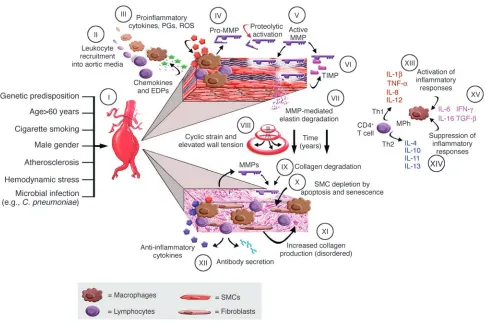
models. Although the specific etiology is still unclear, aneurysms are probably initiat-ed by aortic wall injury couplinitiat-ed with a series of epidemiological risk factors. Recruitment of leukocytes into the aortic media appears to be an early and pivotal event, likely pro-moted by chemokines (2) and elastin degra-dation peptides (3). Mononuclear phagocyte infiltration is associated with production of proinflammatory cytokines (4), prostaglan-din derivatives (5), and reactive oxygen spe-cies (6) as part of an innate inflammatory response. These macrophages are the prin-ciple source of MMPs (7), which can also be secreted by neutrophils, lymphocytes, and resident mesenchymal cells. Gelatinase B (MMP-9) has been extensively studied in human AAAs (8), but many other MMPs and endogenous tissue inhibitors of metal-loproteinases (known as TIMPs) have also been described. Animal models of aortic aneurysm confirm that MMPs produced by chronic inflammatory cells are mediators of elastin and collagen degradation (9–11); moreover, the suppression of experimental aneurysms by MMP inhibitors has led to a promising therapeutic strategy (12). Other enzymes expressed in atherosclerosis and AAAs, particularly plasminogen activators and cathepsins, may also contribute to matrix proteolysis.
Degradation of elastin and interstitial collagen initiates aortic dilatation and tor-tuosity, with changes in aortic wall geom-etry increasing cyclic strain and wall ten-sion over a period of years. At later stages of disease, disorganized interstitial collagen is deposited within the media and adventitia, and collagen degradation becomes more prominent, further weakening the aortic wall. Although medial smooth muscle cells (SMCs) might otherwise promote structur-al repair in the damaged aorta, apoptosis and cellular senescence cause depletion of this cell population (13, 14).
Adaptive immunity in aortic aneurysms
In addition to macrophages, human AAAs demonstrate large numbers of T cells, B lymphocytes, plasma cells, and DCs within the outer media and adventitia (15, 16). AAA tissues also contain large amounts of immunoglobulin protein, and IgG extracted from human AAAs exhibits immunoreactivity with aortic wall matrix proteins (17). This suggests that a humor-al (auto)immune response is a frequent occurrence in AAAs. Recent work has led to identification of several putative antigens that may be novel extracellular matrix pro-teins associated with large arteries (18).
The specificity of the immune response in AAAs is still unclear, as B lymphocytes derived from AAAs exhibit an unrestrict-ed repertoire of immunoglobulin heavy chain genes (19), and T cell receptor diversity reflects a polyclonal response (20). In a recent and comprehensive analysis, Ocana et al. (21) demonstrated that aneurysm-infiltrating lymphocytes consist of activated memory cells
express-Abdominal aortic aneurysms are common and life threatening. Although
CD4
+T cells are abundant in aneurysm tissue, their role in disease
pro-gression remains unclear. A new study (see the related article beginning
on page 300) shows that mouse aortic allografts placed in animals
lack-ing IFN-γ receptors develop a Th2 inflammatory response with aortic
aneurysms, whereas Th1 responses promote intimal hyperplasia. It is
expected that these surprising findings will stimulate further efforts to
clarify whether adaptive cellular immunity in aneurysm disease is
detri-mental or potentially beneficial.
The propensity of abdominal aortic aneu-rysms (AAAs) to rupture is their most important clinical feature, a consequence of increased hemodynamic stresses placed on the dilated wall, diminished tensile strength within the outer media and adven-titia, and dynamic factors influencing the balance between matrix metabolism and repair. Surgical repair of AAAs greater than 5.5 cm in diameter is effective treatment, but repair of smaller aneurysms offers no survival advantage. Effective nonsurgical treatments to prevent aneurysm expansion would therefore be an enticing prospect for patients with small AAAs (1).
Pathophysiology of AAAs
Figure 1 presents a summary of pathophys-iological events currently thought to con-tribute to aneurysmal degeneration, based on studies of human end-stage AAA tissues and several different experimental animal
Nonstandard abbreviations used: abdominal aortic aneurysm (AAA); IFN-γ receptor deficient (GRKO); smooth muscle cell (SMC); tissue inhibitor of metalloproteinase (TIMP).
Conflict of interest: The authors have declared that no conflict of interest exists.
ing costimulatory molecules. More spe-cifically, CD4+ T cells predominated in AAA tissue, with expression of αβ T cell receptors, T cell activation markers (CD69 and DR), a memory cell pheno-type (CD45RO+CD45RA–CD62L–), and a distinct pattern of cell surface molecules (including CD54, CD31, CD11a, CD29, CD44, CD95, and CD27). These results parallel observations in other chronic autoimmune/inflammatory disorders, supporting the existence of a cellular immune response in AAAs.
The triggers of adaptive immunity in AAAs are unknown, but there is often evidence of infection with Chlamydia pneumoniae
in individuals with AAAs, and microbial infection might directly stimulate patho-logical immune responses in AAA tissue (22). Prior microbial infection might also target immune responses to aortic wall proteins through a process of molecular mimicry; alternatively, immune responses against aortic wall structural compo-nents may arise secondary to long-stand-ing inflammation and connective tissue destruction, through proteolytic exposure of neoepitopes within matrix proteins.
The pathophysiologic implications of aortic wall cellular immune responses are an important focus for investigation, because cytokines produced by T cells (as
[image:3.585.50.536.79.403.2]well as direct interactions between T cells and activated macrophages) may have a substantial influence on macrophage production of MMPs and other matrix-degrading proteases. Proinflammatory cytokines associated with Th1 immune responses are elevated in the blood and aortic tissue of patients with AAAs (23), and circulating levels of IFN-γ correlate with aneurysm expansion (24). Advanced human AAA tissues also express Th2-asso-ciated cytokines, particularly IL-4 and IL-10 (25). This is important given that IL-4 and IL-10 suppress human macrophage expression of MMP-9 (26), which suggests that Th2 immune responses might serve
Figure 1
to restrain ongoing aneurysmal degen-eration. In contrast, Schonbeck et al. (27) recently described that Th2 cytokines (IL-4, IL-5, and IL-10) were elevated to a greater extent in AAAs than in atheroma, where Th1 cytokines (IFN-γ, IL-2, IL-12, IL-15, and IL-18) predominated, and that IFN-γ
receptor expression was minimal in AAAs. This led the authors to propose that Th2 immune responses might actually direct atherosclerotic lesions toward aneurysm development rather than the formation of occlusive atheroma.
Experimental studies
Experimental studies are only beginning to help clarify the functional role of adap-tive immunity in AAAs. For example, pre-vious work from our laboratory indicated that mice lacking IL-10 developed larger aneurysms than did wild-type controls when subjected to elastase-induced AAAs, in association with more extensive degra-dation of elastin and collagen (28). Further studies have shown that larger aneurysms also occur in CD4-deficient and IL-4–defi-cient mice, which suggests the existence of a Th2 immune response that might oth-erwise suppress the extent of aneurysmal degeneration, whereas no significant effect on AAAs has been observed in mice with IFN-γ deficiency (R.W. Thompson et al., unpublished results).
Using a calcium chloride–induced mouse model of AAAs, Baxter and colleagues (29) demonstrated that the absence of CD4+ T cells prevents the induction of experimen-tal aneurysms. They also observed that administration of IFN-γ reconstituted AAAs in CD4-deficient mice, that IFN-γ– deficient mice exhibited suppression of AAAs, and that the aneurysm-resistant phenotype was reversed by administration of wild-type splenocytes. These results lend strong support to the notion that Th1-type immune responses are detrimental in aneu-rysm disease. Based on this paradigm, one can envision future efforts to shift the cel-lular immune response from one dominat-ed by Th1 cytokines to one favoring Th2 cytokines (perhaps by mucosal immuniza-tion strategies) in order to suppress aneu-rysmal degeneration.
Upon this background, Shimizu et al. (30) present an intriguing study in this issue of the JCI that examines the diver-gent functional roles of IFN-γ and IL-4 in aortic pathology associated with a mouse model of arterial allotransplantation. The authors demonstrate that
histocompat-ibility-mismatched aortas transplanted into IFN-γ receptor–deficient (GRKO) recipients develop an immune response dominated by IL-4. These vessels subse-quently develop severe inflammation, elas-tin degradation, increased expression of elastolytic MMPs (MMP-9 and MMP-12), and large aortic aneurysms. AAAs were not observed in allotransplants into wild-type recipients, which otherwise devel-oped intimal hyperplasia. Furthermore, aneurysm development in GRKO allograft recipients was prevented by administra-tion of anti–IL-4 blocking antibodies or by concomitant genetic deficiency in IL-4. Shimizu et al. conclude that IL-4 specifi-cally mediates an inflammatory process leading to aneurysmal degeneration in this allograft model — a surprising and potentially important finding that chal-lenges current concepts of how adaptive cellular immune responses might influ-ence arterial disease.
The work by Shimizu et al. (30) has potentially valuable implications for the pathophysiology of allograft arteriopathy, intimal hyperplasia, arteritis, and AAAs and directly contradicts the paradigm that Th1 immune responses contribute to aneurys-mal degeneration. There are several issues complicating interpretation of this study, however, and a number of questions that still need to be addressed. For example, the experimental model involved transplanta-tion of histocompatibility-mismatched aortic tissues into genetically altered recipi-ents. Although the authors note that CD8+ T cells were rarely observed within the transplanted aortic wall, it seems unlikely that the allograft response can be over-looked in assessing the results, since MHC incompatibility would also affect CD4+ T cell responses. Secondly, it is apparent that after wild-type aortas were transplanted into GRKO recipients, resident cells within the graft were still able to respond to IFN-γ, while only the GRKO host (inflammatory) cells were unresponsive. This suggests that development of a Th1 response (as indicat-ed by the large observindicat-ed increase in IFN-γ
expression) might still have had an effect on cells within the aortic graft. It is not known if the activity of other Th1 cytokines might be enhanced in this experimental context, where IFN-γ is unable to exert restraint on inflammatory cell function; indeed, previous studies in IFN-γ–deficient mice demonstrate a marked increase in inflammatory responses (31). A third issue that needs to be considered is the peculiar
role played by IFN-γ in maintaining the immunologic integrity of large vessels. Vir-gin and colleagues have demonstrated that herpesvirus infection of GRKO mice causes a severe and lethal panarteritis restricted to the large elastic arteries (32). In normal and GRKO mice there is persistent infection of aortic medial SMCs due to failure to clear infection from this location, as opposed to other organs or areas of the vessel wall (33). Persistence of disease is also accompa-nied by failure of T cells and macrophages to enter the elastic media, suggesting that the aortic media is an immunoprivileged site normally maintained by IFN-γ. Loss of IFN-γ–mediated immune privilege within the aortic media may therefore have con-tributed to the development of aneurysms observed in GRKO recipients of aortic allografts (30). Thus, it is difficult to deter-mine how aneurysms arising in this experi-mental model might be related to other animal models based on induction of non-specific aortic wall inflammatory responses or, more importantly, how these findings might relate to human AAAs.
Cause, consequence, or context?
aneu-rysmal degeneration, based on the local bal-ance of proinflammatory and anti-inflam-matory molecules, ultimately leading to clinically useful applications.
Acknowledgments
This work was supported by NIH grants HL56701, HL64332, and HL64333 (R.W. Thompson). John A. Curci is supported by a grant from the Barnes-Jewish Hospital Foundation and an American College of Surgeons Faculty Fellowship Award.
Address correspondence to: Robert W. Thompson, Section of Vascular Sur-gery, Washington University School of Medicine, 5101 Queeny Tower, One Barnes-Jewish Hospital Plaza, St. Louis, Missouri 63110, USA. Phone: (314) 362-7410; Fax: (314) 747-3548; E-mail: [email protected].
1. Powell, J.T., and Brady, A.R. 2004. Detection, man-agement, and prospects for the medical treatment of small abdominal aortic aneurysms. Arterioscler. Thromb. Vasc. Biol. 24:241–245.
2. Koch, A., et al. 1993. Enhanced production of the chemotactic cytokines interleukin-8 and monocyte chemoattractant protein-1 in human abdominal aortic aneurysms. Am. J. Pathol. 142:1423–1431. 3. Hance, K.A., Tataria, M., Ziporin, S.J., Lee, J.K., and
Thompson, R.W. 2002. Monocyte chemotactic activity in human abdominal aortic aneurysms: role of elastin degradation peptides and the 67-kD cell surface elastin receptor. J. Vasc. Surg. 35:254–261. 4. Newman, K.M., Jean-Claude, J., Li, H., Ramey,
W.G., and Tilson, M.D. 1994. Cytokines that acti-vate proteolysis are increased in abdominal aortic aneurysms. Circulation. 90:II224–II227.
5. Walton, L.J., et al. 1999. Inhibition of prostaglandin E2 synthesis in abdominal aortic aneurysms: impli-cations for smooth muscle cell viability, inflamma-tory processes, and the expansion of abdominal aortic aneurysms. Circulation. 100:48–54. 6. Miller, F.J., Jr., et al. 2002. Oxidative stress in human
abdominal aortic aneurysms: a potential mediator of aneurysmal remodeling. Arterioscler. Thromb. Vasc. Biol. 22:560–565.
7. Freestone, T., et al. 1995. Inflammation and matrix metalloproteinases in the enlarging abdominal
aortic aneurysm. Arterioscler. Thromb. Vasc. Biol.
15:1145–1151.
8. Thompson, R.W., et al. 1995. Production and local-ization of 92-kilodalton gelatinase in abdominal aortic aneurysms: An elastolytic metalloproteinase expressed by aneurysm-infiltrating macrophages. J. Clin. Invest. 96:318–326.
9. Pyo, R., et al. 2000. Targeted gene disruption of matrix metalloproteinase-9 (gelatinase B) sup-presses development of experimental abdominal aortic aneurysms. J. Clin. Invest. 105:1641–1649. 10. Daugherty, A., Manning, M.W., and Cassis, L.A.
2000. Angiotensin II promotes atherosclerotic lesions and aneurysms in apolipoprotein E-defi-cient mice. J. Clin. Invest. 105:1605–1612. 11. Longo, G.M., et al. 2002. Matrix metalloproteinases
2 and 9 work in concert to produce aortic aneu-rysms. J. Clin. Invest. 110:625–632. doi:10.1172/ JCI200215334.
12. Mosorin, M., et al. 2001. Use of doxycycline to decrease the growth rate of abdominal aortic aneu-rysms: a randomized, double-blind, placebo-con-trolled pilot study. J. Vasc. Surg. 34:606–610. 13. Henderson, E.L., et al. 1999. Death of smooth
muscle cells and expression of mediators of apop-tosis by T lymphocytes in human abdominal aortic aneurysms. Circulation. 99:96–104.
14. Liao, S., Curci, J.A., Kelley, B., Sicard, G.A., and Thompson, R.W. 2000. Accelerated replicative senescence of medial smooth muscle cells derived from abdominal aortic aneurysms as compared to the adjacent inferior mesenteric artery. J. Surg. Res.
92:85–95.
15. Koch, A.E., et al. 1990. Human abdominal aortic aneurysms. Immunophenotypic analysis suggest-ing an immune-mediated response. Am. J. Pathol.
137:1199–1213.
16. Bobryshev, Y.V., and Lord, R.S. 2001. Vascular-asso-ciated lymphoid tissue (VALT) involvement in aor-tic aneurysm. Atherosclerosis. 154:15–21.
17. Gregory, A.K., et al. 1996. Features of autoimmu-nity in the abdominal aortic aneurysm. Arch. Surg.
131:85–88.
18. Hirose, H., Ozsvath, K.J., Xia, S., and Tilson, M.D. 1997. Molecular cloning of the complementary DNA for an additional member of the family of aortic aneurysm antigenic proteins. J. Vasc. Surg.
26:313–318.
19. Walton, L.J., Powell, J.T., and Parums, D.V. 1997. Unrestricted usage of immunoglobulin heavy chain genes in B cells infiltrating the wall of atheroscle-rotic abdominal aortic aneurysms. Atherosclerosis.
135:65–71.
20. Yen, H.C., Lee, F.Y., and Chau, L.Y. 1997. Analysis of the T cell receptor V beta repertoire in human aortic aneurysms. Atherosclerosis. 135:29–36.
21. Ocana, E., Bohorquez, J.C., Perez-Requena, J., Brie-va, J.A., and Rodriguez, C. 2003. Characterisation of T and B lymphocytes infiltrating abdominal aortic aneurysms. Atherosclerosis. 170:39–48. 22. Halme, S., et al. 1999. Chlamydia pneumoniae
reac-tive T lymphocytes in the walls of abdominal aortic aneurysms. Eur. J. Clin. Invest. 29:546–552. 23. Szekanecz, Z., Shah, M.R., Pearce, W.H., and Koch,
A.E. 1994. Human atherosclerotic abdominal aor-tic aneurysms produce interleukin (IL)-6 and inter-feron-gamma but not IL-2 and IL-4: the possible role for IL-6 and interferon-gamma in vascular inflammation. Agents Actions. 42:159–162. 24. Juvonen, J., et al. 1997. Elevated circulating levels of
inflammatory cytokines in patients with abdomi-nal aortic aneurysm. Arterioscler. Thromb. Vasc. Biol.
17:2843–2847.
25. Davis, V.A., et al. 2001. Cytokine pattern in aneu-rysmal and occlusive disease of the aorta. J. Surg. Res. 101:152–156.
26. Lacraz, S., Nicod, L.P., Chicheportiche, R., Welgus, H.G., and Dayer, J.M. 1995. IL-10 inhibits metal-loproteinase and stimulates TIMP-1 production in human mononuclear phagocytes. J. Clin. Invest.
96:2304–2310.
27. Schonbeck, U., Sukhova, G.K., Gerdes, N., and Libby, P. 2002. T(H)2 predominant immune responses prevail in human abdominal aortic aneu-rysm. Am. J. Pathol. 161:499–506.
28. Geraghty, P.J., Starcher, B.C., Borhani, M., and Thompson, R.W. 2001. Interleukin-10 deficiency potentiates aortic collagen and elastin degradation in elastase-induced murine aortic aneurysm forma-tion. Arterioscler. Thromb. Vasc. Biol. 21:676. (Abstr.) 29. Xiong, W., Zhao, Y., Prall, A., Greiner, T.C., and
Baxter, B.T. 2004. Key roles of CD4(+) T cells and IFN-gamma in the development of abdominal aortic aneurysms in a murine model. J. Immunol.
172:2607–2612.
30. Shimizu, K., Shichiri, M., Libby, P., Lee, R.T., and Mitchell, R.N. 2004. Th2-predominant inflam-mation and blockade of IFN-γ signaling induce aneurysms in allografted aortas. J. Clin. Invest.
114:300–308. doi:10.1172/JCI200419855. 31. Dalton, D.K., et al. 1993. Multiple defects of
immune cell function in mice with disrupted inter-feron-gamma genes. Science. 259:1739–1742. 32. Weck, K.E., et al. 1997. Murine
gamma-herpesvi-rus 68 causes severe large-vessel arteritis in mice lacking interferon-gamma responsiveness: a new model for virus-induced vascular disease. Nat. Med.
3:1346–1353.