Ikaros integrates endocrine and immune
system development
Shereen Ezzat, … , Philippe Poussier, Sylvia L. Asa
J Clin Invest.
2005;
115(4)
:1021-1029.
https://doi.org/10.1172/JCI22486
.
Ikaros transcription factors are essential regulators of lymphopoiesis and the development
of the immune system. We now show that Ikaros is expressed in hormone-producing
pituitary corticomelanotroph cells, where it binds the proopiomelanocortin promoter and
regulates endogenous gene expression. Loss of Ikaros in vivo results in contraction of the
pituitary corticomelanotroph population, reduced circulating adrenocorticotrophic hormone
levels, and adrenal glucocorticoid insufficiency. While hemopoietic reconstitution failed to
correct this hormonal deficit, the phenotype of reduced body weight and diminished survival
was rescued by systemic glucocorticoid-hormone administration. Given the established
immunomodulatory properties of glucocorticoid hormones, these findings reveal a novel role
for Ikaros in orchestrating immune-endocrine development and function.
Article
Development
Find the latest version:
Ikaros integrates endocrine and
immune system development
Shereen Ezzat,1,2 Rene Mader,2,3 ShunJiang Yu,1,2 Terry Ning,4 Philippe Poussier,4 and Sylvia L. Asa2,3
1Department of Medicine, Mount Sinai Hospital and University of Toronto, Toronto, Ontario, Canada.
2The Freeman Centre for Endocrine Oncology and The Ontario Cancer Institute, Toronto, Ontario, Canada. 3Department of Pathology,
University Health Network and University of Toronto, Toronto, Ontario, Canada. 4Sunnybrook and Women’s Health Sciences Centre, Toronto, Ontario, Canada.
Ikaros transcription factors are essential regulators of lymphopoiesis and the development of the immune
system. We now show that Ikaros is expressed in hormone-producing pituitary corticomelanotroph cells,
where it binds the proopiomelanocortin promoter and regulates endogenous gene expression. Loss of
Ikaros in vivo results in contraction of the pituitary corticomelanotroph population, reduced circulating
adrenocorticotrophic hormone levels, and adrenal glucocorticoid insufficiency. While hemopoietic
recon-stitution failed to correct this hormonal deficit, the phenotype of reduced body weight and diminished
survival was rescued by systemic glucocorticoid-hormone administration. Given the established
immuno-modulatory properties of glucocorticoid hormones, these findings reveal a novel role for Ikaros in
orches-trating immune-endocrine development and function.
Introduction
Increasing evidence supports the existence of a functional network of distinct signals at the interface between the immune and endo-crine systems that coordinate neuroendoendo-crine stress responses (1). Most of the interconnections between these 2 systems have been identified at the level of cytokine action. Inflammatory cytokines modulate the output of the principal product of pituitary corti-cotrophs, the prohormone proopiomelanocortin (POMC), which is cleaved into adrenocorticotrophic hormone (ACTH), which in turn has immunomodulatory effects (1, 2).
The process of pituitary hormone regulation is dependent on a number of cis- and trans-active elements that are necessary for tissue-specific gene expression. Several transcription-regulating proteins identified in the pituitary have been implicated as key elements in the definition of cell-specific phenotypes and the regulation of hormone genes uniquely expressed by these cells (3, 4). Which of the lineage-regulating factors predominates in determining the lineage and cell population expansion can be decided not only by exogenous regulatory substances (3) but pos-sibly also by epigenetic factors that alter the chromatin accessi-bility at their cognate sites.
Ikaros was initially described as a transcription factor that recognizes regulatory sequences of genes expressed in lymphoid cells (5, 6). The Ikaros gene undergoes alternative splicing to generate at least 8 isoforms, Ik1–Ik8. The N-terminal region includes zinc finger motifs that bind DNA. Alternatively, spliced isoforms lacking the N-terminus but retaining the C-terminus form inactive heterodimers. The various isoforms can act either as activators or repressors in a functionally diverse chromatin remodeling network (5).
We recently identified expression of Ikaros in the mouse pitu-itary (7), where it is thought to play a role in regulation of fibroblast growth factor receptor 4 (FGFR4), a gene also expressed by hemato-poietic elements (8). Altered expression of Ikaros isoforms is also implicated in human pituitary tumorigenesis through its actions on FGFR4 transcription (9, 10). In this report, we identify Ikaros expression by POMC-producing pituitary corticomelanotroph cells and demonstrate a direct role for Ikaros in the regulation of ACTH expression and adrenocortical hormone output. The data unmask a critical contribution of Ikaros in pituitary gland devel-opment and function as part of a network that has previously been regarded as nearly exclusive to the immune system.
Results
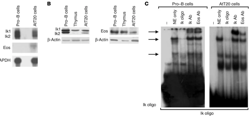
Pituitary corticomelanotroph cells express Ikaros. Using Northern blotting, we detected an Ikaros mRNA doublet of approximately 2.7 kb in mouse corticomelanotroph AtT20 cells. This product corresponds to the expected size of Ik1 and Ik2 mRNA isoforms. It comigrated with and was of a similar level of expression to the product derived from pro–B lymphocytes, which represent the principal source of Ikaros (Figure 1A). The Ikaros mRNA tran-scripts were translated, as demonstrated by Western immunoblot-ting (Figure 1B). Moreover, a weak mRNA transcript encoding the Ikaros-related factor Eos was detected in pituitary corticomelano-troph cells by Northern blotting (Figure 1A), and this protein was also detected in these cells by Western blotting (Figure 1B).
The expression of Ikaros by pituitary corticomelanotroph cells was also examined by EMSA. Using an Ikaros consensus oligonucle-otide as a probe, we found that nuclear extracts from AtT20 corti-cotroph cells formed specific DNA-protein complexes (Figure 1C). These complexes were competed and supershifted in a manner similar to those formed by nuclear extracts from pro–B lympho-cytes (Figure 1C). The same complex in lympholympho-cytes and in pitu-itary AtT20 cells was supershifted by an Eos antibody.
The pituitary POMC gene contains functional Ikaros binding sites. Hav-ing identified Ikaros expression in pituitary corticomelanotrophs, we examined whether the major pituitary hormone gene
Nonstandard abbreviations used: ACTH, adrenocorticotrophic hormone; FGFR4, fibroblast growth factor receptor 4; HDAC, histone deacetylase; POMC, proopiomelanocortin.
Conflict of interest: The authors have declared that no conflict of interest exists.
research article
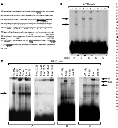
expressed by these cells represents a transcriptional target for Ikaros. To determine whether Ikaros can recognize a specific element in the POMC promoter, we screened potential Ikaros-binding domains of the POMC promoter using EMSA. We used nuclear extracts from corticotroph AtT20 cells with overlapping oligonucleotide fragments containing all 5 potential Ikaros-bind-ing sites in the mouse POMC promoter (Figure 2A). DNA-protein complexes were detected when extracts were incubated with 3 fragments containing putative Ikaros-binding sites, fragments A (–451/–420), B (–163/–137), and C (–118/–89) (Figure 2B). Formation of these DNA-protein complexes was abolished by excess unlabeled wild-type Ikaros oligonucleotide (Figure 2C) but not by mutant Ikaros or non-Ikaros (Sp1, Ap-1, Ap-2) oli-gonucleotides (data not shown). Further, these complexes were supershifted by Ikaros antibody in a dose-responsive manner (Figure 2C) and to a lesser extent by Eos antibody (Figure 2C) but not by other control antibodies, including those against Rb, Pit-1, and Sp1. Mutation of the Ikaros binding sites A, B, and C of the POMC promoter resulted in failure to form specific com-plexes when the probe was incubated with pituitary AtT20 or pro–B lymphocyte nuclear extracts(data not shown).
Ikaros binds the POMC promoter and regulates gene expression. We tested the response of the wild-type P(–543)-Luc POMC promoter to cotransfection with Ik1 or the Ikaros dominant negative (non– DNA-binding) Ik6 isoform. Figure 3A demonstrates the ability of Ik1 to activate the POMC promoter in AtT20 cells, according to a luciferase reporter assay. In contrast, transfection of Ik6 repressed
POMC promoter activity, which is consistent with functional
antagonism between the 2 Ikaros isoforms. Transfection of the related family member Eos also resulted in modest (approximately 25%) attenuation of POMC promoter activity.
To determine the functional significance of the Ikaros-bind-ing sites in the POMC promoter, we individually mutated each of these sites. Disruption of binding site A or B but not site C resulted in appreciable loss of POMC promoter activity (Figure 3A). The effects of Ik1 overexpression were abolished by mutation of site A or B but were not affected by mutation of site C. These data are consistent with the lack of efficient competition by the Ikaros oligonucleotide of the site C complex, which indicates a minimal contribution from site C to POMC promoter activity.
Stable transfection of Ik1 resulted in enhanced endogenous
POMC gene expression as determined by Northern blotting of inde-pendent, stably transfected clones (Figure 3B). In contrast, cells stably transfected with either Ik6 or Eos demonstrated reduced
POMC mRNA levels. Immunoassay measurements of ACTH in conditioned culture media revealed a nearly 5-fold increase in ACTH secretion by AtT20 cells stably transfected with Ik1 com-pared with empty vector-, Ik6-, or Eos-transfected cells (Figure 3C). Taken together, these data provide evidence of a role for Ikaros in pituitary POMC gene regulation.
[image:3.585.59.540.81.307.2]Loss of Ikaros disrupts pituitary corticomelanotroph development. To examine the effect of Ikaros on pituitary corticomelanotroph development, we systematically examined the pituitary glands of mice with disruption of the Ikaros gene. Animals were exam-ined at the earliest stages of expected corticomelanotroph dif-ferentiation and subsequent stages of late fetal development Figure 1
into adulthood. At E15.5, developing corticomelanotroph cells were identified by Tpit expression at the periphery of the gland as previously described (11). At E17.5, POMC expression was reduced in Ikaros-heterozygous mice and was almost undetect-able in Ikaros-null (–/–) mice despite the presence of normal Tpit expression (Figure 4A).
The contraction of the corticomelanotroph population per-sisted into postnatal life with reduction of corticomelanotrophs in Ikaros-null animals noted at birth and at 1 week (Figure 4B) and 2 weeks after birth. Morphometry confirmed reduction in this cell population in the anterior and intermediate lobes to less than 50% of controls at all ages examined.
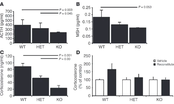
Loss of Ikaros impairs pituitary-directed adrenocortical function. To examine the functional consequences of disrupted Ikaros sig-naling on pituitary corticomelanotroph development, we com-pared the hormonal (Figure 5) and developmental profiles of Ikaros-null mice with heterozygous and wild-type (+/+) control littermates. Homozygous Ikaros-null mice demonstrated the lowest levels of ACTH in the systemic circulation (Figure 5A) and exhibited lighter fur color, consistent with a loss of POMC-derived melanocyte-stimulating hormone (MSH). MSH levels
measured by immunoassay when the mice were 3 weeks of age were also reduced in heterozygous but more significantly in Ikaros-null mice (Figure 5B).
The functional consequences of a diminished population of POMC-producing corticomelanotroph cells were evident in homozygous mice. Consistent with the trophic functions of ACTH on adrenocortical development and function, Ikaros-null mice exhibited reduced circulating corticosterone levels compared with heterozygous and wild-type littermates (Figure 5C). These changes were also reflected in reduction of adrenal gland size with striking loss of adrenocortical mass, especially in homozygous Ikaros-defi-cient mice (not shown).
Pituitary-adrenocortical dysfunction in Ikaros-deficient mice is not reversed by wild-type lymphocyte reconstitution. To examine the pos-sibility that the diminished POMC production and adrenocorti-cal insufficiency in Ikaros-deficient animals may be secondary to their altered lymphocyte development, we examined the impact of wild-type bone marrow reconstitution on the endocrine phe-notype. Five weeks after adoptive transfer in neonatal mice, the proportion of splenic CD4+ and CD8+ T cells in the homozygous
[image:4.585.43.425.75.491.2]Ikaros-null recipients of Ikaros wild-type syngenic bone marrow Figure 2
research article
was significantly restored compared with that of vehicle-treated controls (Table 1). Despite this successful reconstitution with normal lymphocytes, the phenotype of homozygous Ikaros-defi-cient animals was unchanged, with persistent small size, small pituitaries, and reduction in POMC expression, and was indistin-guishable from that of vehicle-treated age-matched homozygous animals. Moreover, neither heterozygous nor homozygous
Ikaros-deficient animals demonstrated a significant change in serum corticosterone levels as a result of lymphocyte reconstitution (Figure 5D). These data demonstrate the direct role of Ikaros in pituitary corticotroph development and function independent of its influence on lymphopoiesis.
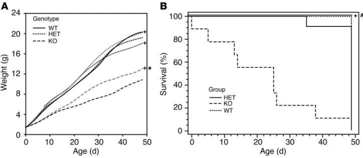
Glucocorticoid treatment rescues Ikaros-deficient mice. Recognizing that Ikaros mutant mice exhibit adrenocortical insufficiency, we examined the impact of systemic hormone replacement on growth and survival of the Ikaros-null mouse. Treatment with glucocorticoid hormone compared with vehicle alone resulted in significant weight increase in Ikaros-null mice; the effect was present but less evident in heterozygous animals (Figure 6A). By comparison, the same treatment paradigm had no significant impact on the growth of wild-type mice.
[image:5.585.46.282.82.406.2]Overall, homozygous Ikaros-null mice exhibit diminished life expectancy with nearly 5% mortality at birth, 40% by 3 weeks of age, and 75% by 6 weeks of age (Figure 6B). Given the recognized functions of Ikaros on immune cell development and function, Figure 3
[image:5.585.282.541.518.740.2]Ikaros regulation of the pituitary POMC gene. (A) The effect of Ikaros on POMC promoter activity was tested in a luciferase (luc) assay. Note activation by wild-type Ik1 and loss of activity by the Ikaros dominant negative isoform (Ik6) and by Eos. The functional role of the putative Ikaros-binding domains at sites A, B, and C in the POMC promoter was also examined. Mutation of 2 Ikaros-binding sites at sites A (mA) and B (mB) resulted in loss of reporter activity in transfected corticotroph AtT20 cells. This effect was not seen with mutation of site C (mC). (B) The effect of Ikaros or Eos transfection on endogenous POMC gene mRNA expression. AtT20 cells stably transfected with Ik1, Ik6, or Eos were examined by Northern blotting using a cDNA probe for the POMC gene. Densitometric analysis from multiple independent clones revealed an approximately 2.5-fold increase in POMC/GAPDH expression in Ik1-transfected clones. In contrast, forced expression of Eos (right) reduces POMC expression. pcDNA, cells transfected with empty vector. (C) Con-ditioned culture media from Ikaros-transfected cells were examined for ACTH content by ELISA. Note significantly enhanced ACTH secretion by Ikaros-transfected cells compared with empty vector in contrast to mild reduction by Ik6 or Eos-transfected cells.
Figure 4
we performed complete organ surveys in search of opportunis-tic infections or malignancy. However, we found no evidence of either of these conditions to explain the observed mortality at these stages of development. Moreover, hormone treatment with glucocorticoids resulted in enhanced survival. In particu-lar, 100% of Ikaros-null mice treated with the adrenocortical hormone (n =7) survived beyond 6 weeks of age. By comparison, none of the vehicle-treated homozygous mice (n =8) survived beyond the same time point.
Discussion
We present here what we believe to be the first evidence that Ikaros is directly involved in the development and function of the endo-crine pituitary-adrenocortical system. Gene targeting experiments have firmly established that nuclear factors encoded by the Ikaros
gene are essential for normal lymphoid development (12). Indeed, mice homozygous for a null mutation in the Ikaros gene exhibit
severely impaired B and T cell differ-entiation (13, 14). In addition to its role in differentiation, Ikaros has also been suspected to play an important role in regulating cell proliferation. Heterozygous Ikaros mice exhibit hyperactive T cell receptor–mediated proliferative responses and eventually develop leukemias and lymphomas (15, 16), consistent with a dose-relat-ed effect of action. Our data indicate that Ikaros is also an essential direct regulator of normal pituitary cortico-troph development, which parallels its role in lymphocytes.
We have previously demonstrated that Ikaros targets the FGFR4 pro-moter, which likely plays a role, along with other FGFRs (17), in normal pitu-itary development. However, we recog-nized that FGFR4 cannot be the only target of Ikaros in the pituitary, since an FGFR4-null mouse model has not shown evidence of significant pituitary dysgenesis (18). This is not surpris-ing, given the marked redundancy of FGFRs and the fact that the pituitary gland expresses FGFR1, -2, and -3 (17).
Consistent with the striking mor-phologic alterations in the pituitaries of Ikaros-null mice, our data show thatIkaros targets the princi-pal hormone gene product of corticomelanotrophs, POMC. The
POMC promoter contains 5 potential Ikaros-binding sites. Two of these, site A (–451/–420) and site B (–163/–137), showed spe-cific binding, complex formation, and interaction with an Ikaros-specific antibody. Mutational analysis confirmed the functional importance of these 2 proximal Ikaros-binding sites to POMC
promoter activity. These sites are within close proximity (see Figure 2A) upstream and downstream of the recently identified Tpit/Pitx1 regulatory element (19).
[image:6.585.46.386.80.284.2]The pituitary gland has provided unique insights into the molecular mechanisms and regulatory factors controlling both gene transcription and cell differentiation. The T-box factor Tpit was found to be restricted to POMC-expressing corticome-lanotroph lineage (19). It activates POMC gene transcription in cooperation with contiguously bound Pitx1 as part of the same regulatory element. Tpit activates transcription by recruiting the Figure 5
Disrupted corticomelanotroph development in Ikaros-deficient mice impairs pituitary-adrenocortical function. Levels of circulating hormones of the pituitary-adrenal axis including the POMC-derived peptides ACTH (A) and MSH (B) and the ACTH-dependent adrenocortical hormone corticosterone (C) were measured by immunoassay in Ikaros-null (n =8), heterozygous (n =31), and wild-type littermates (n =18) at 3 weeks of age. The reductions in ACTH, MSH, and corticosterone levels were most pronounced in Ikaros-null mice, as illustrated by the horizontal-bar comparisons. (D) Neither lymphocyte reconstitution nor treatment with vehicle changed circulating corticosterone levels in Ikaros-deficient animals. Data are from animals described in Table 1 and reflect the impact of recon-stitution with wild-type marrow cells 5 weeks after intrahepatic injection.
Table 1
Impact of bone marrow transplantation on splenic lymphocytes in Ikaros-deficient mice
WT Heterozygous Homozygous
Vehicle Reconstituted Vehicle Reconstituted Vehicle Reconstituted (n = 4) (n = 3) (n = 6) (n = 3) (n = 2) (n = 3)
Spleen T cells (%) CD4+ CD8+ CD4+ CD8+ CD4+ CD8+ CD4+ CD8+ CD4+ CD8+ CD4+ CD8+
Mean 12.97 3.18 13.10 4.47 13.65 4.07 12.88 4.82 4.90 5.85 28.31 22.81 SEM 1.28 0.35 2.65 1.37 1.37 0.50 1.25 1.09 0.52 3.11 2.85 6.63
Newborn mice of different Ikaros genotypes were injected with 106 cells derived from wild-type animal bone marrow and sacrificed 5 weeks later. Control
research article
SRC/p160 coactivators to its cognate DNA target in the POMC
promoter (20). Unlike Ikaros-null mice, Tpit-deficient mice have almost no POMC-reactive cells in the pituitary (11, 19). Cortico-troph differentiation is normal in Ikaros-null mice, as determined by expression of Tpit in a large number of cells; however, the intensity of POMC expression in these cells is markedly reduced from E17.5, and after birth there is an evident reduction of this cell population. Consistent with this finding, the animals have reduced circulating corticosterone, which demonstrates the mag-nitude of the reduction, since as little as 10–20% of pituitary tis-sue mass can maintain hormonal homeostasis (3, 4). The pheno-typic differences between the 2 models suggest that Ikaros, unlike Tpit, is not required for corticomelanotroph cell differentiation but rather plays a modulatory role in hormone gene expression and cell population expansion.
The formation of Ikaros homo- and heterodimers among the DNA-binding Ikaros family members increases their affinity for DNA, whereas heterodimers between the DNA-binding isoforms and non–DNA-binding isoforms are unable to bind DNA. Thus, Ikaros proteins with fewer than 3 N-terminal zinc fingers can negatively interfere with the activity of Ikaros isoforms that bind DNA (21, 22). Histones have been shown to be underacetylat-ed in the vicinity of Ikaros recruitment sites, while the histone deacetylase (HDAC) inhibitor trichostatin abrogates transcrip-tional repression mediated by Ikaros (23). An abundance of the non–DNA-binding Ikaros isoforms could result in deregulated expression of other target genes that are essential for normal pituitary development and maturation. Indeed, when bound to Ikaros-binding sites, only those isoforms with DNA-binding domains in cis are able to activate gene transcription (21, 22, 24). In contrast, Ikaros represses transcription when recruited to DNA through a heterologous DNA-binding domain (23). This repres-sion is mediated through at least 2 main represrepres-sion domains at the N- and C-termini that interact with HDAC 1 and 2 containing
mSin3 (23) and Mi-2 proteins (25). The extent to which this mechanism of gene repression is global or gene specific remains to be determined. Further, other Ikaros family members Aiolos, Helios, and Eos, can associate with all of Ikaros’s corepressors including class II HDACs. Thus, the loss of function noted in the Ikaros-deficient mice may also represent a relative abundance of other family members. The potential for this repression by Ikaros-interacting proteins, at least on the POMC gene, was suggested by the effects of Eos overexpression on the POMC
promoter and the endogenous gene. With the exception of Eos, other Ikaros family related proteins, including Helios and Aiolos, are largely restricted to hematolymphoid tissues (26).
Ikaros has been shown to interact with the HDAC-inde-pendent corepressor CtBP. However, mutations that block Ikaros/CtBP interactions do not abolish Ikaros’s repressive func-tions. Instead, Ikaros also interacts with the CtBP-interacting pro-tein (CtIP) (27). Interestingly, CtIP’s association with Ikaros does not require CtBP but relies heavily on interaction with Rb. The Ikaros/Rb interaction is of potential relevance in the pituitary. Rb is pivotal in suppressing E2F-dependent transcription and con-sequently G1/S cell cycle transition. Mice with heterozygous Rb mutations develop pituitary tumors of the corticomelanotroph lineage (28). These data raise questions about the participation of Ikaros in deregulating key components of pituitary cell cycle control that are implicated in pituitary growth (29).
Ikaros can also function as a transcription activator. It is note-worthy that Ikaros-mediated gene activation with localization in heterochromatin complex has been described (30). In this model, Ikaros is in direct association with its target genes in a predomi-nantly restrictive chromatin environment that houses tightly regulated genes (31). POMC is an example of a tightly regulated gene that is typically repressed but may be activated under unique conditions when specific activators (such as Tpit) as well as other factors including Ikaros are coexpressed. Under such conditions, Ikaros functions as a potentiator by remodeling a densely pack-aged chromatin environment, presumably facilitating activator access during pituitary development.
[image:7.585.47.409.82.240.2]The identification of Ikaros as a factor involved in the regula-tion of the POMC-ACTH- adrenal axis assigns new funcregula-tions to this zinc finger protein. Ikaros’s role as a mediator of lym-phopoietic cell differentiation parallels its effects on pituitary cell differentiation. Given the role of Ikaros in lymphoid cell development, leukemia, and immune functions (31), the cur-rent findings of Ikaros in pituitary development, POMC hor-mone regulation, and corticomelanotroph function provide new insights into the transcriptional mechanisms at the interface of neuro-endocrine-immune interactions.
Figure 6
Methods
Cell culture. Mouse corticomelanotroph AtT20 and pro–B lymphocytic cells
were propagated in DMEM (Life Technologies Inc.) with a high level of glucose supplemented with 10% FBS (Sigma-Aldrich), 2 mM glutamine, 100 IU/ml penicillin, and 100 μg/ml streptomycin.
Plasmids. The expression vectors encoding full-length Ikaros (CDM8-Ik1)
and dominant negative Ikaros 6 (CDM8-Ik6) were generously provided by K. Georgopolous (Harvard Medical School, Charlestown, Massachusetts, USA). The Eos-encoding vector pcDNA3.1-Eos was generously provided by M. Crossley (University of Sydney, New South Wales, Australia). The orien-tation and sequence of all constructs were verified by restriction analysis and nucleotide sequencing.
Promoter analyses. Promoter predictions were performed with the
assis-tance of The Transcription Factor Database TRANSFAC 6.0 (http://www. gene-regulation.com; BIOBASE GmbH). The convention for sequence coordinates with +1 as the first base of the coding sequence in exon 1 was adopted. The mouse POMC –543-bp luciferase reporter was a generous gift of J. Drouin (University of Montreal, Montreal, Quebec, Canada.).
Site-directed mutagenesis.Site-directed mutagenesis was performed using
a QuikChange Site- Directed Mutagenesis Kit (Stratagene) following the manufacturer’s instructions. We introduced mutation of the Ikaros tran-scription binding site -CGGGAA- substituted by -CGTTAA- into the POMC promoter fragment P(–543)-Luc construct by using mutagenic primers for each of the 5 potential Ikaros-binding sites identified in this promoter (Figure 2A). All mutations were confirmed by nucleotide sequencing.
Transfection and luciferase assays. Plasmid reporters were prepared by
col-umn chromatography (QIAGEN) for sequencing and transfections. Cells were transfected by the Lipofectamine Method (Invitrogen Corp.) accord-ing to the manufacturer’s protocol. Cells were plated into 6-well cluster dishes (7 × 10⁵ cells per well), transfected the following day with 3 μl or 5 μl/well of lipofectamine and 1 or 2 μg of DNA per well as indicated. We kept the total amount of transfected DNA constant by adding empty vector. Transfection efficiency was monitored by simultaneous cotransfec-tion with a β-gal control expression plasmid cytomegalovirus (CMV)–β-gal (20 ng/well). Forty-eight hours after transfection, cells were lysed in buffer containing 25 mM glycylglycine, 15 mM MgSO4, 4 mM EGTA, 1% Triton X-100, and 1 mM DTT. Luciferase activity was measured for 20 seconds in a luminometer. We measured β-gal activity to normalize for variations in transfection efficiency. Promoter activity of each construct was expressed as firefly luciferase/β-gal activity. Each experiment was independently per-formed on 3 separate occasions with triplicate wells in each experiment.
Preparation of nuclear extracts.We prepared nuclear extracts by washing
cells in ×1 PBS and lysing them in 100 μl buffer containing 10 mM HEPES, pH 7.9, 1 mM DTT, 1 mM EDTA, 60 mM KCl, 0.5% NP-40, 1 mM PMSF for 5 minutes on ice. The pellet was resuspended into 100 μl of the nuclear resuspension buffer (0.25 mM Tris-HCl, pH 7.8, 60 mM KCl, 1 mM DTT, 1.5 mM PMSF) and lysed with 3 cycles of freezing and thawing to 37°C. After centrifugation at 13,000 rpm for 10 minutes at 4°C, the clear super-natant was collected for further analysis. Protein concentrations were determined by the Bio-Rad method (Bio-Rad Laboratories).
EMSA.We labeled oligonucleotides with [γ-32P]dATP using the T4
poly-nucleotide kinase (Invitrogen Corp.). Five micrograms of nuclear pro-tein extracts and 2 ng of labeled oligonucleotides were allowed to bind for 30 minutes at room temperature in a final volume of 20 μl of binding buffer [20 mM HEPES, pH 7.9, 50 mM KCl, 1 mM EDTA, 1 mM DTT, 0.5 mM MgCl2, 2% glycerol, and 0.5 μg poly(dI-dC)] (Pharmacia Corp). Protein-DNA complexes were resolved in 4% polyacrylamide gels contain-ing ×0.5 tris-borate EDTA (TBE). Five double-stranded oligonucleotide fragments containing the potential Ikaros-binding sites in the POMC promoter were synthesized (Sigma-Aldrich) and used as probes and for
competition in EMSAs as follows: Ikaros binding site A (–448 to –431), 5′-AACACTGGGGAAATCTGA-3′; Ikaros binding site B (–159 to –142), sense: 5′-CCCCGACCGGGAAGCCCC-3′; Ikaros binding site C (–112 to –95), 5′-GTGGCCGGGGATTCGCTT-3′; Ikaros binding site D (–12 to +7), 5′-GTGACAGGGACCAAACGG-3′; Ikaros binding site E (–12 to +16), 5′-ACCAAACGGGAGGCGACG-3′, and their mutant forms (mA, mB, mC, mD and mE), where the core Ikaros binding site (-GGGAA-) was substituted by -GTTAAA-. Other probes and competitor oligonucleotides containing transcription binding sites were as follows: Sp1, 5′ -ATTC-GATCGGGGCGGGGCGAGC-3′ and its complementary strand; Ikaros, 5′-AAGAAGCGGGAGTGACAGG-3′ and its complementary strand; Pit1, 5′-TGTCTTCCTGAATATGAATAAGAAATA-3′ and its complementary strand; Ap-1, 5′-CGC TTGATGAGTCAGCCGGAA-3′ and its complemen-tary strand; Ap-2, 5′-GATCGAACTGACCGCCCGCGGCCCGT-3′ and its complementary strand. The complementary strands were annealed in the annealing buffer (10 mM Tris-Cl, pH 8.0, 50 mM NaCl, 1 mM EDTA).
Gel shift probes were purified with a G50 spin column (Amersham Biosciences) after radiolabeling. For competition assay, 10 or 100 M excess of unlabeled fragment was added as competitor DNA. Supershifting was achieved using 2 Ikaros antibodies: a mouse monoclonal antibody (kindly provided by K. Georgopolous) and a polyclonal antibody (kindly provided by S. Smale, UCLA, Los Angeles, California, USA). Both antibodies recog-nize the C-terminal fragments of Ikaros proteins. Supershifting with Eos was performed using a specific antibody recognizing Eos (kindly provided by M. Crossley). Antibodies to Pit-1, Rb, and Sp1 were used as described previously (7). Antibodies were used at different concentrations and were applied 30 minutes before addition of radiolabeled probe. Samples were electrophoresed and gels dried under vacuum and autoradiographed.
RNA isolation and Northern blotting.PolyA-enriched RNA was prepared
from exponentially growing cells (2–5 × 10⁷) using a FastTrack 2.0 mRNA Isolation Kit (Invitrogen Corp.) following the manufacturer’s instructions. Two micrograms of polyA+ RNA were electrophoresed on a 1% formalde-hyde-agarose gel and transferred to a nylon membrane. Hybridization was performed using probes labeled randomly (Roche Diagnostics Corp.) with [α-32P]-dCTP from the cDNAs of Ikaros, Eos, POMC, and GAPDH. The Ikaros probe consisted of a 1,200-bp fragment encoding the C-terminus BamHI-EcoRV fragment of the Ikaros cDNA. The Eos probe was a 587-bp BamHI-HindIII fragment of the mouse Eos cDNA. The POMC probe was generated by RT-PCR using the specific primers (sense: 5′- ATGCCGAGA-TTCTGCTACAG- 3′; antisense: 5′- TCACTGGCCCTTCTTGTGCG- 3′ to produce a 708-bp fragment from POMC cDNA, and the GAPDH probe was made from RT-PCR with the specific primers to produce a 687-bp fragment. Northern blots were hybridized in the ×1 hybridization buffer (Sigma-Aldrich) at 65°C overnight with rotation in oven, then washed and exposed to film. RNA sizes were calculated using the marker 0.24–9.5 kb RNA Ladder (Invitrogen Corp.).
Western blotting. Forty micrograms of whole lysates were separated on 10%
SDS denaturing polyacrylamide gels, transferred onto nylon membrane (Millipore) at 100 V for 1 hour at room temperature. Blots were incubated with a mouse monoclonal antibody (4E9; kindly provided by K. Georgop-olous) that recognizes the C-terminal fragments of Ikaros proteins (22) or POMC (DakoCytomation) at 1:2,000 dilution in PBS–5% nonfat milk with 0.1% Tween at 4°C overnight, after which they were washed with PBS-Tween-20 four times for 10 minutes each at room temperature and incu-bated with secondary antibody of peroxidase-conjugated goat antimouse IgG (1:2,000) for 1 hour at room temperature with agitation. Proteins were detected using a chemiluminescence method (Amersham Biosciences).
Ikaros-deficient mice.Ikaros-null mice were generated by Georgopolous et
research article
and undetectable protein (13). Mice were propagated on the original C57BL/6 background. We monitored germline allelic transmission by PCR analysis using tail DNA and previously described primers (13). Specimens for blood and tissue analyses were obtained from embryos at various times during gestation and from adult mice. The care and handling of animals were approved by the Institutional Animal Care Facilities at the Ontario Cancer Institute, where the animals were housed.
Morphologic studies of the pituitary.In order to localize prenatal and
neo-natal pituitaries, we took sagittal sections of the intact head of the fetus, using the cartilage of the sphenoid bone as a landmark as previously described (32, 33). Consecutive serial sections from the midline to the lat-eral aspect of the pituitary were collected. Pituitary size was estimated in postnatal mice by the summation of scan area cross-sections starting from the midline portion all the way through the lateral wing of the pituitary. Serial sections were stained with H&E or with antibodies to localize the various pituitary hormones as described previously (34) and to identify transcription factors using anti-sera described above and an antibody to Tpit (kindly provided by J. Drouin). Immunolocalization was detected with the streptavidin-biotin-peroxidase complex technique and 3,3′ -diamino-benzidine (DAB) staining. Colocalization with pituitary hormones was performed with double staining as previously described (34). As a negative control, we used normal mouse ascites or normal rabbit serum, replacing the primary monoclonal or polyclonal antibody, respectively. Additional negative controls included preabsorption of the primary antibody or anti-serum with purified antigen. We carried out morphometry to determine the areas of immunostained cells as a proportion of pituitary area.
Hormone measurements and administration.SecretedACTH was measured
by a 2-site ELISA according to the manufacturer’s (Biomerica Inc.) pro-tocol.A corticosterone enzyme immunoassay was used to reflect adreno-cortical hormone secretion using a commercially available kit (ALPCO Diagnostics). We commenced hormone rescue treatment when mice were 5 days of age using the synthetic glucocorticoid dexamethasone
(Sigma-Aldrich). This was administered intraperitoneally at a dose of 0.05 mg/kg every 2 days for 6 weeks. The dosing regimen was based on titration studies and utilized the maximum dose that was tolerated without weight gain in wild-type control littermates.
Hematopoietic cell repopulation.Bone marrow was prepared from 6-week-old
wild-type Ikaros littermates. Following lysis of red blood cells, the resulting cell suspension was filtered through a 70-μm cell strainer, and 106 cells were injected intrahepatically into newborn Ikaros-deficient and -sufficient ani-mals as described previously (35). Bone marrow recipients were sacrificed 5 weeks after reconstitution for examination of pituitary morphology and cir-culating hormone measurements. Lymphocyte reconstitution was assessed in the spleen and thymus of bone marrow recipients by multicolor cell-sur-face immunofluorescence and FACS analysis, as previously described (36).
Acknowledgments
We thank K. Georgopoulos for generously providing the Ikaros-null mice and Ikaros antibody, S. Smale for Ikaros antiserum, and M. Crossley for Eos reagents. We also thank J. Drouin for the Tpit antibody and for insightful discussions. The technical assis-tance of K. So is also gratefully acknowledged. This work was supported by the Canadian Institutes of Health Research and the Toronto Medical Laboratories.
Rene Mader and ShunJiang Yu contributed equally to this work.
Received for publication June 21, 2004, and accepted in revised form January 11, 2005.
Address correspondence to: Sylvia L. Asa, Department of Pathol-ogy, University Health Network, 610 University Avenue 4-302, Toronto, Ontario M5G-2M9, Canada. Phone: (416) 946-2099; Fax: (416) 946-6579; E-mail: [email protected].
1. Chrousos, G.P. 1995. The hypothalamic-pituitary-adrenal axis and immune-mediated inflammation.
N. Engl. J. Med.332:1351–1362.
2. Turnbull, A.V., and Rivier, C.L. 1999. Regula-tion of the hypothalamic-pituitary-adrenal axis by cytokines: actions and mechanisms of action.
Physiol. Rev.79:1–71.
3. Asa, S.L., and Ezzat, S. 1998. The cytogenesis and pathogenesis of pituitary adenomas. Endocr. Rev.
19:798–827.
4. Asa, S.L., and Ezzat, S. 1999. Molecular determinants of pituitary cytodifferentiation. Pituitary.1:159–168. 5. Molnar, A., et al. 1996. The Ikaros gene encodes a family of lymphocyte-restricted zinc finger DNA binding proteins, highly conserved in human and mouse. J. Immunol.156:585–592.
6. Georgopoulos, K., Winandy, S., and Avitahl, N. 1997. The role of the Ikaros gene in lymphocyte development and homeostasis. Annu. Rev. Immunol.
15:155–176.
7. Yu, S., Asa, S.L., and Ezzat, S. 2002. Fibroblast growth factor receptor 4 is a target for the zinc-fin-ger transcription factor Ikaros in the pituitary. Mol. Endocrinol.16:1069–1078.
8. Armstrong, E., Partanen, J., Cannizzaro, L., Hueb-ner, K., and Alitalo, K. 1992. Localization of the fibroblast growth factor receptor-4 gene to chro-mosome region 5q33-qter. Genes Chromosomes Can-cer.4:94–98.
9. Ezzat, S., Yu, S., and Asa, S.L. 2003. Ikaros isoforms in human pituitary tumors: distinct localization, histone acetylation, and activation of the 5′ fibro-blast growth factor receptor-4 promoter. Am. J. Pathol.163:1177–1184.
10. Yu, S., Asa, S.L., Weigel, R.J., and Ezzat, S. 2003. Pituitary tumor AP-2alpha recognizes a cryptic
promoter in intron 4 of fibroblast growth factor receptor 4. J. Biol. Chem.278:19597–19602. 11. Pulichino, A.M., et al. 2003. Human and mouse
TPIT gene mutations cause early onset pituitary ACTH deficiency. Genes Dev.17:711–716. 12. Cortes, M., Wong, E., Koipally, J., and Georgopoulos,
K. 1999. Control of lymphocyte development by the Ikaros gene family. Curr. Opin. Immunol.11:167–171. 13. Wang, J.H., et al. 1996. Selective defects in the devel-opment of the fetal and adult lymphoid system in mice with an Ikaros null mutation. Immunity.
5:537–549.
14. Winandy, S., Wu, L., Wang, J.H., and Georgopoulos, K. 1999. Pre-T cell receptor (TCR) and TCR-con-trolled checkpoints in T cell differentiation are set by Ikaros. J. Exp. Med.190:1039–1048.
15. Winandy, S., Wu, P., and Georgopoulos, K. 1995. A dominant mutation in the Ikaros gene leads to rapid development of leukemia and lymphoma.
Cell.83:289–299.
16. Avitahl, N., et al. 1999. Ikaros sets thresholds for T cell activation and regulates chromosome propaga-tion. Immunity.10:333–343.
17. Abbass, S.A.A., Asa, S.L., and Ezzat, S. 1997. Altered expression of fibroblast growth factor receptors in human pituitary adenomas. J. Clin. Endocrinol. Metab.82:1160–1166.
18. Weinstein, M., Xu, X., Ohyama, K., and Deng, C.X. 1998. FGFR-3 and FGFR-4 function cooperatively to direct alveogenesis in the murine lung. Develop-ment.125:3615–3623.
19. Lamolet, B., et al. 2001. A pituitary cell-restricted T box factor, Tpit, activates POMC transcription in cooperation with Pitx homeoproteins. Cell.
104:849–859.
20. Maira, M., et al. 2003. The T-box factor Tpit recruits
SRC/p160 co-activators and mediates hormone action. J. Biol. Chem.278:46523–46532.
21. Molnar, A., and Georgopoulos, K. 1994. The Ikaros gene encodes a family of functionally diverse zinc finger DNA-binding proteins. Mol.Cell. Biol.
14:8292–8303.
22. Sun, L., Liu, A., and Georgopoulos, K. 1996. Zinc finger-mediated protein interactions modulate Ikaros activity, a molecular control of lymphocyte development. EMBO J.15:5358–5369.
23. Koipally, J., Renold, A., Kim, J., and Georgopoulos, K. 1999. Repression by Ikaros and Aiolos is mediat-ed through histone deacetylase complexes. EMBO J.
18:3090–3100.
24. Wargnier, A., et al. 1998. Down-regulation of human granzyme B expression by glucocorticoids. Dexamethasone inhibits binding to the Ikaros and AP-1 regulatory elements of the granzyme B pro-moter. J. Biol. Chem.273:35326–35331.
25. Kim, J., et al. 1999. Ikaros DNA-binding proteins direct formation of chromatin remodeling com-plexes in lymphocytes. Immunity.10:345–355. 26. Perdomo, J., Holmes, M., Chong, B., and Crossley,
M. 2000. Eos and pegasus, two members of the ikaros family of proteins with distinct DNA bind-ing activities. J. Biol. Chem.275:38347–38354. 27. Koipally, J., and Georgopoulos, K. 2002.
Ikaros-CtIP interactions do not require C-terminal binding protein and participate in a deacetylase-independent mode of repression. J. Biol. Chem.
277:23143–23149.
28. Jacks, T., et al. 1992. Effects of an Rb mutation in the mouse. Nature.359:295–300.
lambda5 promoter silences transcription through a mechanism that does not require heterochroma-tin formation. EMBO J.20:2812–2822.
31. Georgopoulos, K. 2002. Haematopoietic cell-fate decisions, chromatin regulation and ikaros. Nat. Rev. Immunol.2:162–174.
32. Bennani-Bäiti, I.M., et al. 1998. DNase I-hypersen-sitive sites I and II of the human growth hormone locus control region are a major developmental activator of somatotrope gene expression. Proc.
Natl. Acad. Sci. U. S. A.95:10655–10660.
33. Shewchuk, B.M., Asa, S.L., Cooke, N.E., and Lieb-haber, S.A. 1999. Pit-1 binding sites at the somato-trope-specific DNase I hypersensitive sites I, II of the human growth hormone locus control region are essential for in vivo hGH-N gene activation.
J. Biol. Chem.274:35725–35733.
34. Asa, S.L., Coschigano, K.T., Bellush, L., Kopchick, J.J., and Ezzat, S. 2000. Evidence for growth hormone (GH) autoregulation in pituitary somatotrophs in
GH antagonist-transgenic mice and GH receptor-deficient mice. Am. J. Pathol.156:1009–1015. 35. Nichogiannopoulou, A., Trevisan, M., Neben, S.,
Friedrich, C., and Georgopoulos, K. 1999. Defects in hemopoietic stem cell activity in Ikaros mutant mice. J. Exp. Med.190:1201–1214.
36. Poussier, P., Ning, T., Banerjee, D., and Julius, M. 2002. A unique subset of self-specific intraintes-tinal T cells maintains gut integrity. J. Exp. Med.