Loss of estrogen upregulates
osteoblastogenesis in the murine bone marrow.
Evidence for autonomy from factors released
during bone resorption.
R L Jilka, … , P K Roberson, S C Manolagas
J Clin Invest.
1998;
101(9)
:1942-1950.
https://doi.org/10.1172/JCI1039
.
Loss of sex steroids causes an increase in both the resorption and formation of bone, with
the former exceeding the latter. Based on evidence that the increased bone resorption after
estrogen loss is due to an increase in osteoclastogenesis, we hypothesized that estrogen
loss also stimulates osteoblastogenesis. We report that the number of mesenchymal
osteoblast progenitors in the murine bone marrow was increased two- to threefold between
2 and 8 wk after ovariectomy and returned to control levels by 16 wk. Circulating
osteocalcin, as well as osteoclastogenesis and the rate of bone loss, followed a very similar
temporal pattern. Inhibition of bone resorption by administration of the bisphosphonate
alendronate led to a decrease of the absolute number of osteoblast progenitors; however, it
did not influence the stimulating effect of ovariectomy on osteoblastogenesis or
osteoclastogenesis. These observations indicate that the increased bone formation that
follows loss of estrogen can be explained, at least in part, by an increase in
osteoblastogenesis. Moreover, they strongly suggest that unlike normal bone remodeling,
whereby osteoblast development is stimulated by factors released from the bone matrix
during osteoclastic resorption, estrogen deficiency unleashes signals that can stimulate the
differentiation of osteoblast progenitors in a fashion that is autonomous from the need
created by bone resorption, and therefore, inappropriate.
Research Article
Find the latest version:
J. Clin. Invest.
© The American Society for Clinical Investigation, Inc. 0021-9738/98/05/1942/09 $2.00
Volume 101, Number 9, May 1998, 1942–1950 http://www.jci.org
Loss of Estrogen Upregulates Osteoblastogenesis in the Murine Bone Marrow
Evidence for Autonomy from Factors Released during Bone Resorption
Robert L. Jilka, Kenshiro Takahashi, Medha Munshi, Daniel C. Williams,* Paula K. Roberson, and Stavros C. Manolagas
Division of Endocrinology and Metabolism, the University of Arkansas for Medical Sciences Center for Osteoporosis and Metabolic Bone Diseases, Geriatric Research, Education, and Clinical Center, Veterans Affairs Medical Center, University of Arkansas for Medical Sciences, Little Rock, Arkansas 72205; and *Lilly Research Laboratories, Indianapolis, Indiana 46285
Abstract
Loss of sex steroids causes an increase in both the resorption and formation of bone, with the former exceeding the latter. Based on evidence that the increased bone resorption after estrogen loss is due to an increase in osteoclastogenesis, we hypothesized that estrogen loss also stimulates osteoblasto-genesis. We report that the number of mesenchymal osteo-blast progenitors in the murine bone marrow was increased two- to threefold between 2 and 8 wk after ovariectomy and returned to control levels by 16 wk. Circulating osteocalcin, as well as osteoclastogenesis and the rate of bone loss, fol-lowed a very similar temporal pattern. Inhibition of bone re-sorption by administration of the bisphosphonate alendro-nate led to a decrease of the absolute number of osteoblast progenitors; however, it did not influence the stimulating ef-fect of ovariectomy on osteoblastogenesis or osteoclastogen-esis. These observations indicate that the increased bone formation that follows loss of estrogen can be explained, at least in part, by an increase in osteoblastogenesis. Moreover, they strongly suggest that unlike normal bone remodeling, whereby osteoblast development is stimulated by factors re-leased from the bone matrix during osteoclastic resorption, estrogen deficiency unleashes signals that can stimulate the differentiation of osteoblast progenitors in a fashion that is autonomous from the need created by bone resorption, and therefore, inappropriate. (J. Clin. Invest. 1998. 101:1942– 1950.) Key words: osteoblast formation• osteoclast formation • estrogen deficiency • bone remodeling • postmenopausal os-teoporosis
Introduction
Both osteoblasts and osteoclasts are derived from progenitors originating in the bone marrow: osteoblasts from the mesen-chymal lineage and osteoclasts from the hematopoietic lineage (1, 2). The differentiation of these progenitors to either cell type is controlled by a network of growth factors and cytokines
produced in the bone microenvironment, adhesion molecules that mediate cell–cell and cell–matrix interactions, as well as systemic hormones.
The multipotent stem cell that gives rise to osteoblasts, the colony-forming unit fibroblast (CFU-F),1 can also give rise to
fibroblastic stromal cells, chondrocytes, adipocytes, and mus-cle cells. The existence of this common ancestor is supported by the evidence that fibroblastic colonies formed in cultures of adherent bone marrow cells can differentiate, under the ap-propriate stimuli, into each cell type (1). The CFU-F–derived committed osteoblast progenitors, designated as CFU-osteo-blast (CFU-OB), can be identified by their ability to form a mineralized bone nodule in the presence of b -glycerophos-phate (3, 4). Cells derived from the CFU-F colonies also pro-vide essential support for osteoclast development (5). Al-though the precise stage of differentiation of the cells that support osteoclast development remains unknown, the osteo-clast support cells exhibit similarities to both bone marrow stromal cells and osteoblasts; and for lack of a better term, are referred to as stromal/osteoblastic cells.
Loss of estrogens in rodents and humans stimulates bone resorption as well as bone formation. The former of course ex-ceeds the latter, thus causing loss of bone (6–10). Earlier stud-ies have revealed that loss of estrogen in mice stimulates osteo-clast formation. This phenomenon is apparently mediated by increased production and/or action of IL-6, and is probably re-sponsible for the increased bone resorption that ensues after loss of estrogen, as blockade of IL-6 prevents the upregulation of osteoclastogenesis, and IL-6–deficient mice fail to exhibit increased osteoclastogenesis or bone loss (8, 11). IL-6–medi-ated upregulation of osteoclast development and bone loss oc-cur also as a result of orchidectomy in mice (12). Consistent with this evidence, estrogen and androgen suppress the expres-sion of the genes encoding IL-6 (12–18) as well as the two sub-units of the IL-6 receptor, gp80 and gp130 (19); and removal of the suppressive effect of estrogen upregulates the expression of these genes in the murine bone marrow (19).
The increase in bone formation after menopause or acute sex steroid deficiency due to gonadectomy in animals and hu-mans has been well documented by histomorphometric stud-ies, and can be monitored by the elevated level of circulating osteoblast-derived proteins such as osteocalcin and bone-spe-cific alkaline phosphatase (6–9, 20). Based on the evidence that the increase in bone resorption is due to an increase in os-teoclastogenesis in the marrow, we have investigated here the
Address correspondence to Robert L. Jilka, Division of Endocrinol-ogy and Metabolism, University of Arkansas for Medical Sciences, Slot 587, 4301 W. Markham, Little Rock, AR 72205.
Received for publication 25 June 1997 and accepted in revised form 26 February 1998.
possibility that ovariectomy causes an increase in osteoblasto-genesis as well. We present evidence that ovariectomy in mice causes an increase in the number of CFU-F and CFU-OB in the bone marrow, and that these changes are temporally as-sociated with increased osteoblast activity as measured by changes in serum osteocalcin. Furthermore, the changes in teoblastogenesis occur at approximately the same time that os-teoclastogenesis is increased and bone loss occurs. Finally, us-ing alendronate as a means of preventus-ing osteoclastic bone resorption (9, 21), we show that the upregulation of osteoblas-togenesis and osteoclasosteoblas-togenesis after loss of estrogen is inde-pendent of bone resorption.
Methods
Mice. Swiss Webster female mice (Taconic Farms Inc., Germantown, NY, or Harlan Sprague Dawley, Inc., Indianapolis, IN) weighing 26– 30 g and z 10–12-wk-old, were sham-operated, or ovariectomized. In some experiments, mice received daily injections (subcutaneously) of 0.25 mg/kg of alendronate (4-amino-1-hydroxybutylidene-1,1-bisphos-phonate, obtained from C.W.G.M. Löwik, University Hospital, Leiden, The Netherlands) dissolved in saline, beginning 1 d before surgery. Animals were killed by heart puncture at various times after the operation, as indicated in the figure legends. Serum was obtained from the tail vein or during heart puncture, and was frozen at 2208C for later determination of osteocalcin by radioimmunoassay (Bio-medical Technologies, Inc., Stoughton, MA). In some experiments, urine was collected between 5 p.m. and 8 a.m. using a metabolic cage, and frozen for later determination of creatinine (Sigma Chemical Co., St. Louis, MO) and deoxy-pyridinoline cross-links (Pyrilinks-D; Metra Biosystems, Mountain View, CA). In all experiments, the success of ovariectomy was confirmed by establishing a decrease in uterine wet weight. The femurs were removed for the harvest of bone marrow cells or for analysis of cancellous bone. Marrow cells were obtained by cutting the ends of the femur and flushing the marrow with 5 ml of phenol red–free aMEM (GIBCO BRL, Gaithersburg, MD) contain-ing 10% FBS (Hyclone, Logan, UT), uscontain-ing a syrcontain-inge fitted with a 25 gauge needle. After the cells were rinsed and resuspended using a 23 gauge needle to obtain a single cell suspension, the nucleated cell count was determined using a Coulter Counter (Coulter Corp., Mi-ami, FL).
Assay of osteoblast progenitors. Freshly isolated marrow cells were seeded at 1.5 3 106 or 2.5 3 106 cells per 10-cm2 well for the
de-termination of CFU-F and CFU-OB, respectively, and maintained in phenol red–free aMEM containing 15% preselected FBS, 50 mM ascorbic acid, and 10 mM b-glycerophosphate; one-half of the me-dium was replaced every 5 d. For the determination of CFU-F, cells were cultured for 10 d, and then stained for alkaline phosphatase and counterstained with hematoxylin. Colonies of cells containing a mini-mum of 20 cells were designated as CFU-F. For the determination of CFU-OB, the cultures were maintained for 25–28 d, fixed in 50% eth-anol and 18% formaldehyde, and then stained for boney deposits us-ing 2% alizarin red or Von Kossa stainus-ing. Where indicated, 1 mM ascorbate-2-phosphate was used to stimulate osteoblast differentia-tion and mineralizadifferentia-tion, instead of the combinadifferentia-tion of ascorbic acid and b-glycerophosphate. Preliminary studies indicated that there was a linear correlation between the number of marrow cells seeded and the number of CFU-F or CFU-OB colonies formed.
Assay of individual CFU-F colonies. The number of cells and al-kaline phosphatase activity in individual CFU-F colonies was quanti-fied as previously described (22). Briefly, between 6,000 and 20,000 marrow cells per well were seeded in a 96-well plate (n5 168 wells per assay), and cultured as described above for the determination of CFU-F. After fixation, lactate dehydrogenase activity (LDH) was de-termined by the rate of reduction of 3-(4,5-dimethylthiazol-2-yl)-5-(3-carboxymethoxyphenyl)-2-(4-sulfophenyl)-2H-tetrazolium, inner
salt (MTS). LDH activity was used as an index of the number of cells present in the well, and was expressed as MTS units, which were de-fined as the change in OD490 (3103) per minute. Alkaline
phos-phatase activity was then determined using kit No. 104 (Sigma Chem-ical Co.). 1 mU of activity was defined as that amount of enzyme capable of hydrolyzing 1 pmol of p-nitrophenylphosphate per minute at room temperature. Data on cell number (LDH activity) and alkaline phosphatase content were analyzed only in the case of wells that contained a single colony, determined by microscopic ex-amination.
Assay of osteoclast progenitors. For the determination of CFU-granulocytes/macrophages (CFU-GM), triplicate cultures of marrow cells were established by plating 105 cells in 35-mm tissue culture
dishes containing 1.5 ml of semisolid methycellulose medium with pokeweed mitogen–stimulated spleen cell conditioned medium (No. M3430; Stem Cell Technologies, Inc., Vancouver, British Columbia, Canada). After 7 d, CFU-GM colonies (. 50 cells) were enumerated. The number of CFU-GM formed per femur was calculated using the marrow cell yield. The number of CFU-GM in S-phase (“cycling” CFU-GM) was determined after killing cells in S-phase by incubation for 30 minutes at 378C in medium containing 50 mC/ml
[methyl-3H]thymidine (75 Ci/mmol) before establishing the culture. The
num-ber of cycling CFU-GM per femur was determined by subtracting the number of CFU-GM colonies per femur formed after preincubation with [methyl-3H]thymidine from the number of colonies formed per
femur without preincubation.
Osteoclastogenesis in ex vivo cultures of bone marrow cells was carried out as previously described (11, 13). Briefly, marrow cells were cultured at 1.5 3 106 per 2-cm2 well on 13-mm round
Ther-manox disks and maintained for 8 d in the presence of 10% FBS in
aMEM supplemented with 1028 M 1,25(OH)
2D3 (provided by Dr.
Mi-lan Uskokovic, Hoffman-LaRoche, Nutley, NJ). At the end of the ex-periment, cells were processed for autoradiographic detection of bound 125I-labeled calcitonin and stained for tartrate-resistant acid
phosphatase (TRAPase). All cells expressing both TRAPase and au-toradiographic grains due to bound 125I-calcitonin were enumerated,
including mononucleated and multinucleated cells. In view of the fact that many osteoclasts in murine bone possess only one nucleus (22), it is impossible to distinguish between preosteoclasts and mononuclear osteoclasts in ex vivo cultures of murine bone marrow cells. There-fore, TRAPase-positive and CT receptor cells were designated as os-teoclastic cells.
Analysis of trabecular bone. Femurs were fixed in neutral buff-ered formalin, and cut at the midshaft and on either side of the patel-lar groove, using a precision cutting diamond saw. Bones were dehy-drated using ethanol and embedded in glycol methacrylate. Then a 100-mm ground section was prepared in the mid-sagittal plane through the distal metaphysis. Calcified areas were detected by Von Kossa staining. The area of the central portion of the metaphysis, “to-tal volume” (TV), and the area of this region occupied by calcified cancellous bone in the secondary spongiosa, “bone volume” (BV), was determined using a Bioquant System IV image analysis system (Nashville, TN). These data were used to calculate the percentage of cancellous bone in the distal femur (BV/TV).
cyto-kines had to be used. The standard error of the ratio of means was calculated using established methods (23).
Results
The results of experiments examining the effect of estrogen deficiency on progenitors able to form CFU-F and CFU-OB
[image:4.612.58.298.58.224.2]colonies in ex vivo bone marrow cell cultures are shown in Fig. 1. As compared to cultures from sham-operated control animals, cultures of bone marrow cells obtained 3 wk after ovariectomy exhibited two- to threefold higher numbers of CFU-F determined after 10 d of culture, or CFU-OB deter-mined after 28 d of culture. In a previous study, we observed a similar increase in osteoblast progenitors in orchidectomized mice (24). When expressed per 106 cells seeded, the absolute
number of CFU-OB colonies was approximately one half of the number of CFU-F colonies. More important, the ratio of CFU-OB colonies to CFU-F colonies was similar in cultures from sham-operated and ovariectomized mice (0.37 and 0.52, respectively, not significantly different by z test). This suggests that the difference in absolute numbers of colonies was due to a different number of progenitors at the beginning of the cul-ture.
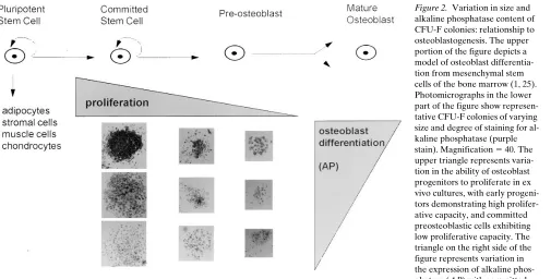
To examine whether the changes induced by ovariectomy in colony numbers in ex vivo cultures were the result of an in vivo effect on the commitment of mesenchymal stem cells to-ward the osteoblast lineage, we proceeded to determine the level of alkaline phosphatase activity, an early marker of os-teoblast differentiation (1), within individual colonies. Analy-sis of features exhibited by a single CFU-F colony was accom-plished using an assay we have recently developed that allows the estimation of the number of cells within a colony simulta-neously with determination of alkaline phosphatase activity (22). As seen in Fig. 2, CFU-F colonies exhibit considerable variation in size and the intensity of staining for alkaline phos-phatase. Based on extensive studies of other cell lineages that undergo proliferation and differentiation during development from multipotent progenitors, many aspects of which have been confirmed in the case of osteoblastogenesis (1, 25), we reasoned that small colonies develop from more committed progenitors, which have less proliferative potential, and that
Figure 1. Effect of ovariectomy on osteoblast progenitors. Mice were
sham-operated or ovariectomized. 4 wk after the operation, animals were killed, femoral marrow cells were isolated, and cells from each treatment group were pooled (n 5 4 per group). Quadruplicate paral-lel cultures were established for the determination of F or CFU-OB, as described in Methods. Bars represent the mean (6SD) num-ber of colonies formed per 106 marrow cells seeded in the cultures.
[image:4.612.60.565.447.703.2]Data were analyzed by Student’s t test. *P , 0.05 versus sham-oper-ated animals. There was no difference in the ratio of the number of CFU-F to CFU-OB colonies in cultures from sham-operated and ovariectomized mice as determined by z test.
Figure 2. Variation in size and
large colonies arise from less differentiated progenitors with high proliferation capacity (Fig. 2). Thus, smaller CFU-F colo-nies with the most intense staining for alkaline phosphatase must contain more differentiated osteoblastic cells as com-pared to small colonies with less intense alkaline phosphatase staining.
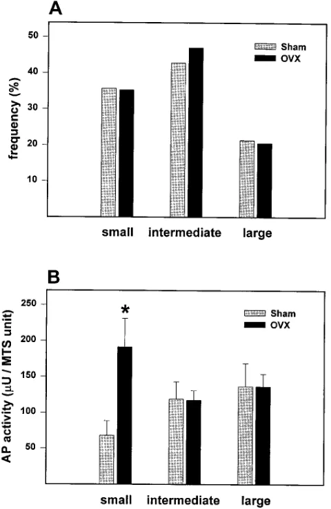
Cultures from ovariectomized mice had the same micro-scopic cell features when compared to cultures from sham-oper-ated controls. Moreover, the distribution of different colony sizes (i.e., small, medium, and large) was indistinguishable
be-tween the two groups, suggesting that ovariectomy did not cause a change in the proliferative potential of the progenitors (Fig. 3 A). This finding was unlike our previous observation of smaller CFU-F colonies in ex vivo cultures from marrow of mice with defective osteoblast development and osteopenia, compared to CFU-F colonies from control mice (22).
Alkaline phosphatase activity was increased by two- to threefold in small colonies from ovariectomized mice, as com-pared to small colonies from sham-operated animals (Fig. 3
B). On the other hand, alkaline phosphatase activity in inter-mediate size and large colonies was unaffected by the estrogen status of the donor animal. These findings are consistent with the idea that acute estrogen deficiency causes a shift in the commitment of mesenchymal progenitors toward the osteo-blast phenotype.
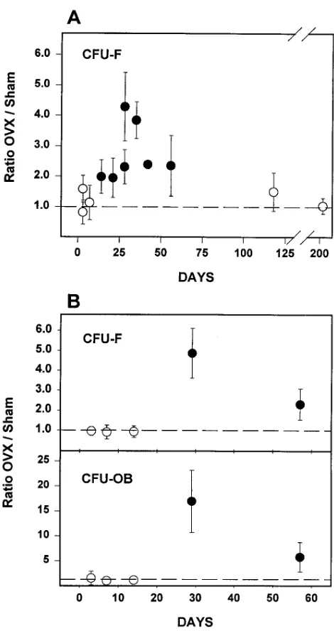
Evidence from both animal and human studies indicates that the effect of estrogen loss on bone metabolism changes with time (26–28). Therefore, we investigated the time course of the changes in CFU-F numbers after ovariectomy over a pe-riod of 210 d, by determining CFU-F numbers in ex vivo bone marrow cultures obtained from ovariectomized animals at pro-gressively longer time intervals after the operation (Fig. 4 A). CFU-F numbers were unchanged at 3 and 7 d, but were signif-icantly elevated at 14 d after ovariectomy. They remained sig-nificantly higher than the sham-operated controls as long as 56 d, but at later time points (115 and 210 d after ovariectomy), CFU-F numbers from ovariectomized animals were indistin-guishable from those of sham-operated mice. Similar results were obtained in a second time course experiment, in which both CFU-F and CFU-OB numbers were examined over a pe-riod of 57 d after sham operation or ovariectomy (Fig. 4 B). However, unlike the experiment shown in Fig. 4 A, an in-creased number of CFU-F at 2 wk after ovariectomy was not observed in this second experiment. The effect of ovariectomy on CFU-F and CFU-OB numbers was the same regardless of whether ascorbate-2-phosphate or the combination of ascorbic acid and b-glycerophosphate was used to stimulate osteoblast differentiation and mineralization (data not shown).
Taking advantage of the finding that CFU-F and CFU-OB numbers change with time after ovariectomy, we next sought to determine whether these changes have an impact on osteo-blast biology in vivo by searching for parallel temporal changes in osteoblast activity in the whole animal. To this end, we de-termined the circulating level of osteocalcin, a protein pro-duced by osteoblasts and used extensively as a marker of os-teoblastic activity, at various time points after ovariectomy, roughly covering the same period of time that we had deter-mined CFU-F numbers. Similar to our observations on bone marrow progenitors, we found that ovariectomy caused an ap-proximately twofold increase in the level of osteocalcin in the serum as early as 14 d after the operation. Osteocalcin levels were elevated as late as 56 d after ovariectomy (Fig. 5). How-ever, by 180 d, the levels of osteocalcin in ovariectomized mice were indistinguishable from sham-operated controls.
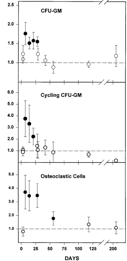
[image:5.612.58.298.61.430.2]In view of the fact that ovariectomy leads to an increase in rate of bone remodeling and that, as in the case of the bone-forming effects of estrogen deficiency, the rate of resorption may be changing with time after loss of ovarian function, we determined changes in osteoclast progenitors in the bone mar-row and their temporal relationship with the rate of bone loss. Early hematopoietic osteoclast progenitors, assessed as CFU-GM, increased by approximately twofold as early as 7 d after
Figure 3. Effect of ovariectomy on colony size and alkaline
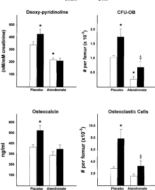
the operation, and returned to levels similar to that of sham-operated animals by 28–35 d (Fig. 6). The number of CFU-GM in S-phase (cycling CFU-GM), a sensitive indicator of expo-sure to IL-6 (12), was also increased after ovariectomy and fol-lowed the same temporal pattern as CFU-GM. Ovariectomy also induced a three- to fourfold increase in osteoclastogenesis in ex vivo marrow cultures, as we and others had shown previ-ously (11, 29). This change followed the same time course as the increase in CFU-GM; however, unlike CFU-GM, osteo-clastic cell formation in ex vivo cultures remained elevated at 28 and 56 d after the operation, before declining to the level exhibited by sham-operated animals at 115 and 210 d. Over the same period of time, cancellous bone volume was significantly reduced in ovariectomized mice, compared to that of sham-operated controls (Fig. 7). A significant decrease in trabecular bone volume was detected as early as 7 d after the operation and reached a plateau by 4 wk after ovariectomy. Sham-oper-ated mice also exhibited a decline in femoral cancellous bone during this experiment, albeit much smaller than that seen in ovariectomized mice. This later phenomenon has been ob-served previously in the rat, but its cause remains unclear (27). Finally, we investigated whether increased osteoblastogen-esis was dependent on factors released from the bone matrix during the increased resorption caused by increased osteoclas-togenesis. To do this, sham-operated or ovariectomized mice were treated with either vehicle or the potent antiresorptive agent alendronate (0.25 mg/kg per day) beginning 1 d before the operation, and the number of osteoblast and osteoclast progenitors in the marrow was determined 4 wk later. Adminis-tration of alendronate suppressed the level of urinary deoxy-pyridinoline, a measure of bone resorption (20), in sham-operated animals; moreover, alendronate prevented the ovariectomy-induced increases in urinary deoxy-pyridinoline and serum osteocalcin (Fig. 8). Strikingly however, the stimu-latory effect of ovariectomy on osteoblastogenesis or
osteo-Figure 4. Time course of change in osteoblast progenitors in the bone
[image:6.612.59.294.55.499.2]marrow after ovariectomy. (A) Mice were either sham-operated or ovariectomized (n 5 4–6 per group), and killed 3, 7, 14, 21, 28, 35, 42, 56, 118, or 205 d later. Replicate experiments were carried out at 3 and 28 d. Marrow cell cultures were established for the determina-tion of CFU-F as described in Methods. (B) Mice were either sham-operated or ovariectomized (n 5 6 per group), and killed 3, 7, 14, 28, or 56 d later. Marrow cells were established for the determination of CFU-F and CFU-OB as described in Methods. The data shown are the ratio of the mean number of CFU-F or CFU-OB per femur found in ovariectomized mice and that found in sham-operated mice, at each time point. The mean number of osteoblast progenitors per fe-mur in sham-operated animals among the experiments shown ranged between 400 and 2,500 for CFU-F, and 100 and 800 for CFU-OB. Er-ror bars represent the calculated SEM of the ratio, derived from the SEM values of the means used to calculate the ratio (23). A statisti-cally significant effect of ovariectomy on the number of CFU-F per femur, determined using Student’s t test, is indicated by the closed cir-cles (P , 0.05); and lack of a statistically significant effect is indicated by open circles.
Figure 5. Time course of change in serum osteocalcin in mice after
[image:6.612.319.553.58.249.2]clastogenesis, as evidenced by changes in CFU-OB and osteo-clast progenitor numbers, was not affected by administration of alendronate. Nonetheless, the absolute number of CFU-OB was decreased by alendronate in both sham-operated and ovariectomized animals.
Discussion
The maintenance of a balance between bone resorption and formation during normal bone remodeling requires that the proper number of mature osteoblasts and osteoclasts is pro-duced to meet the needs of the remodeling process. The results of the present studies demonstrate that estrogen loss increases the number of mesenchymal progenitors that are capable of committing to the osteoblast lineage in the murine bone mar-row, and that these changes are temporally linked with in-creased osteoblast activity. These findings are consistent with evidence suggesting that estrogen deficiency affects osteoblast differentiation in the rat (30, 31). Moreover, they elucidate the cellular changes underlying the increased bone formation asso-ciated with loss of estrogen. Taken together with earlier obser-vations that increased osteoclastogenesis leads to increased bone resorption in estrogen deficiency (11), and that de-creased osteoblastogenesis is associated with dede-creased bone formation and bone mass in a murine model of osteopenia (22), our results provide compelling evidence that the orderly births of osteoclasts and osteoblasts from their respective pro-genitors in the bone marrow are essential determinants of the rate of bone remodeling (32). Whether the increased number of CFU-F observed in the bone marrow of estrogen-deficient mice leads to an increase in other cell types derived from this progenitor (e.g., adipocytes or stromal cells) remains un-known.
[image:7.612.313.554.58.234.2]The increased osteoblastogenesis and osteoclastogenesis that followed ovariectomy subsided with time in these studies,
Figure 6. Time course of change in osteoclast progenitors in the bone marrow after ovariectomy. Marrow cells obtained from the same animals (n 5 4–6 per group) used for the experiments shown in Fig. 4 A were placed into ex vivo culture for the determination of CFU-GM, cycling CFU-GM, and osteoclastic cell formation, as de-scribed in Methods. Replicate experiments were carried out at 3 and 28 d, and at some time points the osteoclastic cell formation assay was omitted. The data shown are the ratio of the mean number of progen-itors per femur found in ovariectomized mice and that found in sham-operated mice. The mean number of progenitors per femur in sham-operated animals among the experiments shown ranged be-tween 10,000 and 60,000 for CFU-GM, 2,000 and 13,000 for cycling CFU-GM, and 2,500 and 7,600 for osteoclastic cells. Error bars repre-sent the calculated SEM of the ratio, derived from the SEM values of the means used to calculate the ratio (23). A statistically significant effect of ovariectomy on the number of osteoclast progenitors per fe-mur, determined using Student’s t test, is indicated by the closed circles (P , 0.05). Lack of a statistically significant effect is indicated by the open circles.
Figure 7. Time course of bone-loss mice after ovariectomy. Femurs
[image:7.612.80.298.63.511.2]as did osteocalcin levels in the serum and the rate of bone loss. These findings are in agreement with previous studies in rats, indicating that the increased bone remodeling and bone loss caused by ovariectomy subsides with time (26, 27). Moreover, they are in agreement with studies in humans indicating that the rapid bone loss associated with increased bone remodeling subsides with time after loss of sex steroids, and is followed by a later stage of slower bone loss (33), and with evidence that the increased bone remodeling at the menopause is attenuated within 10 yr (28). Nevertheless, other studies have indicated that bone loss after menopause may continue unabated throughout life (34).
During normal bone remodeling, the provision of the ap-propriate number of mature osteoblasts is thought to be con-trolled by factors, such as TGF-b, that are released from the bone matrix during its resorption by osteoclasts (35, 36). Our finding that administration of alendronate reduced the num-ber of CFU-OB in the bone marrow of sex steroid–sufficient mice is entirely consistent with this idea. Nonetheless, the ob-servation that increased osteoblastogenesis in ovariectomized mice was still present when bone resorption (and thereby re-lease of factors from the bone matrix) was blocked by alendro-nate, indicates that this cellular change could not have been brought about by this mechanism. Instead, our results strongly
indicate that estrogen loss unleashes signals that can stimulate early osteoblast progenitors. However, the ovariectomy-induced increase in osteoblast progenitors in mice receiving alendro-nate did not lead to increased bone formation, as evidenced by the lack of change in circulating osteocalcin. This is most prob-ably due to the fact that bone formation occurs only at sites of previous resorption in remodeling bone; and because the latter was blocked by alendronate, bone formation could not occur (37). We recognize that we have not excluded the possibility that alendronate might have exerted additional effects on cells of the bone marrow. However, this possibility is very unlikely, as an identical dose of alendronate had no effect on the num-ber of osteoclast precursors in the murine bone marrow in an earlier study (38).
[image:8.612.57.363.67.442.2]The source and nature of the signals leading to increased osteoblastogenesis unleashed by ovariectomy is only a matter of conjecture at this stage. IL-6–type cytokines have been shown to stimulate the early stages of osteoblast differentia-tion (39–41); overexpression of leukemia inhibitory factor or oncostatin M in mice causes increased bone formation (42, 43); and IL-6–type cytokines exert antiapoptotic effects on osteo-blastic cells (44, 45). Thus, it is reasonable to propose, that be-sides osteoclastogenesis, the increased IL-6 production and in-creased IL-6–type cytokine responsiveness caused by estrogen
Figure 8. Effect of alendronate on
ovariectomy-induced increase in bone cell activity and bone cell progenitors. Mice were injected daily with vehicle or alendronate (0.25 mg/kg body weight) begin-ning 1 d before sham-operation or ovariectomy. Urinary deoxy-pyridinoline was determined at 2 wk after the operation when ovariectomy-induced bone loss is rapid (Fig. 7). Animals were killed at 4 wk after the operation and serum osteo-calcin, osteoclastogenesis, and osteoblastogenesis (CFU-OB) were determined, as described in Methods, using ascorbate-2-phosphate to stimu-late osteoblast differentiation and formation of mineralized bone nodules. Data shown are the mean (6SEM) values obtained from 7–10 animals per group. Data were analyzed by ANOVA. *P , 0.05 versus sham-operated vehicle-treated mice,
‡P , 0.05 versus sham-operated
deficiency (46) is also responsible for stimulating osteoblasto-genesis (39).
Because osteoclastogenesis is critically dependent on stro-mal/osteoblastic cells (5), the increased osteoblastogenesis and osteoclastogenesis in the bone marrow of ovariectomized mice may be interdependent. Studies of mice with defective osteo-blast development (SAMP6) have revealed that osteoclasto-genesis was also decreased, thus providing evidence for the im-portance of this dependency in vivo (22). More important, orchidectomy failed to stimulate osteoblastogenesis in these mice; and the expected increase in osteoclastogenesis and the bone loss that occur in mice with normal osteoblastogenesis were blunted in SAMP6 (24). Hence, paradoxical as it may seem, it is possible that the ovariectomy-induced stimulation of mesenchymal cell differentiation towards the osteoblastic lineage plays a role in the increased osteoclastogenesis. Obvi-ously, the increased osteoblast development described here must not be sufficient to meet the demand imposed by the in-creased osteoclastogenesis and bone resorption that occurs af-ter estrogen loss. Therefore, cellular changes that favor resorp-tion over formaresorp-tion must occur.
In conclusion, the results of this study provide strong sup-port for the contention that the rates of osteoclastogenesis and osteoblastogenesis in the bone marrow are critical determi-nants of the rates of bone resorption and bone formation, and thereby responsible for the increased rate of bone remodeling in estrogen deficiency. In addition, these studies reveal for the first time that a normally operating mechanism, which assures the coordinated production of osteoblasts in response to the needs generated by osteoclastic bone resorption, is overridden in acute estrogen deficiency by the unleashing of factors that stimulate osteoblastogenesis in a fashion autonomous from the need created by bone resorption. Therefore, the upregulation of osteoblastogenesis after acute estrogen loss is inappropriate for the normal remodeling process.
Acknowledgments
The authors wish to thank Michael Parfitt (University of Arkansas for Medical Sciences, Little Rock, AR) for advice, and Catherine Smith (UAMS) and Don Paul (Lilly Research Labs, Indianapolis, IN) for technical assistance in the conduct of this work.
This work was supported by Public Health Service grants (P01 AG13918 and AR43003), and the Department of Veterans Affairs.
References
1. Aubin, J.E., K. Turksen, and J.N.M. Heersche. 1993. Osteoblastic cell lin-eage. In Cellular and Molecular Biology of Bone. M. Noda, editor. Academic Press, San Diego, CA. 1–45.
2. Roodman, G.D. 1996. Advances in bone biology: the osteoclast. Endocr. Rev. 17:308–332.
3. Bellows, C.G., J.E. Aubin, and J.N.M. Heersche. 1991. Initiation and pro-gression of mineralization of bone nodules formed in vitro: the role of alkaline phosphatase and organic phosphate. Bone Miner. 14:27–40.
4. Van Vlasselaer, P., N. Falla, H. Snoeck, and E. Mathieu. 1994. Character-ization and purification of osteogenic cells from murine bone marrow by two-color cell sorting using anti-Sca-1 monoclonal antibody and wheat germ aggluti-nin. Blood. 84:753–763.
5. Suda, T., N. Takahashi, and T.J. Martin. 1992. Modulation of osteoclast differentiation. Endocr. Rev. 13:66–80.
6. Parfitt, A.M., C.H.E. Mathews, A.R. Villanueva, M. Kleerekoper, B. Frame, and D.S. Rao. 1983. Relationships between surface, volume and thick-ness of iliac trabecular bone in aging and in osteoporosis: implications for the microanatomic and cellular mechanism of bone loss. J. Clin. Invest. 72:1396– 1409.
7. Eriksen, E.F., S.F. Hodgson, R. Eastell, S.L. Cedel, W.M. O’Fallon, and
B.L. Riggs. 1990. Cancellous bone remodeling in type I (postmenopausal) os-teoporosis: quantitative assessment of rates of formation, resorption, and bone loss at tissue and cellular levels. J. Bone Miner. Res. 5:311–319.
8. Poli, V., R. Balena, E. Fattori, A. Markatos, A. Yamamoto, H. Tanaka, G. Ciliberto, G.A. Rodan, and F. Costantini. 1994. Interleukin-6 deficient mice are protected from bone loss caused by estrogen depletion. EMBO (Eur. Mol. Biol. Organ.) J. 13:1189–1196.
9. Balena, R., B.C. Toolan, M. Shea, A. Markatos, E.R. Myers, S.C. Lee, E.E. Opas, J.G. Seedor, H. Klein, D. Frankenfield, et al. 1993. The effects of 2-year treatment with the aminobisphosphonate alendronate on bone metabo-lism, bone histomorphometry, and bone strength in ovariectomized nonhuman primates. J. Clin. Invest. 92:2577–2586.
10. Kalu, D.N. 1991. The ovariectomized rat model of postmenopausal bone loss. Bone Miner. 15:175–192.
11. Jilka, R.L., G. Hangoc, G. Girasole, G. Passeri, D.C. Williams, J.S. Abrams, B. Boyce, H. Broxmeyer, and S.C. Manolagas. 1992. Increased osteo-clast development after estrogen loss: mediation by interleukin-6. Science. 257: 88–91.
12. Bellido, T., R.L. Jilka, B.F. Boyce, G. Girasole, H. Broxmeyer, S.A. Dalrymple, R. Murray, and S.C. Manolagas. 1995. Regulation of interleukin-6, osteoclastogenesis and bone mass by androgens: the role of the androgen re-ceptor. J. Clin. Invest. 95:2886–2895.
13. Girasole, G., R.L. Jilka, G. Passeri, S. Boswell, G. Boder, D.C. Williams, and S.C. Manolagas. 1992. 17b-estradiol inhibits interleukin-6 production by bone marrow-derived stromal cells and osteoblasts in-vitro: a potential mecha-nism for the antiosteoporotic effect of estrogens. J. Clin. Invest. 89:883–891.
14. Pottratz, S.T., T. Bellido, H. Mocharla, D. Crabb, and S.C. Manolagas. 1994. 17b-estradiol inhibits expression of human interleukin-6 promoter-repor-ter constructs by a receptor-dependent mechanism. J. Clin. Invest. 93:944–950.
15. Ray, A., K.E. Prefontaine, and P. Ray. 1994. Down-modulation of inter-leukin-6 gene expression by 17 beta-estradiol in the absence of high affinity DNA binding by the estrogen receptor. J. Biol. Chem. 269:12940–12946.
16. Stein, B., and M.X. Yang. 1995. Repression of the interleukin-6 pro-moter by estrogen receptor is mediated by NF-kappa B and C/EBP beta. Mol. Cell Biol. 15:4971–4979.
17. Kassem, M., S.A. Harris, T.C. Spelsberg, and B.L. Riggs. 1996. Estrogen inhibits interleukin-6 production and gene expression in a human osteoblastic cell line with high levels of estrogen receptors. J. Bone Miner. Res. 11:193–199.
18. Galien, R., and T. Garcia. 1997. Estrogen receptor impairs interleukin-6 expression by preventing protein binding on the NF-kappaB site. Nucleic Acids Res. 25:2424–2429.
19. Lin, S.-C., T. Yamate, Y. Taguchi, V.Z.C. Borba, G. Girasole, C.A. O’Brien, T. Bellido, E. Abe, and S.C. Manolagas. 1997. Regulation of the gp80 and gp130 subunits of the IL-6 receptor by sex steroids in the murine bone mar-row. J. Clin. Invest. 100:1880–1890.
20. Delmas, P.D. 1993. Biochemical markers of bone turnover. J. Bone Miner. Res. 8:S549–S554.
21. Hughes, D.E., K.R. Wright, H.L. Uy, A. Sasaki, T. Yoneda, G.D. Rood-man, G.R. Mundy, and B.F. Boyce. 1995. Bisphosphonates promote apoptosis in murine osteoclasts in vitro and in vivo. J. Bone Miner. Res. 10:1478–1487.
22. Jilka, R.L., R.S. Weinstein, K. Takahashi, A.M. Parfitt, and S.C. Manol-agas. 1996. Linkage of decreased bone mass with impaired osteoblastogenesis in a murine model of accelerated senescence. J. Clin. Invest. 97:1732–1740.
23. Stuart, A., and J.K. Ord. 1987. Kendall’s Advanced Theory of Statistics. Oxford University Press, New York. 223.
24. Weinstein, R.S., R.L. Jilka, A.M. Parfitt, and S.C. Manolagas. 1997. The effects of androgen deficiency on murine bone remodeling and bone mineral density are mediated via cells of the osteoblastic lineage. Endocrinology. 138: 4013–4021.
25. Triffitt, J.T. 1996. The stem cell of the osteoblast. In Principles of Bone Biology. J.P. Bilezikian, L.G. Raisz, and G.A. Rodan, editors. Academic Press, San Diego, CA. 39–50.
26. Wronski, T.J., L.M. Dann, K.S. Scott, and M. Cintrön. 1989. Long-term effects of ovariectomy and aging on the rat skeleton. Calcif. Tissue Int. 45:360–366. 27. Li, M., Y. Shen, H. Qi, and T.J. Wronski. 1996. Comparative study of skeletal response to estrogen depletion at red and yellow marrow sites in rats. Anat. Rec. 245:472–480.
28. Stepan, J.J., A. Tesarova, T. Havranek, J. Jodl, J. Formankova, and V. Pacovsky. 1985. Age and sex dependency of the biochemical indices of bone re-modeling. Clin. Chim. Acta. 151:273–283.
29. Most, W., L. Schot, A. Ederveen, L. Van der Wee-Pals, S. Papapoulos, and C. Löwik. 1995. In vitro and ex vivo evidence that estrogens suppress in-creased bone resorption induced by ovariectomy or PTH stimulation through an effect on osteoclastogenesis. J. Bone Miner. Res. 10:1523–1530.
30. Scutt, A., U. Kollenkirchen, and P. Bertram. 1996. Effect of age and ovariectomy on fibroblastic colony-forming unit numbers in rat bone marrow. Calcif. Tissue Int. 59:309–310.
31. Modrowski, D., L. Miravet, M. Feuga, and P.J. Marie. 1993. Increased proliferation of osteoblast precursor cells in estrogen-deficient rats. Am. J. Physiol. 264:E190–E196.
pathophys-iology of osteoporosis. N. Engl. J. Med. 332:305–311.
33. Parfitt, A.M. 1992. The two-stage concept of bone loss revisited. Trian-gle. 31:99–110.
34. Eastell, R., P.D. Delmas, S.F. Hodgson, E.F. Eriksen, K.G. Mann, and B.L. Riggs. 1988. Bone formation rate in older normal women: concurrent as-sessment with bone histomorphometry, calcium kinetics, and biochemical markers. J. Clin. Endocrinol. Metab. 67:741–748.
35. Oreffo, R.O.C., G.R. Mundy, S.M. Seyedin, and L.F. Bonewald. 1989. Activation of the bone-derived latent TGF beta complex by isolated osteo-clasts. Biochem. Biophys. Res. Commun. 158:817–823.
36. Hayden, J.M., S. Mohan, and D.J. Baylink. 1995. The insulin-like growth factor system and the coupling of formation to resorption. Bone. 17(Suppl):93S–98S.
37. Hattner, R., B.N. Epker, and H.M. Frost. 1965. Suggested sequential mode of control of changes in cell behavior in adult bone remodelling. Nature. 206:489–490.
38. van Beek, E.R., C.W. Löwik, and S.E. Papapoulos. 1997. Effect of alen-dronate treatment on the osteoclastogenic potential of bone marrow cells in mice. Bone. 4:335–340.
39. Manolagas, S.C., R.L. Jilka, T. Bellido, C.A. O’Brien, and A.M. Parfitt. 1996. Interleukin-6-type cytokines and their receptors. In Principles of Bone Bi-ology. J.P. Bilezikian, L.G. Raisz, and G.A. Rodan, editors. Academic Press, San Diego, CA. 701–713.
40. Bellido, T., V.Z.C. Borba, P. Roberson, and S.C. Manolagas. 1997.
Acti-vation of the JAK/STAT signal transduction pathway by IL-6 type cytokines promotes osteoblast differentiation. Endocrinology. 138:3666–3676.
41. Taguchi, T., T. Yamate, H. Mocharla, S.-C. Lin, A. Vertino, P. DeTogni, E. Abe, and S.C. Manolagas. 1996. Interleukin-6 induces osteoblast differentia-tion in uncommitted embryonic fibroblasts (EF). J. Bone Miner. Res. 11: S101(Abstr.).
42. Metcalf, D., and D.P. Gearing. 1989. Fatal syndrome in mice engrafted with cells producing high levels of the leukemia inhibitory factor. Proc. Natl. Acad. Sci. USA. 86:5948–5952.
43. Malik, N., H.S. Haugen, B. Modrell, M. Shoyab, and C.H. Clegg. 1995. Developmental abnormalities in mice transgenic for bovine oncostatin M. Mol. Cell Biol. 15:2349–2358.
44. Bellido, T., L. Han, R.L. Jilka, and S.C. Manolagas. 1997. gp130/STAT3 activation stimulates the transcription of the cyclin dependent kinase inhibitor p21WAF1,CIP1 gene in osteoblasts: a prerequisite for the biologic effects of IL-6
type cytokines. J. Bone Miner. Res. 12:S159(Abstr.).
45. Jilka, R.L., R.S. Weinstein, T. Bellido, A.M. Parfitt, and S.C. Manola-gas. 1998. Osteoblast programmed cell death (apoptosis): modulation by growth factors and cytokines. J. Bone Miner. Res. In press.