INTRODUCTION
Stem cell homeostasis is the set of processes by which growth and differentiation are dynamically regulated so as to allow long-term persistence of the stem cell population at its normal size (Reddy 2008; Spradling et al., 2008). Shoot apical meristems (SAMs) of higher plants that are located at the growing tip harbor sets of stem cells/initials within the central zone (CZ) (Fig. 1A) (Steeves and Sussex, 1989). Progeny of stem cells differentiate as leaves or flowers within the flanking peripheral zone (PZ) (Fig. 1A). The CZ also provides cells to the rib meristem (RM) located beneath the CZ, where cells of the stem form (Fig. 1A). The relative ratios of cells in each functional domain of SAMs remain largely constant, despite cells being continually diverted to differentiation pathways (Steeves and Sussex, 1989). The SAM stem cell niche, thus, represents a dynamic cellular network that requires coordination between gene expression dynamics and cell proliferation patterns to maintain homeostatic balance between distinct functional domains (Meyerowitz, 1997). However, cell proliferation patterns and cell identity transitions are spatiotemporally coupled, which makes it difficult to analyze the mutual influence they exert on each
other in static studies. Therefore, understanding real-time gene expression and cell division dynamics will be crucial in deciphering the regulatory principles that mediate stem cell homeostasis.
Earlier studies, in Arabidopsis, have revealed that WUS, which is expressed in the RM cells, provides cues to the overlying cells of the CZ to help to specify them as stem cells (Laux et al., 1996; Mayer et al., 1998). The spatial confinement of WUStranscription to a small domain is crucial for maintaining a constant number of stem cells and it is in part mediated by the CLAVATA (CLV) class of genes (Clark et al., 1997; Jeong et al., 1999; Fletcher et al., 1999; Schoof et al., 2000). CLV3, which is expressed in the stem cells of the CZ, encodes a small secreted peptide that binds and activates the CLV1 transmembrane receptor kinase that is active in the RM (Ogawa et al., 2008). CORYNE, an another transmembrane receptor kinase, has been shown to function with CLV1 to regulate WUSexpression and SAM size (Muller et al., 2008; Bleckmann et al., 2010). At the same time, WUS not only specifies stem cells but also activates CLV3expression. As CLV3 restricts the domain of WUS expression, the two proteins participate in a self-correcting feedback loop between cells of the CZ and the RM (Schoof et al., 2000; Brand et al., 2000; Rieu and Laux, 2009). Although the self-correcting feedback loop restricts WUS expression, the mechanisms by which WUS ensures spatio-temporal coordination of gene expression and growth patterns in distinct cell types of the shoot apical meristem are not well understood (Reddy et al., 2007; Barton, 2010). This is largely because of the static analysis of terminal mutant phenotypes, which fails to reveal the dynamics of the meristem system.
Development 137, 3581-3589 (2010) doi:10.1242/dev.054973 © 2010. Published by The Company of Biologists Ltd
2150 Batchelor Hall, Center for Plant Cell Biology (CEPCEB), Department of Botany and Plant Sciences, University of California, Riverside, CA 92521, USA.
*Author for correspondence (venug@ucr.edu)
Accepted 31 August 2010 SUMMARY
Plant stem cell populations, unlike their animal counterparts, do not use cell migration and oriented cell divisions to maintain their size, and therefore require a precise coordination between self-renewing divisions of stem cells, and rates of cell division and differentiation among stem cell progenitors. Shoot apical meristems (SAMs) of higher plants harbor a set of stem cells within the central zone (CZ) that divide infrequently. Stem cell daughters that are displaced towards the surrounding peripheral zone (PZ) divide at a faster rate and enter into differentiation at specific locations to form leaves or flowers. The relative ratios of cells in the CZ and the PZ are maintained, despite a constant displacement of cells from the CZ into the PZ, and subsequent allocation of cells within the PZ to form organ primordia. The mechanisms that mediate this homeostatic balance are not well understood. A homeodomain transcription factor WUSCHEL, expressed in the rib meristem (RM), located beneath the CZ, has been shown to provide nonautonomous cues for stem cell specification. By employing transient spatial manipulation and live imaging, we show that an elevated level of WUS not only induces expansion of the CZ, but also results in increased cell division rates in cells of the PZ; conversely, decreases in WUS level lead to a smaller CZ and are associated with a reduction in cell division rate. Moreover, low levels of WUS lead to enlarged organ primordia, by elevating the responsiveness of the PZ cells to the plant hormone auxin. This reveals a function of WUS in mediating the balance between differentiating and non-differentiating cells of the PZ. Regulation of stem cell numbers, growth and differentiation patterns by a single transcription factor forms a interconnected and self-correcting feedback loop to provide robustness to stem cell homeostasis in a dynamic cellular environment.
KEY WORDS: CLAVATA3, Shoot apical meristem, WUSCHEL, Arabidopsis
WUSCHEL mediates stem cell homeostasis by regulating stem
cell number and patterns of cell division and differentiation
of stem cell progenitors
Ram Kishor Yadav, Montreh Tavakkoli and G. Venugopala Reddy*
D
E
V
E
LO
P
M
E
N
An earlier live-imaging study has revealed that CLV3 functions in maintaining a constant number of CZ stem cells by preventing de-differentiation of stem cell daughters that have been displaced by CZ cell divisions into the surrounding PZ, and that CLV3 maintains the overall size of the SAM by repressing cell division rates in cells of the PZ (Reddy and Meyerowitz, 2005) (Fig. 1A). A related study, using an inducible system, has shown that overexpression of CLV3 results in decrease of mitotic activity (Muller et al., 2006). However, it is not clear whether both of these functions of CLV3 are mediated through the spatial regulation of WUSalone. This is because the increased cell division rates observed in cells of the PZ upon transient down-regulation of CLV3 levels could be an indirect consequence of the expanding CZ (Reddy and Meyerowitz, 2005), as well as a consequence of the expanded RM. It has also been suggested that although WUS functions in specifying stem cells, another transcription factor, SHOOTMERISTEMLESS, regulates amplification of stem cell daughters (Lenhard et al., 2002). In order to distinguish between these possibilities, we have transiently altered WUS levels by employing both dexamethasone-inducible downregulation and ectopic expression of WUSin distinct spatial domains of the shoot apical meristem, and examined the dynamic effects on CZ size, meristem cell division rates and differentiation patterns in real time by use of live imaging. We show here that WUS can perform multiple functions depending upon its levels and places of expression. Elevated levels of WUS within the CZ not only induce expansion of the CZ, but also result in increased cell division rates in cells of the PZ, whereas a decrease in WUS levels lead to a smaller CZ and associated reduction in cell division rate. Our analysis also reveals that transient decrease in WUS levels leads to enlarged organ primordia, by elevating the responsiveness of the PZ cells to plant hormone auxin. Regulation of stem cell numbers, growth rates and differentiation patterns of stem cell progeny by a single transcription factor forms a self-correcting feedback loop that may impart robustness to stem cell homeostasis.
MATERIALS AND METHODS Transgenic lines
For generating WUSfoldback (double stranded RNAi) construct, first exon
of WUS gene amplified using primer pair (forward) WUS-RNAi
5-CACCAAAATCTCTTTACTACCAGCAAGTTG-3 and (reverse) WUS-RNAi 5-CGTTATTGAAGCTGGGATATGGCTTGTTAT-3, and cloned in
to pENTR D/TOPO vector (Invitrogen). To create 6XOP::WUSdsRNAi
vector, LR reaction of pENTR D/TOPO was setup with popoff2 binary
vector (Wielopolska et al., 2005). To generate 6XOP::CLV3, a CLV3open
reading frame was amplified by PCR using primer pair CLV3-F CACCATGGATTCTAAAAGCTTTGT and CLV3R TTAATTAATCAA -GGGAGCTGAAAGTTGTTTCTTGG, and cloned into pENTR D/TOPO
vector. LR reaction was set up with 6Xop pzp222binary vector to create
6Xop::CLV3. 35S::WUS-GRwas assembled in pGREEN vector by placing
glucocorticoid coding sequences at the C terminus of WUS. The WUS-GR
region was amplified with BamHI sites by using PCR and cloned into
6XOP promoter housed in pARTbinary vector to generate 6XOP::
WUS-GR. pCLV3::mGFP5-ER, p35S::YFP29-1transgenes have been described in an earlier study (Reddy and Meyerowitz, 2005) and
pDR5rev::3XVENUS-N7, pPIN1::PIN1:GFP have also been described in
a different study (Heisler et al., 2005). The pCLV3:;LhG4 and
PWUS::LHG4have been described previously (Lenhard et al., 2002). The
pSTM::LhG4and pLAS::LhG4were kindly provided by Yuval Eshed; their expression patterns were determined by performing RNA in situ
hybridization using LhG4gene probe.
Plant growth and live imaging
Arabidopsisplants were maintained in continuous light. Soil and growth conditions were maintained as described earlier (Reddy and Meyerowitz, 2005). Plant growth and preparation for live imaging, dexamethasone
treatment, the optics, microscopy platform, image acquisition and image reconstruction have been described in earlier studies (Reddy et al., 2004; Heisler et al., 2005; Reddy and Meyerowitz, 2005).
In situ hybridization
In situ hybridization was carried out using the protocol posted at
http://www.its.caltech.edu/~plantlab/html/protocols.html. Full-length LhG4
was amplified by using PCR and used for probe synthesis.
RT-PCR
RNA was isolated from finely dissected 4-week-old shoot apices that were
either treated with 10 M dexamethasone or mock-treated for 4 days before
harvesting. Semi-quantitative reverse transcription PCR was performed according to a protocol using WUS-specific PCR primer sequences described previously (Yadav et al., 2009).
Cell division analysis
Cell division activity was analyzed by reconstructing individual cell lineages of the L1 layer cells from time-lapse data acquired between 72 and 120 hours. Cell division rate was expressed as the size of individual lineages (the number of daughter cells at 120 hours that originated from a single cell present at 72 hours). In each case, cell division analysis was carried out on time-lapse imagery obtained from at least three different plants. Lineages were represented on SAM surface by marking individual lineages in separate colors (same color has been assigned to different lineages as long as they did not abut each other) on a 3D-reconstructed surface layer. Only the lineages that are part of the SAM region have been included in the quantitative analysis represented in Fig. 2G; lineages that are part of the primordia have been excluded.
Quantitative analysis of differentiation patterns
The number of pDR5-positive cells in each one of the early stage primordia
was counted. Early stage primordia that are not clearly separated from SAMs (for example, stages P1 and P2 in Fig. 6K and stages P0, P1 and P2
in Fig. 6Q were considered for counting). In parallel, the number of pDR5
-negative cells (the cells located in between discrete groups of pDR5
-positive cells) were also counted for comparison. All counting was carried out on images taken 96 hours after dexamethasone or mock treatment. Three independent plants were considered for analysis in each treatment
case. The numbers of pDR5-positive or pDR5-negative cells were averaged
over three plants and expressed as number of cells/SAM.
RESULTS
Ubiquitous expression of WUS leads to sequential expansion of the CZ
To understand the immediate role of WUSin stem cell specification and growth control, we constructed an inducible form of WUS consisting of the WUS protein-coding region fused to sequences coding for the ligand-binding domain of the rat glucocorticoid receptor (GR), expressed under a cauliflower mosaic virus coat protein promoter that causes ubiquitous expression of adjacent genes (35S::WUS-GR). In the absence of the ligand dexamethasone, the plants carrying the 35S::WUS-GRtransgene were found to develop normally (Fig. 1B). However, when these plants were grown in the presence of 10 M dexamethasone, the cotyledons failed to expand and overall growth of the seedlings was inhibited, as described in an earlier WUSoverexpression study (Lenhard et al., 2002) (Fig. 1C). To follow the reorganization patterns of the CZ and SAM size by live imaging, 35S::WUS-GR plants were crossed to transgenic plants carrying both the CZ reporter pCLV3::mGFP5-ER, which expresses an endoplasmic reticulum-localized green fluorescent protein under CLV3promoter control, and 35S::YFP 29-1, encoding a plasma membrane-localized yellow fluorescent protein expressed under a promoter that causes ubiquitous expression and which highlights cell boundaries of all SAM cells, thus allowing for detection of cell
D
E
V
E
LO
P
M
E
N
division events. Plants carrying all three constructs were treated either with dexamethasone or a control (mock treatment) solution and then they were imaged at 12-hour intervals. Mock-treated
plants showed very little variation in CZ size or SAM size (Fig. 1D-F). However, dexamethasone-treated plants showed an increase in CLV3promoter activity in the native domain within 12 hours (data not shown) and radial expansion of pCLV3::mGFP5-ER expression within 24 hours (Fig. 1H; see Fig. S5 in the supplementary material). In the next 24- to 48-hour window pCLV3::mGFP5-ERexpression intensified in the native CLV3 expression domain and further expanded radially into the adjacent PZ (Fig. 1I). A comparison of high-resolution time-lapse images of pCLV3::mGFP5-ERand 35S::YFP29-1revealed that the expansion of the CZ is due to the acquisition by PZ cells of pCLV3::mGFP5-ERexpression, suggesting a transition of the multipotent PZ cells into the pluripotent stem cells of the CZ (Fig. 1J,K). This analysis also revealed that uniform expression of WUS does not lead to simultaneous CLV3 activation and to the pluripotent stem cell pattern of gene expression in all SAM cells. Instead, the stem cell gene expression pattern spreads sequentially from the CZ, suggesting that the WUS-mediated stem cell factor is active only in the CZ and the PZ cells on its expanding border with the PZ.
Ubiquitous expression of WUS does not result in increased mitotic activity
It has been shown that transient decrease in CLV3 levels results in radial and sequential expansion of the CZ within 24-48 hours of dexamethasone treatment followed by an increase in SAM size owing to the higher cell division rates in cells of the PZ (Reddy and Meyerowitz, 2005). Upon ubiquitous WUS expression, we observed radial expansion of the CZ; however, we did not observe a striking increase in the overall SAM size even after 5 days of dexamethasone treatment (Fig. 2B). We analyzed cell division rates by reconstructing individual cell lineages of the L1 layer cells from time-lapse data acquired between 72 and 120 hours after the dexamethasone treatment. Cell division rate was expressed as the size of individual lineages (the number of daughter cells at 120 hours that originated from a single cell present at 72 hours). The lineage sizes in dexamethasone-treated plants indicated that only a few cells divided just once, resulting in lineages consisting of two cells (Fig. 2G). However, most of the cells in mock-treated controls divided at least once during this time window (Fig. 2G). In order to visualize the spatial patterns of mitotic activity, the individual lineages were superimposed on a reconstructed L1 layer of the SAM. This analysis revealed that a large proportion of cells on the SAM surface remain undivided, suggesting a negative impact of ubiquitous WUS expression on cell division activity (Fig. 2B). This is in contrast to the earlier observations, wherein downregulation of CLV3 resulted not only in expansion of the CZ but also in increased cell division rates in cells in the PZ (Reddy and Meyerowitz, 2005). This suggested either that the CLV3 effect on meristem size is not mediated through WUS, or that ubiquitous WUS expression does not have the same consequences for meristem size as expanded expression in the RM domain.
Inducible activation of WUS within the CZ results in both sequential expansion of stem cell domain and enlarged SAMs
[image:3.612.51.283.58.426.2]In order to further characterize the spatial requirements for the different effects of WUS overexpression, we expressed dexamethasone-inducible WUS (WUS-GR) in distinct spatial domains of the SAM by using bacterially derived two component expression systems. To activate WUS in the CZ, plants carrying pCLV3::LhG4 (the CLV3 promoter driving the expression of bacterial transcription factor LhG4) were crossed to Fig. 1. Ubiquitous ectopic WUS expression induces sequential
expansion of the CZ.(A)Schematic representation of functional domains of the ArabidopsisSAM. The CZ harbors a set of about 35 stem cells, stem cell progenitors in the inner peripheral zone (IPZ) that are capable of conversion into stem cells, while cells of the outer peripheral zone (OPZ) fail to de-differentiate as stem cells upon downregulation of CLV3. The cells of the rib meristem (RM) express WUS, which provides cues to overlying cells of the CZ to specify them as stem cells. Clusters of a few cells at specified locations within the PZ differentiate as organ primordia owing to the accumulation of higher levels of the plant hormone auxin. (B)A 35S::WUS-GRseedling grown on MS agar plate for 10 days. (C)Ten-day-old 35S::WUS-GRseedlings grown on MS agar plate supplemented with 10M dexamethasone. (D-F)Three-dimensional reconstructed view of L1 layer of wild-type SAM time series showing the stem cell domain highlighted by
pCLV3::mGFP5-ER (green) and cell outlines marked by plasma membrane-localized YFP driven by an ubiquitously expressed promoter,
35S::YFP pm29-1(red). (G-I)Three-dimensional reconstructed view of L1 layer time series showing sequential expansion of the CZ domain in
35S::WUS-GRplants treated with 10M dexamethasone. (J,K)Higher magnification images of G,H, respectively, showing the re-specification of the peripheral zone (PZ) cells into stem cells. Subtle changes in the CZ size are also observed in mock-treated plants. Arrows indicate the same cells or siblings of the same cells within the PZ across time points. Total elapsed time after dexamethasone or mock treatment is marked on individual panels. Scale bar: 25m.
D
E
V
E
LO
P
M
E
N
6Xop::WUS:GRplants (multimerized operator sequences that bind LhG4 and drive the expression of glucocorticoid-inducible WUS). The F1 seedlings in the presence of dexamethasone developed enlarged SAMs with complete failure of organ formation on the flanks of the SAM (see Fig. S3A in the supplementary material) (Brand et al., 2002). To visualize these effects, pCLV3::mGFP5-ER and 35S::YFP29-1 transgenes were introduced into this background. Plants carrying four constructs pCLV3::LhG4, 6Xop::WUS:GR, pCLV3::mGFP5-ER and 35S::YFP29-1 were treated either with dexamethasone or mock-treated and imaged at 12-hour intervals. In the first 12-24 hours, we observed an increase in CLV3::GFP fluorescence within the native CZ domain, followed by a radial and sequential expansion of the CZ in the next 24-120 hours (Fig. 3I-N). High resolution time-lapse imagery of pCLV3::mGFP5-ER and 35S::YFP29-1 allowed cell-by-cell examination of CZ expansion behavior and it revealed that the CZ expansion is due to the acquisition by PZ cells of pCLV3::mGFP5-ERexpression, suggesting respecification of the PZ into the CZ (Fig. 3O,P). In addition, between 72 and 120 hours, a dramatic increase in SAM size was observed along with a lack of differentiation and outgrowth of new organs (Fig. 3L-N). Enlargement in SAM size could be due either to an increase in cell division rates or to decreased departure of cells from the SAM because of inhibition of differentiation program. In order to distinguish between these two possibilities we analyzed cell division rates by reconstructing individual cell lineages of the L1 layer cells from time-lapse data acquired between 72 and 120
hours. The lineage sizes in dexamethasone-treated plants were much larger than those in mock-treated plants, revealing that activation of WUS throughout the CZ results in increased cell division (Fig. 2G). In order to visualize the spatial patterns of mitotic activity, the individual lineages were superimposed on a reconstructed L1 layer of the SAM. This analysis revealed a higher mitotic activity in cells of the PZ, demonstrating that control of WUS expression within the CZ is crucial to regulate both the size of the stem cell domain and also to control mitotic activity in the adjacent cells of the PZ (Fig. 2A,C).
Inducible activation of WUS within the CZ results in transformation of outer peripheral zone cells (OPZ) into stem cells
[image:4.612.54.540.60.293.2]It has been shown that transient downregulation of CLV3results in sequential expansion of the CZ as a result of de-differentiation of PZ cells (Reddy and Meyerowitz, 2005). However, a thin band of PZ cells located on the outer edge of the PZ (OPZ; Fig. 1A) retain their identity and failed to de-differentiate as stem cells (Reddy and Meyerowitz, 2005). We introduced stem cell marker pCLV3::mGFP5-ERinto clv3-2, a putative null allele of CLV3, in order to test whether the OPZ identity is maintained in these mutants. Analysis of pCLV3::mGFP5-ERexpression in 25-day-old clv3-2SAMs revealed a massive enlargement of the stem cell domain; however, a distinct lack of pCLV3expression in the OPZ cells was observed (Fig. 4B). This region retains the ability to differentiate and to continue to produce organs continuously, Fig. 2. Elevated levels of WUS in the central part of the SAM alone result in increased cell division rates in differentiating daughters of the PZ. (A-F)Cell division analysis was carried out in various Arabidopsisbackgrounds that express WUSin distinct regions of the SAM. Cell division activity is expressed as lineage size of individual cells that have divided in a time window between 72 and 120 hours after WUSactivation. (G)Quantified data in various backgrounds are expressed as average number of lineages/SAM versus lineage size, numbers in parentheses indicate number of SAMs considered for analysis. The spatial patterns of cell division activity was depicted by superimposing individual cell lineages, 120 hours after WUSactivation, on 3D-reconstructed L1 layer of SAMs of mock-treated control plants (A), p35S::WUS-GR (B),pCLV3::LhG4;
6XOP::WUS-GR(C), pSTM::LhG4;6XOP::WUS-GR(D), pLAS::LhG4; 6XOP::WUS-GR(E) and p35S::GR-LhG4; 6XOP::CLV3(F). Individual lineages are color coded. In a few cases, the same color has been assigned to more than one lineage as long as they are not located next to each other. Lineages that are part of the SAM region have been included in the quantitative analysis represented in G; lineages that are part of the primordia are excluded. Arrows in C indicate lineages with four or five cells located outside the expanding CZ, indicating an elevated cell division activity. Arrows in D indicate large regions of the PZ showing no cell division activity. No elevated cell division activity was observed in A,D,E, and a considerable decrease in cell division activity and a reduction in SAM size in F.
D
E
V
E
LO
P
M
E
N
revealing a sharp boundary between the PZ cells that are capable of differentiation into the CZ and the PZ cells that fail to de-differentiate, even in massively disorganized clv3mutants. Upon overexpression of WUSwithin the CZ, we noticed a dramatic and complete conversion of the entire PZ into cells with pCLV3 activity, including cells in the OPZ (Fig. 4C), which is in contrast to clv3 null mutants in which the OPZ identity is maintained. WUS activation in the CZ therefore has effects in addition to those of WUS in the RM, even more than in clv3 mutants where the RM is expanded.
Direct mis-expression of WUS within the PZ results in sequential expansion of the CZ and inhibition of cell division activity
Thus far, our analysis suggested that spatial regulation of WUS levels within the SAM center is crucial to regulate cell division rates of the PZ cells. In order to understand the nature of the WUS-mediated cell division-inducing factor, we next tested whether ectopic expression of WUSdirectly in cells of the PZ can enhance their mitotic activity. We introduced 6Xop::WUS:GRtransgene into pSTM::LhG4 andpLAS::LhG4driver lines, which are active in the PZ, in independent experiments (see Fig. S1A-C in the supplementary material). Both sets of transgenic plants were examined for CZ identity and cell division patterns upon dexamethasone treatment in live-imaging experiments. Upon
[image:5.612.52.550.62.335.2]ectopic expression of WUSwithin the PZ, radial expansion of the CZ occurred abutting the native CZ (Fig. 5B-G,I-N). Cell-by-cell comparison of higher magnification images of the time-lapse series revealed that the expansion of the CZ is due to the re-specification of PZ cells (Fig. 5O,P,Q,R). Misexpression of WUS by using the LASpromoter, which is active only in cells of the boundary regions that form between SAM and the developing primordia resulted in the CZ pattern of expression in the rest of the PZ cells that are located between the pre-existing CZ and the boundary regions. This confirms the non-cell autonomous function of WUS in stem cell specification. It further demonstrates that either WUS or WUS-mediated non-cell-autonomous signal functions with additional factors that are localized to the CZ or the PZ cells on its border. However, cell division analysis revealed no increase in mitotic activity (Fig. 2A,D,E,G), rather SAM size becomes smaller with prolonged dexamethasone treatment. Taken together, our results demonstrate that WUS-mediated mitotic signal is separable from the sequential acquisition of CZ identity. Such a signal might originate from the central regions of the SAM, while the surrounding PZ cells respond to such a signal. Alternatively, WUS-mediated signal may form a gradient with the highest concentration at the SAM center, which is inhibitory to cell division activity, and the lowest concentration in the PZ inducing higher mitotic activity. This notion is consistent with reduced cell division activity in the PZ upon direct misexpression of WUSin these cells (Fig. 2D,E). Fig. 3. Effect of WUS misexpression in the CZ on stem cell organization and growth patterns.(A)SAM of a 30 day-old wild-type
Arabidopsisplant. (H)SAMs of a 30-day-old pCLV3::LhG4; 6XOP::WUS-GRplant treated with dexamethasone 20 days after germination. Arrows indicate enlarged SAM in H. (B-G,I-N) Time-lapse series consisting of reconstructed 3D views of the L1 layer of pCLV3::LhG4; 6XOP::WUS-GRSAMs either mock treated (B-G) or dexamethasone treated (I-N), showing CZ marker-pCLV3::mGFP5-ER expression (green) and plasma membrane-localized YFP29-1 (red). In the first 24 hours of dexamethasone treatment, activation of WUSin the CZ leads to elevation of pCLV3::mGFP-ER
expression in its native domain followed by a sequential radial expansion of the CZ over time (I-N), arrows indicate the same cells or siblings of the same cells at different time points showing dedifferentiation of PZ cells into cells where pCLV3is active. Blue arrowheads indicate those few cells in the PZ that are not re-specified as stem cells. Primordia at different stages of development are marked as P0 to P3, with P0 being the youngest and the P3 the oldest. There is a lack of primordia development in L-N. (O,P)Higher-magnification views of SAMs represented in I,J, respectively, with arrows indicating the same cells or siblings of same cells at different time points. Scale bar: 25m.
D
E
V
E
LO
P
M
E
N
Inducible downregulation of WUSalters
differentiation and cell division patterns of stem cell progenitors
Stem cell homeostasis not only depends on maintenance of balance between the CZ and the PZ but is also influenced by events that regulate patterns and rates of differentiation within the PZ. In order to examine the dynamics of the effects of WUSdownregulation, we employed a dexamethasone-inducible two-component system to activate both a double-stranded RNAi (dsRNAi) construct that is specific to WUSand, in separate experiments, overexpression of
[image:6.612.54.484.60.173.2]CLV3, which has been shown to rapidly downregulate WUS expression (Muller et al., 2006). We introduced both 6Xop::WUSdsRNAi and 6Xop::CLV3 constructs into 35S::GR-LhG4driver plants in independent experiments. The T1 plants, which resembled wusmutants after dexamethasone application (see Fig. S3C in the supplementary material), were crossed to a plant carrying the differentiation marker pDR5rev::3XVENUS-N7(auxin responsive promoter driving a nuclear-localized variant of YFP) and pPIN1::PIN1:GFP (auxin efflux transporter PIN1::GFP protein fusion driven by the native PIN1promoter) to reveal the Fig. 4. Inducible activation of WUS within the CZ results in transformation of outer peripheral zone cells into stem cells. (A-C) Three-dimensional reconstructed view of L1 layer of ArabidopsisSAMs showing stem cell domain highlighted by pCLV3::mGFP5-ER (green) and cell outlines marked by plasma membrane-localized YFP driven by a ubiquitously expressed promoter 35S::YFP pm29-1(red). (A)Wild-type, (B) clv3-2
and (C) pCLV3::LhG4; 6XOP::WUS-GRplants 5 days after dexamethasone treatment. A thin layer of the PZ is present in clv3-2SAM (B) and there is a complete transformation of the outer peripheral zone into stem cells in dexamethasone-treated pCLV3::LhG4; 6XOP::WUS-GRplants (C). P0, P1 and P2 represent organ primordia at different stages of growth, with P0 being the youngest. PZ refers to the peripheral zone. Arrows indicate cells of the outer peripheral zone that have acquired stem cell identity.
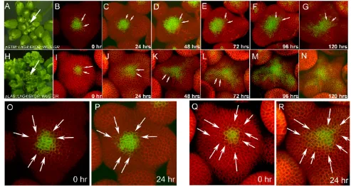
Fig. 5. Effect of WUS misexpression in the PZ on stem cell organization and growth patterns.SAMs of pSTM::LhG4;6XOP::WUS-GR(A) and pLAS::LhG4; 6XOP::WUS-GR(H) Arabidopsisplants treated with dexamethasone 20 days after germination. Arrows indicate SAMs. (B-N) Time-lapse series consisting of reconstructed 3D views of the L1 layer of SAMs of pSTM::LhG4; 6XOP::WUS-GR(B-G) and pLAS::LhG4; 6XOP::WUS-GR
(I-N) upon dexamethasone treatment. Refer to Fig. 3B-G for mock-treated controls. Direct expression of WUSin the PZ using pSTM::LhG4; 6XOP::WUS-GR(B-G) and pLAS::LhG4;6XOP::WUS-GR(I-N) leads to the activation of pCLV3::mGFP5-ERin CZ cells and sequential radial expansion of the CZ over time. There is directional expansion of pCLV3activity towards the boundary regions in I-N compared with a radially symmetric expansion in B-G. (O,P)Higher magnification images of B,C. (Q,R)Higher magnification images of I,J, respectively. Arrows point to same cells or
siblings of same cells at different time points. Total time elapsed after dexamethasone treatment is indicated on each panel. Scale bar in: 25m.
D
E
V
E
LO
P
M
E
N
[image:6.612.58.551.389.651.2]direction of auxin flow (Heisler et al., 2005). Plants carrying either 6Xop::WUSdsRNAi, 35S::GR:LhG4, pDR5rev::3XVENUS-N7, PIN1::PIN1:GFP or 6Xop::CLV3; 35S::GR:LhG4; pDR5rev::3XVENUS-N7; pPIN1::PIN1:GFPwere imaged every 12 hours after dexamethasone application. In mock-treated plants, the pDR5::Venus-YFP expression appears as a discrete set of three or four cells of the organ primordia at defined locations within the PZ. Upon downregulation of WUS, either by activation of WUSdsRNAi(Fig. 6P-R) or by overexpression of CLV3(Fig. 6J-L), the expression domain of pDR5rev::3XVENUS-N7 contained a substantially larger number of cells when compared with mock-treated plants. The proportion of pDR5-positive cells to pDR5 -negative cells was significantly higher upon both transient WUS downregulation and CLV3 overexpression (Fig. 6X). However, a more dramatic increase in pDR5-positive cells was observed in the case of WUS downregulation than CLV3 overexpression. This suggests that overexpression of CLV3could influence other aspects of primordia differentiation in addition to its primary role of
[image:7.612.55.553.62.382.2]downregulating WUS expression. Furthermore, over time, the pDR5 expression progressively initiated closer and closer to the CZ. This demonstrates a long-range organizing activity of WUS in allocating a definite number of PZ cells to differentiation pathways by delimiting either the domain of auxin accumulation, or the domain of auxin response. Thus, WUS ensures the homeostatic balance between the number of cells that enter into differentiation and non-differentiating cells within the PZ. We noticed lack of initiation of pDR5rev::3XVENUS-N7 expression and failure of specification of new primordia beyond 72 hours of dexamethasone treatment, possibly owing to an increased sink capacity of larger primordia initiated earlier (which can efficiently sequester auxin from adjacent regions within the PZ), but also possibly due to other causes, such as respecification of PZ cells or changes in their responsiveness to auxin. In addition, we observed a progressive decrease in CZ size and overall SAM size (Fig. 6S-V), although a more dramatic effect was observed in the case of overexpression of CLV3 compared with WUSdsRNAi, possibly owing to a partial Fig. 6. Stem cell and differentiation dynamics upon transient downregulation of WUS. Time-lapse images consisting of 3D views of reconstructed L1 layer of ArabidopsisSAMs showing the expression of the auxin responsive marker pDR5rev::3XVENUS-N7(red) and auxin transporter pPIN1:PIN1-GFP(green). (A-F)Mock-treated SAMs. (G-L)Plants carrying the dexamethasone-inducible CLV3overexpression construct treated with 10M dexamethasone showing reduction in the SAM size in a time window of 48-72 hours. A similar reduction in SAM size was observed in plants carrying the dexamethasone-inducible WUSdsRNAi construct (M-R). There is an increase in the size of the pDR5 expression domain with time upon both CLV3overexpression (J-L) and WUSdsRNAi activation (P-R). Arrows in K,L,R indicate predicted position of pDR5
expression in incipient primordia. (S-W)three-dimensional reconstructed surface views of SAMs carrying the dexamethasone-inducible CLV3
overexpression construct, showing gradual reduction in the pCLV3(green) expression domain (arrows indicate a decreasing pCLV3domain) and SAM size as revealed by p35S::YFP29-1(red). Total time elapsed after dexamethasone treatment is indicated on each panel. Scale bar: 25m. (X)Comparison of number of pDR5-positive and pDR5-negative cells/SAM 96 hours after dexamethasone treatment. The number of pDR5-positive (+ve) and pDR5-negative (–ve) cells/SAM are represented side by side for each treatment. Error bars represent standard deviation. P0, P1 and P2 represent organ primordia at different stages of growth with P0 being the youngest. The initiation of new pDR5expression is absent 96 hours (and later) post dexamethasone application in both CLV3overexpression and WUSdsRNAi lines.
D
E
V
E
LO
P
M
E
N
loss of WUS activity in WUSdsRNAi transgenic lines (Fig. 6V; see Fig. S2A,C-H in the supplementary material). Cell division analysis on SAMs experiencing higher levels of CLV3expression revealed a dramatic reduction in cell division activity wherein most of the cells remain undivided between 72 and 120 hours (Fig. 2F,G). This is opposite to the increased cell division activity observed upon increase in WUS levels, supporting the notion that WUS-mediated homeostatic balance between the CZ and the PZ involves re-adjustment of cell division rates in addition to the regulation of gene expression domains.
DISCUSSION
We have shown that WUScan have multiple functions depending upon its levels and places of expression. WUS levels can determine (1) stem cell number within the CZ, (2) growth rates of differentiating PZ cells and (3) size of differentiating organ primordia within the PZ.
WUSCHEL and stem cell numbers
Earlier studies have shown that wus mutants fail to maintain functional meristems (Laux et al., 1996). Ectopic expression of WUSin AINTEGUMENTA (ANT)-expressing cells of the PZ has been shown to induce stem cell identity (Schoof et al., 2000). Therefore, it has been proposed that WUS, which is expressed in cells of the RM, is necessary and sufficient to specify stem cell fate in overlying cells of the CZ. WUS, apart from specifying stem cells, also activates CLV3 expression, thereby regulating its own expression pattern by forming a negative-feedback loop. Consistent with this notion, the transient downregulation of CLV3has been shown to result in sequential and radial expansion of the CZ in which the PZ cells reacquire the stem cell fate (Reddy and Meyerowitz, 2005). The sequential and radial expansion of the CZ suggested that the stem cell-promoting principle could be a short-range diffusible factor that is negatively regulated by CLV3. It is possible that, upon transient downregulation of CLV3, the WUS expression also expands sequentially in cells of the RM, which in turn can activate stem cell promoting factor. However, our study reveals that either the uniform WUSexpression from a ubiquitously active promoter or ectopic WUS expression in cells of the PZ results in radial expansion of the CZ by re-specifying the PZ cells that abut the native CZ. This suggests either that the WUS-mediated stem cell-promoting factor is localized to the cells of the CZ or WUS activity itself is modified by signals that are specifically localized to the central parts of the meristem. Whatever the mechanism by which WUS mediates stem cell specification, WUS levels and spatial expression are expected to vary in wild-type SAMs, as it enters into mutual feedback regulation with the CLV class of genes. Therefore, keeping the PZ cells in a state where they can be re-specified as stem cells may provide the system enough flexibility to re-adjust stem cell numbers to varying amounts of WUS.
WUSCHEL and cell division patterns
The cell cycle length of stem cells located within the CZ is approximately three times that of the PZ cells (Reddy et al., 2004). Mechanisms underlying this differential mitotic activity across different regions of SAM are largely unknown. Here, we have shown that regulation of WUS level, or spatial expression, in central regions of SAMs is crucial for maintaining cell division rates of the PZ cells. However, how WUS expressed in cells of the RM can regulate cell division rates in cells of the PZ is not apparent. It is possible that a WUS-mediated signal, which would
be expected to be at its highest concentration in central regions of the meristem, is inhibitory to mitotic activity, while the lower concentrations in cells of the PZ promotes cell division activity. This is consistent with the reduction in mitotic activity in cells of the PZ upon misexpression of WUS directly in these cells. Alternatively, the CZ cells may be less responsive to the WUS-mediated mitogenic factor than the PZ cells, and this difference may be linked to their cellular identities. It has been documented, in most cases, that stem cells divide at a slower rate than their differentiating siblings, suggesting that cell identity-dependent mechanisms regulate cell division rates (Morrison and Spradling, 2008). In fact we have observed the expansion of the central domain of lower mitotic activity with the expansion of the CZ (Fig. 2C) and similar observations have been made on clv3mutants in an earlier study (Laufs et al., 1998). Therefore, both a higher concentration of WUS in the meristem center and cellular identity-dependent control of cell division machinery may converge to ensure the differential mitotic activity observed in the CZ and the PZ. This could be the reason for not observing increased mitotic activity upon ubiquitous misexpression of WUSbecause the CZ expands more rapidly (see Fig. S5A-C in the supplementary material) than in the case of CZ-driven WUS(Fig. 3I-K). It has been shown that WUS regulates components of signaling by the plant hormone cytokinin (a cell division regulator) and cytokinin levels regulate the size of the WUS expression domain (Leibfried et al., 2005; Guilini et al., 2004; Gordon et al., 2009). Transient and spatially restricted manipulation of cytokinin signaling and cytokinin-responsiveness of SAM cells may provide further insights into the interplay between stem cell specification and their growth control. Nevertheless, WUS-dependent adjustment of CZ size and cell division rates of the PZ cells may ensure maintenance of relative ratios of the CZ and the PZ, thereby adding an additional layer of robustness to stem cell homeostasis.
WUSCHEL and differentiation patterns
An earlier study based on the quantitative analysis of surface growth patterns has revealed an increase in the size of early flower buds upon transient overexpression of CLV3 (Muller et al., 2006). Here, we show that transient downregulation of WUS results in higher proportion of the PZ cells entering into differentiation pathways, as revealed by auxin-responsive pDR5 reporter expression, one of the earliest markers of differentiation (Heisler et al., 2005). Therefore, WUS could influence auxin responsiveness of the PZ cells or even early events that could lead up to auxin accumulation at specific sites within the PZ. This suggests that WUS balances the differentiation program within the PZ by suppressing differentiation to keep auxin responsiveness of the PZ cells to a narrow domain. Such a mechanism could ensure that sufficient number of PZ cells are available for re-specification as CZ cells, a process also controlled by WUS levels. WUS-mediated suppression of the differentiation program shares similarities with other stem cell niches wherein suppression of differentiation has emerged as one of the main mechanisms that mediates stem cell maintenance (Morrison and Spradling, 2008). Though WUS suppresses the differentiation program within the PZ, we have not observedpDR5expression in centrally located cells of the SAMs upon both transient downregulation of WUS and transient overexpression of CLV3(Fig. 6L,R). This suggests that depletion of stem cell fate from cells of the CZ is not sufficient to make them respond to auxin and differentiate as organ primordia. Therefore, non-responsiveness of the CZ to auxin should be regulated through
WUS-independent mechanisms. SHOOTMERISTEMLESS
D
E
V
E
LO
P
M
E
N
(STM), a homeodomain-containing transcription factor has been shown to play a key role in SAM development (Barton and Poethig, 1993; Long et al., 1996). STM function has been implicated both in maintaining cytokinin levels and also in preventing misexpression of differentiation-associated genes such as ASYMMETRIC LEAVES1 (AS1) in meristematic cells (Jasinski et al., 2005; Byrne et al., 2000). In this context, it will be illuminating to test the function of STM, in transient experiments, in regulating auxin non-responsiveness of the CZ. However, key to understanding these diverse functions of WUS is to elucidate the WUS-mediated gene network at a single cell type resolution. Recent advances in cell type-specific genomics should not only enable identification of WUS-mediated stem cell-promoting factors and may also provide network view of stem cell homeostasis (Yadav et al., 2009).
Acknowledgements
We thank Yuval Eshed for pSTM::LhG4, pLAS::LhG4seeds, Thomas Laux for
pCLV3::LhG4seeds, Rudiger Simon for sharing pCLV3::ALCR;ALCA::CLV3 seeds and Frank Wellmer for 35S::WUS-GRseeds. Live imaging work was carried out at the microscopy core facility of the Center for Plant Cell Biology (CEPCEB) and Institute of Integrative Genome Biology (IIGB), UCR. Initial observations on
35:WUS-GRwas made by G.V.R. in Elliot Meyerowitz’s laboratory and we thank him for allowing us to use it and also for his comments on the manuscript. This work was funded by National Science Foundation grant IOS-0718046 to G.V.R.
Competing interests statement
The authors declare no competing financial interests.
Supplementary material
Supplementary material for this article is available at
http://dev.biologists.org/lookup/suppl/doi:10.1242/dev.054973/-/DC1
References
Barton, M. K.(2010). Twenty years on: the inner workings of the shoot apical meristem, developmental dynamo. Dev. Biol. 341, 95-113.
Barton, M. K. and Poethig, R. S.(1993). Formation of the shoot apical meristem in Arabidopsis thaliana: an analysis of development in the wild- type and shoot meristemless mutant. Development 119, 823-831.
Bleckmann, A., Weidtkamp-Peters, S., Seidel, C. A. and Simon, R.(2010). Stem cell signaling in Arabidopsis requires CRN to localize CLV2 to the plasma membrane. Plant Physiol.152, 166-176.
Brand, U., Fletcher, J. C., Hobe, M., Meyerowitz, E. M. and Simon, R.(2000). Dependence of stem cell fate in Arabidopsis on a feedback loop regulated by CLV3 activity. Science289, 617-619.
Brand, U., Grunewald, M., Hobe, M. and Simon, R.(2002). Regulation of CLV3 expression by two homeobox genes in Arabidopsis. Plant Physiol.129, 565-575. Byrne, M. E., Barley, R., Curtis, M., Arroyo, J. M., Dunham, M., Hudson, A.
and Martienssen, R. A.(2000). Asymmetric leaves1 mediates leaf patterning and stem cell function in Arabidopsis. Nature408, 967-971.
Clark, S. E., Williams, R. W. and Meyerowitz, E. M.(1997). The CLAVATA1 gene encodes a putative receptor kinase that controls shoot and floral meristem size in Arabidopsis. Cell89, 575-585.
Fletcher, J. C., Brand, U., Running, M. P., Simon, R. and Meyerowitz, E. M. (1999). Signaling of cell fate decisions by CLAVATA3 in Arabidopsis shoot meristems. Science283, 1911-1914.
Giulini, A., Wang, J. and Jackson, D.(2004). Control of phyllotaxy by the cytokinin-inducible response regulator homologue ABPHYL1. Nature430, 1031-1034.
Gordon, S. P., Chickarmane, V. S., Ohno, C. and Meyerowitz, E. M.(2009). Multiple feedback loops through cytokinin signaling control stem cell number within the Arabidopsisshoot apical meristem. Proc. Natl. Acad. Sci. USA106, 16529-16534.
Heisler, M. G., Ohno, C., Das, P., Sieber, P., Reddy, G. V., Long, J. A. and Meyerowitz, E. M.(2005). Patterns of auxin transport and gene expression
during primordium development revealed by live imaging of the Arabidopsis inflorescence meristem.Curr. Biol. 15, 1899-1911.
Jasinski, S., Piazza, P., Craft, J., Hay, A., Woolley, L., Rieu, I., Phillips, A., Hedden, P. and Tsiantis, M.(2005). KNOX action in Arabidopsis is mediated by coordinate regulation of cytokinin and gibberellin activities.Curr. Biol. 15, 1560-1565.
Jeong, S., Trotochaud, A. E. and Clark, S. E.(1999). The Arabidopsis CLAVATA2 gene encodes a receptor-like protein required for the stability of the CLAVATA1 receptor-like kinase. Plant Cell11, 1925-1934.
Laufs, P., Grandjean, O., Jonak, C., Kieu, K. and Traas, J.(1998). Cellular parameters of the shoot apical meristem in Arabidopsis. Plant Cell10, 1375-1390.
Laux, T., Mayer, K. F., Berger, J. and Jurgens, G.(1996). The WUSCHEL gene is required for shoot and floral meristem integrity in Arabidopsis. Development
122, 87-96.
Leibfried, A., To, J. P., Busch, W., Stehling, S., Kehle, A., Demar, M., Kieber, J. J. and Lohmann, J. U.(2005). WUSCHEL controls meristem function by direct regulation of cytokinin-inducible response regulators. Nature438, 1172-1175.
Lenhard, M., Jurgens, G. and Laux, T.(2002). The WUSCHEL and
SHOOTMERISTEMLESS genes fulfil complementary roles in Arabidopsis shoot meristem regulation. Development129, 3195-3206.
Long, J. A., Moan, E. I., Medford, J. I. and Barton, M. K.(1996). A member of the KNOTTED class of homeodomain proteins encoded by the STM gene of Arabidopsis. Nature379, 66-69.
Mayer, K. F., Schoof, H., Haecker, A., Lenhard, M., Jürgens, G. and Laux, T. (1998). Role of WUSCHEL in regulating stem cell fate in the Arabidopsis shoot meristem. Cell95, 805-815.
Meyerowitz, E. M.(1997). Genetic control of cell division patterns in developing plants. Cell88, 299-308.
Morrison, S. J. and Spradling, A. C.(2008). Stem cells and niches: mechanisms that promote stem cell maintenance throughout life. Cell 132, 598-611. Muller, R., Borghi, L., Kwiatkowska, D., Laufs, P. and Simon, R.(2006).
Dynamic and compensatory responses of Arabidopsis shoot and floral meristems to CLV3 signaling. Plant Cell18, 1188-1198.
Müller, R., Bleckmann, A. and Simon, R.(2008). The receptor kinase CORYNE of Arabidopsis transmits the stem cell-limiting signal CLAVATA3 independently of CLAVATA1. Plant Cell20, 934-946.
Ogawa, M., Shinohara, H., Sakagami, Y. and Matsubayashi, Y.(2008). Arabidopsis CLV3 peptide directly binds CLV1 ectodomain. Science319, 294. Reddy, G. V.(2008). Live-imaging stem cell homeostasis in the Arabidopsis shoot
apex.Curr. Opin. Plant Biol. 11, 88-93.
Reddy, G. V., Heisler, M. G., Ehrhardt, D. W. and Meyerowitz, E. M. (2004). Real-time lineage analysis reveals oriented cell divisions associated with morphogenesis at the shoot apex of Arabidopsis thaliana. Development131, 4225-4237.
Reddy, G. V. and Meyerowitz, E. M.(2005). Stem cell homeostasis and growth dynamics can be uncoupled in the Arabidopsis shoot apex. Science310, 663-667.
Reddy, G. V., Gordon, S. P. and Meyerowitz, E. M.(2007). Unravelling developmental dynamics: transient intervention and live imaging in plants.Nat. Rev. Mol. Cell Biol. 8, 491-501.
Rieu, I. and Laux, T.(2009). Signaling pathways maintaining stem cells at the plant shoot apex. Semin. Cell Dev. Biol. 20, 1083-1088.
Schoof, H., Lenhard, M., Haecker, A., Mayer, K. F., Jürgens, G. and Laux, T. (2000). The stem cell population of Arabidopsis shoot meristems in maintained by a regulatory loop between the CLAVATA and WUSCHEL genes. Cell100, 635-644.
Spradling, A. C., Nystul, T., Lighthouse, D., Morris, L., Fox, D., Cox, R., Tootle, T., Frederick, R. and Skora, A.(2008). Stem cells and their niches: integrated units that maintain Drosophila tissues. Cold Spring Harbor Symp. Quant. Biol. 73, 49-57.
Steeves, T. A. and Sussex, I. M.(1989). Patterns in Plant Development: Shoot Apical Meristem Mutants of Arabidopsis thaliana. New York: Cambridge University Press.
Wielopolska, A., Townley, H., Moore, I., Waterhouse, P. and Helliwell, C. (2005). A high-throughput inducible RNAi vector for plants. Plant Biotechnol. J.
3, 583-590.
Yadav, R. K., Girke, T., Pasala, S., Xie, M. and Reddy, G. V.(2009). Gene expression map of the Arabidopsis shoot apical meristem stem cell niche. Proc. Natl. Acad. Sci. USA106, 4941-4946.