IJPSR (2012), Vol. 3, Issue 08 (Research Article)
Received on 24 April, 2012; received in revised form 14 May, 2012; accepted 21 July, 2012
IN-VITRO EVALUATION OF ANTIBACTERIAL ACTIVITY AND CYTOTOXICITY EFFECT OF CHEMICALLY SYNTHESIZED ZnO NANOPARTICLE
K. Renugadevi*, P. Raji and M. Bavanilatha
Department of Biotechnology, Sathyabama University, Rajiv Gandhi Salai, Chennai- 600 119, Tamil Nadu, India ABSTRACT
Nanoparticles, generally considered as particle with a size of upto 100nm, exhibit completely new or improved properties as compared to the larger particles of the bulk material that they are composed of based on specific characteristics such as size, distribution and morphology. In this study ZnO nanoparticle was synthesized by a simple and easy chemical method from ZnCl2. The synthesized nanoparticle was characterized by SEM and EDAX.
Antibacterial activity of ZnO was observed against E. coli, Enterobacter sp., Bacillus subtilis, Bacillus cereus, Klebsiella pneumoniae, Staphylococcus aureus, Salmonella typhi, Vibrio cholerae by disc diffusion method. Preliminary Cytotoxicity of nanoparticle was studied by MTT assay against vero cell line. ZnO nanoparticle had shown antibacterial activity against all the tested bacteria and maximum activity was observed against salmonella typhi. ZnO nanoparticles has shown less cytotoxicity effect on vero cell than ZnCl2.
INTRODUCTION: Nanotechnology deals with the
nanomaterial and nanoparticle which are in the size of about 100nm. Nanoparticle has wide application in various fields like chemical industry electronics, biomedicine, cosmetics, etc. 1, 2, due to its novel properties. Novel property is due to the alteration in their physical, chemical, electrical and magnetic property 3. When reduced to nanosize, unique size-dependent properties of nanoparticles are manifested
4
. The reason believed to cause this property change from their bulk is increase in relative surface area, a greater percentage of atoms at the material’s surface, quantum effects which can affect chemical reactivity, and other physical and chemical property 4, 5. Many nanoparticles have been synthesized and used in various field due to this altered properties. The metal nanoparticles are most widely used in the field of medicine.
ZnO is a conventional wide band-gap semiconductor 6, 7 that has been highly explored in multiple areas of science. ZnO nanomaterials have been used as semiconductor in microelectronic device and for accelerating degradation of water pollutants via photocalalytic activity.
ZnO are used in cosmetic industry, due to its UV irradiation absorbing ability and optical transparency 8, 9 and also used in photodetectors 10, surface acoustic wave devices 11, ultraviolet nanolaser 12, varistors 13, solar cells 14, gas sensors 15, biosensors 16, ceramics 17, field emission 18, and nanogenerator 19.
ZnO nanoparticles were synthesized by various methods like spray pyrolysis, thermal decomposition, molecular beam epitaxy, chemical vapor deposition, laser ablation etc. Chemical synthesis is one of the important techniques. It can be performed using a
Keywords: ZnO, Nanoparticle, Antibacterial activity,
Characterization, Cytotoxicity Correspondence to Author:
K. Renugadevi
Assistant professor, Department of Biotechnology, Sathyabama University, Rajiv Gandhi Salai (OMR), Chennai-600119, Tamil Nadu, India
temperature, time, concentration of reactants etc. Variation of these parameters leads to different size selection and geometries of resulting particles 20. ZnO nanoparticle synthesized using capping agents like thioglycerol have been reported by Shriwas 20.
In this study, a simple and easy method to synthesis ZnO nanoparticle from ZnCl2 without any capping
agent was employed and the synthesized nanoparticle was focused for its application in field of biomedicine by evaluating its antibacterial and cytotoxic effect. MATERIALS AND METHODS
Materials: Chemicals used for the study were bought from Himedia. Microorganisms (E. coli, Enterobacter sp., Bacillus subtilis, Bacillus cereus, Klebsiella pneumonia, Staphylococcus aureus, Salmonella typhi,
and Vibrio cholerae) used for antibacterial activity study were purchased from MTCC, India. Vero cell line was bought from NCCS, Pune. Research work was carried out during the year 2011 at Sathyabama University, Chennai, India-119
Methods:
1. Synthesis of ZnO nanoparticle: To prepare ZnO
nanoparticle, 0.5M aqueous solution of ZnCl2 and
1M of NaOH were prepared in MilliQ water. The method for the preparation of ZnO nanoparticle is a slight modified method of Abdolmajid et al 21. 1M NaOH solution was taken in a beaker and heated to 50oC. After 5 mins of heating the NaOH solution, ZnCl2 was added dropwise to the above
heated solution under high magnetic stirring. After the complete addition of ZnCl2, the beaker was
sealed and kept at stirring condition for 10min at 50oC. The white precipitate formation was observed at above said condition. The precipitation was collected and washed with milliQ water and with 100% ethanol then air dried at 50oC.
2. Characterization of ZnO nanoparticle: The
synthesized ZnO nanoparticle was characterized by SEM, EDAX analysis.
a. SEM analysis of ZnO nanoparticles: Scanning
Electron Microscopic (SEM) analysis was done in Karunya University using Hitachi S-4500 SEM machine. Thin films of the sample were prepared
on a carbon coated copper grid by just dropping a very small amount of the sample on the grid, extra solution was removed using a blotting paper and then the film on the SEM grid were allowed to dry by putting it under a mercury lamp for 5 min.
b. EDAX measurements: In order to carry out EDAX
analysis, the white precipitate ZnO nanoparticles were dried and drop coated on to carbon film and performed on Hitachi S-3400 NSEM instrument equipped with a Thermo EDAX attachments.
3. Antibacterial analysis: The antibacterial analysis
was done for the ZnO against the following microorganisms: E.coli, Enterobacter sp., Bacillus subtilis, Bacillus cereus, Klebsiella pneumoniae, Staphylococcus aureus, Salmonella typhi, Vibrio cholerae by disc diffusion method.
4. Cytotoxicity study: Cell line maintenance and
growth conditions. Vero cell line was purchased from NCCS (National Centre for Cell Sciences) Pune, India. The cell lines were maintained at 37oC at 5% CO2 in CO2 incubator. Cultures were viewed using
an inverted microscope to assess the degree of confluency and the absence of bacterial and fungal contaminants was confirmed.
a. Cytotoxicity test: MTT Cell Proliferation Assay was
employed to estimate the cytotoxicity of synthesized nanoparticle. Cells were first transferred (200µL/well at 7.5×10⁴mL) into 96 well plates and incubated for 24 hr. The original media was removed and 100 µL fresh media was added. Synthesized nanoparticle was added at concentration of 20-160 µg and then final volume was made to 200µl with the media and incubated for 4 hr.
After incubation media containing nanoparticle was removed. MTT reagent 20 µL was added to each well containing media and incubated for 3.5 hr at 37°C under an atmosphere of 5% CO2 until a
RESULTS AND DISCUSSION:
1. Synthesis of ZnO nanoparticle: The ZnO
nanoparticle was synthesized by chemical method from ZnCl2. The synthesized nanoparticle was
characterized by SEM (fig. 1) and EDAX (fig. 2). The SEM analysis of the particle was found to be in nanometer size and the EDAX result confirmed that the particles were ZnO.
FIG. 1: SEM ANALYSIS OF CHEMICALLY SYNTHESIZED ZnO NANOPARTICLE
FIG. 2: EDAX ANALYSIS OF THE CHEMICALLY SYNTHESIZED ZnO NANOPARTICLE
2. Antibacterial activity of ZnO nanoparticle: The
antibacterial activity of nanoparticle was assessed for the following microorganism E.coli, Enterobacter sp., Bacillus subtilis, Bacillus cereus, Klebsiella pneumoniae, Staphylococcus aureus, Salmonella typhi, Vibrio cholereae by disc diffusion method. Zone of inhibition was recorded and results were tabulated in Table 1. The nanoparticle and ZnCl2 has shown antibacterial activity. The
maximum antibacterial activity was found against
salmonella typhi with the zone of inhibition of 25mm and second maximum of 24 mm against
[image:3.612.26.591.137.728.2]Bacillussubtilis for ZnO nanoparticle.
TABLE 1: ANTIBACTERIAL ACTIVITY OF ZNO NANOPARTICLE
Microorganism Zone of inhibition in mm
ZnO nanoparticle (10µg) ZnCl2 (10µg)
E. coli 16 14
Enterobacter sp. 14 12
Bacillus subtilis 24 24
Klebsiella pneumoniae 15 14
Staphylococcus aureus 20 15
Salmonella typhi 25 16
Vibrio cholrea 22 20
Bacillus cereus 13 12
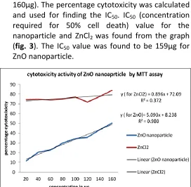
3. Cytotoxicity (MTT) assay: The Cytotoxicity of the ZnO nanoparticle and ZnCl2 was studied In-vitro by
MTT assay against Vero cell line at different concentration (20, 40, 60, 80, 100, 120, 140, 160µg). The percentage cytotoxicity was calculated and used for finding the IC50. IC50 (concentration
required for 50% cell death) value for the nanoparticle and ZnCl2 was found from the graph
(fig. 3). The IC50 value was found to be 159µg for
ZnO nanoparticle.
[image:3.612.315.587.458.724.2]DISCUSSION: In this study, a simple and rapid method was employed to synthesis ZnO nanoparticle. The NaOH was subjected to 50oC and the ZnCl2 solution
was added drop by drop under stirrer condition at 50oC. While adding the ZnCl2 solution to the heated
NaOH white precipitate formation was noted. After complete addition of ZnCl2 solution, set up was kept under same condition for another 15min. White precipitate formation indicates the formation of ZnO nanoparticle. This indicates the Zn ions oxidized to ZnO. The entire process took around 20min. The white precipitate was washed, dried and stored for further analysis.
The ZnO nanoparticle was characterized by SEM and EDAX (Fig. 1 and Fig. 2). The SEM results of the ZnO nanoparticle revealed the size of the particles were in nanosize and its ranges from 44nm-60nm. The elements of the nanoparticle were found to be Zn and O by EDAX analysis. From the EDAX, the weight % was found to be 28.19 and 71.81 for O and Zn respectively. The antibacterial activity of ZnO nanoparticle was assessed against E.coli, Enterobacter sp., Bacillus subtilis, Bacillus cereus, Klebsiella pneumoniae, Staphylococcus aureus, Salmonella typhi, Vibrio cholerae for the ZnO nanoparticle and ZnCl2 solution.
When compared to ZnCl2, ZnO nanoparticle has shown
maximum activity against all the tested bacteria and the maximum activity was found to be against
salmonellatyphi (Table 1).
The cytotoxicity effect of synthesized ZnO nanoparticle and ZnCl2 was studied on vero cell at different
concentration (20-160μg) by MTT assay. After 4 hours of treatment, the ZnO nanoparticles were found to less toxic than ZnCl2 to normal cell. Both the ZnO
nanoparticle and ZnO has shown different pattern of dose-dependent cytotoxicity response (Fig. 3).
The ZnO has shown less toxicity of 11.68% at 20µg but ZnCl2 at same concentration has shown toxicity of
74.53%. IC 50 value for the ZnO was found to be 159µg. The ZnCl2 has shown more toxic at the tested
minimum quantity than ZnO. So, ZnO can be used for in vivo studies like drug carrier and also in wound healing studies.
CONCLUSION: In this study, a simple and rapid method
was employed for the synthesizing of ZnO nanoparticle from 0.5M ZnCl2. Nanoparticle were subjected to SEM and EDAX analysis to confirm the nanosize and element composition. The SEM and EDAX analysis revealed the particles are Zn and size of particles were in the range of 40-60nm. Synthesized nanoparticles were subjected to antibacterial assay by disc diffusion method against the following bacteria E. coli, Enterobacter sp., Bacillus subtilis, Bacillus cereus, Klebsiella pneumoniae, Staphylococcus aureus, Salmonella typhi and Vibrio cholereae.
Antibacterial activity was observed against the tested microorganism and maximum activity was observed against salmonellatyphi. The cytotoxicity study by MTT assay revealed that the particle was very less toxic to normal cell than the ZnCl2. This preliminary cytotoxicity
study of ZnO nanoparticle might contribute to the comprehensive of this compound in vivo study like in drug delivery system.
ACKNOWLEDGEMENT: We thank the Chancellor and
Director, Sathyabama University for their great support. We sincerely thank Karunya University, Coimbatore, for the instrumental facilities. We also, thank faculty of Dept. of Biotechnology, Sathyabama University for their support in finishing this project. REFERENCE:
1. Ronan Baron, Fallyn W. Campbell, Ian Streeter, Lei Xiao and Richard G. Compton: Facile Method for the Construction of Random Nanoparticle Arrays on a Carbon Support for the Development of Welldefined Catalytic Surfaces. Int. J. Electrochem. Sci 2008; 3: 556 – 565.
2. Bayandori Moghaddam A, Kazemzad M, Nabid MR and Dabaghi HH: Improved voltammograms of hydrocaffeic Acid on the single- walled carbon nantube/ graphite-film surfaces. Int. J. Electrochem. Sci2008; 3:291-301.
3. John W. Rasmussen, Ezequiel Martinez, Panagiota Louka and Denise G. Wingett: Zinc oxide nanoparticles for selective destruction of tumor cells and potential for drug delivery applications. Expert Opin Drug Delivery 2010; 7(9):1063-1077. 4. Nel A, Xia T, Madler L and Li N: Toxic potential of materials at
the nanolevel, thorough review of experimental toxicological effects from nanoparticle-treated assays including ZnO nanoparticles. Science 2006; 311:622-7.
5. Lanone S and Boczkowski J: Biomedical applications and potential health risks of nanomaterials: molecular mechanisms. Curr Mol Med 2006; 6: 651-63.
7. Yi GC, Wang C, and Park WI: ZnO nanorods: synthesis, characterization and applications. Semiconductor Science and Technology 2005;20 (4):S22–S34.
8. Nohynek GJ, Dufour EK and Robert MS: Nanotechnology, cosmetics and the skin: is there a health risk? Skin Pharmacol Physiol 2008; 21:136-49.
9. Nohynek GJ, Lademann J, Ribaud C and Robert MS: Grey goo on the skin? Nanotechnology, cometic and sunscreen safety- a study on the use and safety of nanoparticles in cosmetic products. Crit Rev Toxicol 2007; 37: 251-77.
10. Jos A. Rodriguez, Tomas Jirsak, Joseph Dvorak, Sharadha Sambasivan and Daniel Fischer: Reaction of NO with Zn and ZnO: Photoemission, XANES, and Density Functional Studies on the Formation of NO. J. Phys. Chem. B 2000; 104 (2): 319-328. 11. Shih WC and Wu MS: Growth of ZnO films on GaAs substrates
with a SiO2 buffer layer by RF planar magnetron sputtering for
surface acoustic wave applications. J. Cryst. Growth 1994; 137: 319-325.
12. Huang MH, Samuel Mao, Henning Feick, Haoquan Yan, Yiying Wu, Hannes Kind, Eicke Weber, Richard Russo and Peidong Yang: Room-Temperature Ultraviolet Nanowire Nanolasers. Science 2001; 292(5523): 1897-1899.
13. Hung NT, Quang ND, and Bernik S: Electrical and microstructural characteristics of ZnO-Bi2O3-based varistors
doped with rare-earth oxides. Journal of Materials Research 2001; 16(10): 2817–2823.
14. Cooray NF, Kushiya K and Fujimaki A: Optimization of Al-doped ZnO window layers for large-area Cu(InGa)Se2-based modules
by RF/DC/DC multiple magnetron sputtering. Japanese Journal of Applied Physics 1999; 38(11): 6213–6218.
15. Paneva R and Gotchev D: Non-linear vibration behavior of thin multilayer diaphragms. Sensors and Actuators A 1999; 72(1): 79–87
16. Topoglidis E, Cass AEG, O'Regan B, and Durrant JR: Immobilisation and bioelectrochemistry of proteins on nanoporous TiO2 and ZnO films. Journal of Electroanalytical
Chemistry 2001; 517(1-2): 20–27.
17. Gao L, Li Q, Luan W, Kawaoka H, Sekino T and Niihara K: Preparation and electric properties of dense nanocrystalline zinc oxide ceramics. Journal of the American Ceramic Society 2002; 85(4): 1016–1018.
18. Xu CX and Sun XW: Field emission from zinc oxide nanopins. Applied Physics Letters 2003; 83(18): 3806–3808.
19. Gao PX, Ding Y, Mai W, Hughes WL, Lao CS and Wang ZL: Materials science: conversion of zinc oxide nanobelts into superlattice-structured nanohelices. Science 2005; 309(5741): 1700–1704.
20. Shriwas S Ashtaputre, Aparna Deshpande, Sonali marathe, Wankhede ME, Jayashree Chimanpure, Renu Pasricha, Urban J, Haram SK, Gosavi SW and kulkarni SK: Synthesis and analysis of zno and cdse nanoparticles. Pramana - Journal of Physics 2005; 65(4):615-620
21. Abdolmajid Bayandori Moghaddam, Tayebe Nazari, Jalil Badraghi, Mahmood Kazemzad: Synthesis of ZnO Nanoparticles and Electrodeposition of Polypyrrole/ZnO Nanocomposite Film. Int. J. Electrochem. Sci 2009; 4: 247 – 257.
How to cite this article: