RESEARCH ARTICLE
The Wnt/JNK signaling target gene
alcam
is required for
embryonic kidney development
Wiebke Cizelsky1,2,*, Aleksandra Tata1,2,3,*, Michael Kühl1,‡and Susanne J. Kühl1,‡
ABSTRACT
Proper development of nephrons is essential for kidney function. β-Catenin-independent Wnt signaling through Fzd8, Inversin, Daam1, RhoA and Myosin is required for nephric tubule morphogenesis. Here, we provide a novel mechanism through which non-canonical Wnt signaling contributes to tubular development. UsingXenopus laevisas a model system, we found that the cell-adhesion molecule Alcam is required for proper nephrogenesis and functions downstream of Fzd3 during embryonic kidney development. We foundalcamexpression to be independent of Fzd8 or Inversin, but to be transcriptionally regulated by theβ-Catenin-independent Wnt/JNK pathway involving ATF2 and Pax2 in a direct manner. These novel findings indicate that several branches of Wnt signaling are independently required for proximal tubule development. Moreover, our data indicate that regulation of morphogenesis by non-canonical Wnt ligands also involves direct transcriptional responses in addition to the effects on a post-translational level.
KEY WORDS: DM-GRASP,Xenopus laevis,alcam, Kidney, Pronephros
INTRODUCTION
The Xenopusembryonic kidney has become an attractive model system with which to examine nephron formation and to model human kidney diseases. The pronephros has a simple structure and contains one single functional nephron at each body side. The pronephros is formed by inductive signals from adjacent tissues, mesenchymal-epithelial transition (MET), epithelial tubulogenesis, morphogenetic movements and vasculogenesis. Genes that are necessary forXenopuspronephros development are also required for the development of the more-complex mammalian meso- and metanephros (Carroll and Vize, 1999; Dressler, 2006; Ryffel, 2003). Based on molecular studies, theXenopuspronephros can be subdivided into different segments that are homologous to the segments of the metanephric nephrons of mammals (Raciti et al., 2008). In Xenopus, the pronephric tubule branches at its most proximal end to generate three ciliated structures called nephrostomes that receive fluid derived from the kidney filter: the glomerulus, which inXenopusis called glomus.
Wnt proteins are extracellular glycoproteins that bind to Frizzled (Fzd) receptors, thereby activatingβ-Catenin-dependent (canonical) and β-Catenin-independent (non-canonical) signaling pathways
(Kestler and Kuhl, 2008), including the Wnt/ROCK, the Wnt/JNK and the Wnt/Ca2+branches (Schulte, 2010). Wnt signaling is crucial
for kidney development (Merkel et al., 2007) and misregulation of Wnt signaling contributes to congenital kidney diseases. Mutations in the Wnt signaling mediator Inversin, for example, are the cause of nephronophthisis type II (Otto et al., 2003), and polycystic kidney disease has been linked to gain and loss of canonical, as well as changes in non-canonical, Wnt signaling (Lancaster and Gleeson, 2010; Luyten et al., 2010).
Wnts regulate different aspects of nephrogenesis, including differentiation, proliferation, MET, tubulogenesis and morphogenesis. Fzd8 and Inversin have been shown to be required for morphogenetic movements of the proximal and intermediate tubule (Lienkamp et al., 2010; Satow et al., 2004). This process also involves Daam1, WGEF and RhoA, and regulation of the cytoskeleton (Miller et al., 2011). Recent data indicated that morphogenesis of the tubule requires convergent extension movements that are reached by mediolaterally oriented cell intercalations. This process involves Myosin, a protein regulated by the RhoA-activated kinase ROCK (Lienkamp et al., 2012). Wnt4is expressed in the metanephric mesenchyme that develops into the mature nephron through several intermediates (Diez-Roux et al., 2011; Stark et al., 1994). Cultures of metanephric mesenchyme treated with Wnt4 generate nephrons in the absence of the ureteric bud (Kispert et al., 1998). In Xenopus, Wnt4 has been shown to be required for proximal tubule development (Naylor and Jones, 2009; Saulnier et al., 2002). Recent evidence indicated that Wnt4 induces nephrogenesis by β-Catenin-independent signaling (Burn et al., 2011; Tanigawa et al., 2011). It remained unclear whether regulation ofβ-Catenin-independent Wnt signaling in this context also involves transcriptional responses in addition to its effects at the post-translational level.
Alcam is a member of the neuronal immunoglobulin-like domain superfamily of cell-adhesion molecules, and promotes cell adhesion and signaling (Corbel et al., 1996; DeBernardo and Chang, 1996). We previously reportedalcamexpression duringXenopusembryogenesis (Gessert et al., 2008), including its expression in the developing pronephros. With respect to kidney development,Alcamexpression was also reported in the metanephric mesenchyme and the developing tubules in chicken (Tsukamoto et al., 2006). Publicly available microarray data indicate thatAlcamis also expressed in the developing metanephros of mouse E15.5 embryos (http://www.gudmap.org/). Taken together, these data indicate a conserved expression ofalcam during nephrogenesis. However, analyses regarding a potential function of Alcam during pronephros development are not yet available. In addition, earlier experiments suggested thatalcam is regulated by non-canonical Wnt signaling (Gessert et al., 2008; Lapointe et al., 2012; Prieve and Moon, 2003). It remained elusive howalcamis regulated byβ-Catenin-independent Wnt signaling and whetheralcamis involved in Wnt mediated aspects of nephrogenesis. By using theXenopus laevispronephros as a model system, we here show for the first time that Alcam is essential for proper
Received 10 January 2014; Accepted 10 March 2014 1
Institute for Biochemistry and Molecular Biology, Ulm University, Albert-Einstein-Allee 11, Ulm 89081, Germany.2International Graduate School in Molecular Medicine Ulm, Ulm 89081, Germany.3Department of Neurobiology, Harvard Medical School, 220 Longwood Ave, Boston, MA 02115, USA.
*These authors contributed equally to this work
‡
Authors for correspondence (michael.kuehl@uni-ulm.de; susanne.kuehl@uni-ulm.de)
DEVEL
O
tubulogenesis. Alcam functions downstream of Fzd3 and is regulated by Wnt/JNK signaling involving ATF2 and Pax2 in a direct manner. These data suggest a new mechanism by which Wnt signaling coordinates pronephric tubulogenesis.
RESULTS
Alcam is required for nephron development in theXenopus embryonic kidney
Alcamexpression inXenopuspronephric tissue starts around stage 22/23 during epithelial tubule formation. Thereafter, alcam transcripts were detected throughout the whole pronephric tubule (Fig. 1A; supplementary material Fig. S1B).
To study the potential function of Alcam duringXenopuspronephros development, we performed knock down experiments using a well-characterized Alcam antisense morpholino oligonucleotide (MO). This MO is able to block the translation of analcam-GFP fusion construct (Gessert et al., 2008) and reduced the amount of endogenous Alcam proteinin vivo (supplementary material Fig. S1A). In all following experiments,gfpRNA was co-injected as a lineage tracer (Tecza et al., 2011) and manipulation was performed on only one side of the embryo,
leaving the other side unaffected as an internal control. Control MO was injected in all experiments as an injection control.
Alcam-depleted embryos revealed a pronephric phenotype affecting the proximal part of the pronephros, as shown by whole mount in situhybridization against theγ-subunit of the Na+/K+
[image:2.612.52.557.297.635.2]-ATPase,fxyd2, at stage 36 (Fig. 1B) (Raciti et al., 2008). About 51% of embryos revealed a significant reduction of thefxyd2expression domain in the anterior part of the tubule. This phenotype was restored by co-injection of an alcam RNA that is not targeted by the Alcam MO (Gessert et al., 2008), indicating the specificity of the Alcam MO-induced effect. A more-detailed quantification of the total tubular surface indicated that the proximal and intermediate tubule convolute was significantly reduced upon Alcam depletion (Fig. 1C). This reduction was similar in extent to that recently observed upon knock down of Inversin (Lienkamp et al., 2010). Again, this phenotype was rescued byalcamRNA co-injection. In addition, the three nephrostomes did not properly form and were not separated upon Alcam downregulation, as revealed bylhx1staining (Fig. 1D). Furthermore, expression offoxc1, a marker gene for the proximal pronephros, was reduced in about 63% of Alcam morphant
Fig. 1. Alcam is essential for embryonic kidney development inXenopus.(A)Alcamis expressed duringXenopus laevispronephros development in particular in the anterior proximal part (arrows).Lateral views with anterior towards the right are shown. Dotted lines indicate the level of the transverse sections. (B)In situhybridization at stage 36 indicates that Alcam is required for normal expression offxyd2in the proximal tubule (arrow). Loss offxyd2
upon Alcam depletion is rescued byalcamRNA co-injection. (C) Quantification of the tubule convolute area demonstrates a significant reduction in size upon loss of Alcam function compared with Control MO-injected embryos. This phenotype is significantly restored byalcamco-injection. Measurements of individual embryos are indicated. Median values are also shown. Embryos were analyzed from three independent experiments. (D) At stage 36, injection of Alcam MO leads to a fusion of the three nephrostomes (arrow), which is rescued byalcamRNA co-injection. (E) Embryos injected with Alcam MO show a severe reduction infoxc1expression (arrow), which is rescued by co-injection ofalcamRNA. Quantitative representations are shown. Lateral views with anterior towards the left (injected side) or towards the right (uninjected side) are shown. ap, anterior-posterior; dv, dorsal-ventral; n, number of independent batches of embryos; N, number of analyzed embryos in total. *P≤0.05, **P≤0.01, ***P≤0.001 and ****P≤0.0001.
DEVEL
O
embryos (Fig. 1E). Both phenotypes were restored upon co-injection ofalcamRNA (Fig. 1D,E). Taken together, these data indicate that a loss of Alcam results in disturbed morphogenesis of the proximal and intermediate tubules. The rescue experiments revealed that the phenotype observed upon Alcam MO injections is specific.
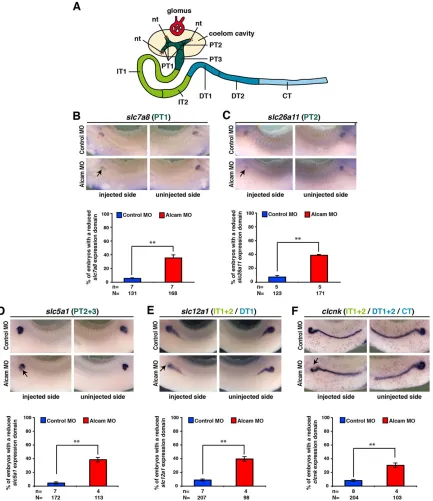
We next analyzed the Alcam MO-induced phenotype in more detail. At an earlier time point of development, expression of pronephros-specific marker genes such aslhx1,dll1,wt1,wnt4and fzd3 was not affected in Alcam-depleted embryos (supplementary material Fig. S2).These data indicate that Alcam is not required for general specification and early differentiation of the pronephros. Next, we investigated the segmentation of the pronephros. Based on a large scale whole-mountin situ hybridization screen, theXenopus pronephros can be subdivided into different segments that are homologous to the different segments of the mammalian metanephric nephron (Raciti et al., 2008) (Fig. 2A). To this end, we usedslc7a8to label PT1,slc26a11 to label PT2, slc5a1 to label PT2 and PT3, slc12a1to stain IT1, IT2 and DT1, andclcnkto label IT1, IT2, DT1, DT2 and CT at stage 36 (Fig. 2B-F). Alcam downregulation led to reduced domains of gene expression, particularly in proximal (PT1-3), intermediate (IT1 and IT2) and distal parts (DT1) of the pronephros, indicating that these segments are established but are smaller in size. The expression ofclcnkin the connecting tubule was not affected. As expression of all genes was not completely abolished, we conclude that the gross segmentation of the pronephros into proximal, intermediate, distal and connecting tubules is not disturbed in Alcam-deficient embryos.
Loss of Fzd3 phenocopies the loss of Alcam
Alcamexpression was recently shown to be affected by Wnt signaling (Gessert et al., 2008; Prieve and Moon, 2003). Therefore, we next searched for Wnt ligands and Fzd receptors that are expressed in the pronephros similar toalcam. For further experiments, we focused on wnt4,fzd3andfzd8as they are prominently expressed in the anterior part of theXenopuspronephros (Maurus et al., 2005; Satow et al., 2004; Saulnier et al., 2002; Shi et al., 1998) (supplementary material Fig. S1B). Wnt4 has been shown to be required for proximal tubule development inXenopus(Naylor and Jones, 2009; Saulnier et al., 2002). Furthermore, studies identified Fzd3 as a likely receptor for Wnt4 (Lyuksyutova et al., 2003; Maurus et al., 2005) and Fzd8 has been implicated in proximal tubule morphogenesis (Lienkamp et al., 2010; Satow et al., 2004).
Alcam-depleted embryos revealed an abnormal ventral extension of the tubule convolute (Fig. 1B,C) similar to embryos with depletion of the non-canonical Wnt mediators Fzd8 and Inversin (Lienkamp et al., 2010, 2012; Satow et al., 2004), implicating a potential relationship between Fzd8, Inversin and Alcam. Interestingly, knock down of neither Fzd8 nor Inversin alteredalcam orfoxc1 (as one selected other pronephric marker) expression, despite their ability to affectfxyd2expression (supplementary material Fig. S3), indicating that Alcam is not downstream of Fzd8 or Inversin.
However, depletion of either Wnt4 or Fzd3 using published MOs (Deardorff et al., 2001; Saulnier et al., 2002) led to a downregulation of alcam, as indicated by whole-mount in situ hybridization (Fig. 3A) and RT-PCR analyses of animal caps induced towards a pronephric fate by treatment with activin A and retinoic acid (RA) (Fig. 3B). Whereas Fzd3 depletion affected onlyalcamexpression in the proximal pronephros, Wnt4 MO injection led to a more severe phenotype – deleting alcam in the entire pronephros (Fig. 3A). Similarly, Wnt4 depletion resulted in a strong reduction of lhx1 expression (supplementary material Fig. S4). By contrast, loss of Fzd3 resulted in a fusion of the three nephrostomes but lhx1
expression was otherwise normal (Fig. 3E; supplementary material Fig. S4) similar to the phenotype observed upon Alcam depletion (compare with Fig. 1D). These data suggest thatalcamexpression in the pronephros depends on Wnt4 and Fzd3 function. As the phenotype observed upon loss of Fzd3 function phenocopied the loss of Alcam, whereas downregulation of Wnt4 resulted in a broader phenotype, we focused in the rest of our study on characterizing the correlation between Fzd3 and Alcam.
To further validate our observation that alcam expression is downstream of Fzd3, we analyzed the Fzd3 knock down phenotype in more detail. Loss of Fzd3 led to defects in proximal tubules at stage 36, as indicated byfxyd2expression (Fig. 3C). This phenotype was restored by co-injection ofalcamRNA. Measurements of the tubular surface area revealed that the proximal and intermediate tubule convolute was significantly reduced in size (Fig. 3D). Again, this phenotype was rescued byalcamRNA co-injection. Moreover, Fzd3 depletion led to fused nephrostomes (Fig. 3E) and a downregulation of foxc1 expression (Fig. 3F), similar to the situation observed upon Alcam MO injections (compare with Fig. 1D,E). Both phenotypes were restored by alcam RNA co-injection. Analyses of segmentation markers indicated that Fzd3 depletion most strongly affected proximal, intermediate and distal tubule development (supplementary material Fig. S5), similar to the Alcam depletion phenotype (compare with Fig. 2).
Taken together, these loss of function and rescue data indicate that alcamis downstream of Fzd3 and that this regulation is of functional significance. These observations raised the question of which Wnt signaling pathway isalcamregulated through?
Fzd3 regulatesalcamexpression through JNK1
To investigate whether regulation of alcam by Fzd3 occurs through aβ-Catenin-dependent or -independent Wnt pathway, we made use of two well-described Dishevelled (dsh) deletion constructs designated dshΔDIX and dshΔDEP. DshΔDEP activates
β-Catenin-dependent Wnt signaling, whereas dshΔDIX acts through
β-Catenin-independent Wnt signaling branches (Boutros et al., 1998; Itoh et al., 2000; Kishida et al., 1999; Li et al., 1999).
We injected Fzd3 MO, together with eitherdshΔDIXordshΔDEP RNA, and monitored expression of alcam. Interestingly, alcam downregulation by Fzd3 depletion could only be restored by co-injectingdshΔDIXbut notdshΔDEPRNA (Fig. 4A), indicating a regulation ofalcamthroughβ-Catenin-independent Wnt signaling. AsdshΔDIXactivates the non-canonical Wnt signaling mediator jun N-terminal kinase JNK (Boutros et al., 1998), we aimed to rescue the Fzd3 MO phenotype through introducing a constitutively active version of JNK (caJNK1) (Lei et al., 2002).caJNK1RNA injection led to a modest but significant rescue ofalcamexpression (Fig. 4B). Injection of higher caJNK1 amounts is not possible due to gastrulation defects. These data indicate that alcam regulation by Fzd3 occurs throughβ-Catenin-independent Wnt/JNK signaling.
The Alcam promoter contains a Fzd3 response element
We next analyzed the 5′upstream regulatory region ofalcam. We determined the transcriptional start site by 5′RACE and isolated a genomic region upstream of the transcription initiation site (supplementary material Fig. S6). We started our analysis with a 3.1 kb fragment including the 5′UTR and the regulatory region 2.7 kb upstream of the transcription start site that we cloned in front of the luciferase reporter gene (−2.7 kb-luc) (Fig. 5A). This construct revealed transcriptional activity in untreated animal cap cells (ACs) of Xenopus embryos (Fig. 5B). Upon inducing a pronephric fate in ACs by treatment with activin A and RA (retinoic
DEVEL
O
acid), we observed a strong upregulation of promoter activity in comparison with untreated ACs (Fig. 5B).
We next tested whether thealcamregulatory region responds to a loss of Fzd3 and co-injected the−2.7 kb-luc construct together with Fzd3 MO to measure luciferase activity in pronephric ACs. Inhibiting Fzd3 function led to a significant reduction of luciferase activity (Fig. 5C). These results indicate that the isolated−2.7 kb
[image:4.612.92.525.57.558.2]upstream fragment contains regulatory elements required foralcam regulation by Fzd3. We next deleted parts of the fragment generating −2.2 kb and −0.8 kb fragments fused to the reporter gene (−2.2 kb-luc and −0.8 kb-luc; Fig. 5A). Both fragments revealed transcriptional activity in pronephric ACs, although at lower levels (Fig. 5B). Whereas the −2.2 kb-luc fragment responded to Fzd3 downregulation (Fig. 5D), the −0.8 kb-luc Fig. 2. Alcam depletion has no effect on pronephric segmentation.(A) AXenopuspronephros (based on Raciti et al., 2008). nt, nephrostome; PT, proximal tubule; IT, intermediate tubule; DT, distal tubule; CT, connecting tubule. (B-F) At stage 36, the embryos injected with Alcam MO reveal a reduction in the expression domain of: (B) the amino acid transporterslc7a8(a marker for PT1 of the proximal tubule); (C) the anion exchangerslc26a11(a marker for PT2 of the proximal tubule); (D) the sodium/glucose co-transporterslc5a1(a marker for PT2 and PT3 of the proximal tubule); (E) the sodium/potassium/chloride co-transporterslc12a1(a marker for IT1 and IT2 of the intermediate and DT1 of the distal tubule); (F) the voltage sensitive chloride channelclcnk(a marker for IT1 and IT2 of the intermediate, DT1 and DT2 of the distal, and the connecting tubule) on the injected side. Whereas Alcam depletion results in reduced expression domains of the marker genes in PT1-3, IT1-2 and DT1 (arrows), the uninjected or control MO-injected side of the embryos display no pronephric phenotype. Quantitative representations are shown. Lateral views with anterior towards the left (injected side) or towards the right (uninjected side) are shown. n, number of independent batches of embryos; N, number of analyzed embryos in total. **P≤0.01.
DEVEL
O
construct did not (Fig. 5E). These data indicate that the Fzd3 response element is likely located between−2152 and−781 of the alcampromoter.
We then tested whether the−2.7 kb-luc construct responds to canonical or non-canonical Wnt signaling. As inXenopusACs, the
−2.7 kb-luc construct showed an activity in MDCK (Madin Darby canine kidney) cells (Fig. 6A). Treatment of MDCK cells with Wnt11 (Fig. 6C), a non-canonical Wnt ligand, but not Wnt3a (Fig. 6B), a canonical Wnt ligand, resulted in a significant upregulation of reporter activity. Also in HEK293 cells, treatment with Wnt3a or the GSK3β inhibitor BIO did not result in any activation of thealcamreporter, although both treatments activate the Wnt/β-Catenin reporter TOPFlash (supplementary material
[image:5.612.52.562.65.454.2]Fig. S7A,B). Transfecting MDCK cells with caJNK1, however, activated −2.7 kb-luc (Fig. 6C). Similarly, caJNK1 activated the reporter in HEK293 cells (supplementary material Fig. S7C). Other non-canonical Wnt signaling mediators such as constitutively active versions of CamKII (Kühl et al., 2000) or NF-AT (Borchers et al., 2006) had no effect on −2.7 kb-luc (supplementary material Fig. S7C). To confirm these observations inXenopus, we used the pronephric AC system and investigated the influence of modulating JNK activity on thealcampromoter. Indeed, inhibition of JNK by SP600125 treatment led to a significant downregulation (Fig. 6D), whereas overexpression of caJNK1 resulted in an upregulation of promoter activity (Fig. 6E). Taken together, these results obtained in different cell lines Fig. 3. Fzd3 depletion phenocopies the loss of Alcam phenotype.(A) Loss of Wnt4 function results in a severe reduction ofalcamexpression in the entire pronephros (arrow). Fzd3 depletion leads toalcamdownregulation only in the proximal part of the pronephros (arrowhead). (B) Animal cap assays show that the expression ofalcamandfoxc1are impaired upon loss of Wnt4 or Fzd3. The effect of Wnt4 MO injection is stronger compared with Fzd3 MO injection. (C) Whole-mountin situhybridization at stage 36 indicates that Fzd3 is required for the normalfxyd2expression in the proximal tubule (arrow). Loss offxyd2upon Fzd3 depletion is rescued byalcamRNA co-injection. (D) Quantification of the tubule convolute area demonstrates a significant reduction in size upon loss of Fzd3 function compared with Control MO-injected embryos. This phenotype is significantly restored byalcamco-injection. Measurements of individual embryos are indicated. Median values are also shown. Embryos were analyzed from three independent experiments. Data for Control MO are the same as in Fig. 1C, as these experiments were carried out in parallel. (E) At stage 36, injection of Fzd3 MO leads to a fusion of the three nephrostomes (arrow), which is rescued byalcamRNA co-injection. (F) Embryos injected with Fzd3 MO show a severe reduction infoxc1expression (arrow), which is rescued by co-injection ofalcamRNA. Quantitative representations are shown. Lateral views with anterior towards the left (injected side) or towards the right (uninjected side) are shown. ap, anterior-posterior; dv, dorsal-ventral; n, number of independent batches of embryos; N, number of analyzed embryos in total. *P≤0.05, **P≤0.001, ****P≤0.0001.
DEVEL
O
supported our findings in whole embryos that thealcamreporter responds to non-canonical Wnt/JNK signaling.
Pax2 regulates the Fzd3 response element
Our experiments identified a Fzd3 responsive element (Fzd3RE) located between−2152 and−781 of thealcampromoter (Fig. 5). A close inspection of this region revealed the presence of seven AP1/ATF2 and two Pax2 sites clustered in a small region of 265 nucleotides (supplementary material Fig. S6). Deleting this region within −2.7 kb-luc to generate −2.7 kbΔ-luc resulted in a dramatically reduced promoter activity (Fig. 5B,F). Moreover
−2.7 kbΔ-luc no longer responded to Fzd3 depletion (Fig. 5F). Previous work by others has linked ATF2 to β -Catenin-independent Wnt signaling (Schambony and Wedlich, 2007). Chromatin immunoprecipitation indicated that ATF2 binds in the 265 bp region that depends on Fzd3 function, supporting a role for ATF2 inalcamregulation (Fig. 7A). Pax2 is another transcription factor that is activated byβ-Catenin-independent Wnt signaling at the post-translational level by phosphorylation through JNK. These earlier studies additionally indicated that phosphorylation of Pax2 by JNK correlates with an increased transactivation of a pax2-dependent reporter (Cai et al., 2002). These observations and the identification of Pax2-binding sites in the alcam regulatory region raised the possibility that Pax2 is a positive regulator ofalcam. We tested this possibility by co-transfecting the alcam reporter together with pax2 in MDCK cells. Pax2 activated −2.7 kb-luc, whereas−2.7 kbΔ-luc did not respond to pax2 transfection (Fig. 7B), suggesting a direct regulation of alcamby Pax2. In line with this idea, we could show Pax2 binding to the regulatory region by chromatin immunoprecipitation. As in the case of ATF2, Pax2 binding was at least in part dependent on Fzd3 function (Fig. 7A). This outcome raised the issue of whether the loss ofalcamexpression upon Fzd3 depletion could be rescued by forced overexpression of Pax2in vivo,thereby overcoming the reduced binding affinity of Pax2 to the promoter as well as its
reduced transcriptional activity. We therefore injected Fzd3 MO together withpax2RNA and monitoredalcamexpression. Indeed, we observed a rescue ofalcamdownregulation (Fig. 7C). Taken together, our data indicate regulation ofalcamexpression by Wnt/ JNK signaling that involves ATF2 and Pax2 (Fig. 7D).
In combination with the functional analyses, our data suggests a novel mechanism how non-canonical Wnt signaling regulates alcam expression and thereby tubular development in the embryonic kidney.
DISCUSSION
Our data make the following novel contributions: (1) we show for the first time, that the cell-adhesion molecule Alcam is required for nephrogenesis; (2) we demonstrate thatalcamexpression in the pronephros depends on Fzd3 function and the activation of a
β-Catenin-independent Wnt signaling pathway; and (3) we identify for the first time a response element in the regulatory region of a direct target gene of non-canonical Wnt signaling.
Requirement of Alcam for nephrogenesis
[image:6.612.50.349.55.342.2]Tubule formation during nephrogenesis requires the transition of mesenchymal cells into epithelial cells (mesenchymal-epithelial transition, called MET). Previous findings suggested Alcam to be involved in adherens junction formation (Choe et al., 2013; Jannie et al., 2012) and in the establishment of epithelial apical-basal polarity, two processes occurring during MET. Later during tubulogenesis, the proximal part enlarges the surface area that is required for proper and efficient reabsorption of water, ions and small compounds. This is achieved by proliferation of tubular cells and morphogenetic movements (Fischer et al., 2006; Karner et al., 2009; Lienkamp et al., 2012). These convergent extension movements require the action of several Wnt components, such as Fzd8 or Inversin (Lienkamp et al., 2010). The similarity between our Alcam and Fzd8/Inversin MO phenotypes suggests that Alcam might also be required for these movements. A detailed analysis of Fig. 4. Fzd3 regulatesalcamexpression through JNK1. (A) Fzd3 depletion leads to a downregulation ofalcam
expression (arrow), which was rescued bydshΔDIXbut not bydshΔDEP(arrow). (B) Depletion of Fzd3 results in a reduction ofalcam(arrow). This phenotype is significantly rescued by co-injection of a constitutively active form of the Wnt signaling mediatorJNK(caJNK1). Lateral views with anterior towards the left (injected side) or towards the right (uninjected side) are shown. Quantitative representations are shown. n, number of independent experiments; N, number of analyzed embryos in total; n.s., not significant. *P≤0.05, **P≤0.01.
DEVEL
O
cell migration in Alcam morphant embryos will shed light onto this issue. It is also tempting to speculate that a loss of epithelial polarity upon loss of Alcam causes disturbed morphogenetic movements of the tubule, finally resulting in the observed phenotype. A detailed analysis of epithelial cell polarity will be required in the future to fully understand the Alcam MO phenotype at the cellular level.
Wnt signaling during nephrogenesis
[image:7.612.89.519.57.348.2]β-Catenin-dependent and -independent Wnt pathways are important for kidney development.β-Catenin-dependent Wnt signaling was previously shown to be required for specification, differentiation and proliferation of pronephric cells, as well as ureteric bud formation (Iglesias et al., 2007; Lyons et al., 2009; Maretto et al., Fig. 5. Identification of a Fzd3-responsive element in thealcampromoter.(A)alcamreporter constructs generated in this study. Regulatory regions are linked to the luciferase gene as reporter. (B) The−2.7 kb fragment is active in untreated and in activin A/retinoic acid (RA)-treatedXenopusanimal cap explants. The−2.7 kb fragment is active in pronephric animal cap cells, whereas the−2.2 kb,−0.8 kb and−2.7 kbΔfragments show considerable lower activities. (C) The activity of the−2.7 kb fragment is reduced upon Fzd3 MO injection. (D) The−2.2 kb fragment reacts to loss of Fzd3. (E) The−0.8 kb fragment does not react to Fzd3 depletion. (F) Deleting a small part of the−2.7 kb fragment is sufficient for loss of promoter activity. luc, luciferase reporter gene; n, number of independent experiments; RLU, relative light units; n.s., not significant. *P≤0.05, **P≤0.01, ****P≤0.0001.
Fig. 6. Thealcampromoter is regulated throughβ-Catenin-independent Wnt in MDCK and AC cells.(A) The−2.7 kb fragment is active in MDCK cells. (B) The promoter does not respond to treatment of MDCK cells with Wnt3A. (C) The promoter responds to transfection of MDCK cells with Wnt11. Activity of the−2.7 kb-luc reporter is increased upon co-transfection withcaJNK1. (D,E) The promoter activity of the−2.7 kb fragment is reduced by JNK inhibition (SP600125 treatment) (D), whereas the JNK overexpression leads to a significant upregulation of promoter activity (E) in activin A/retinoic acid-treated
pronephric animal cap cells (ACs). n, number of independent experiments; luc, luciferase reporter gene; RLU, relative light units. *P≤0.05, **P≤0.01.
DEVEL
O
[image:7.612.49.562.514.683.2]2003; McCoy et al., 2011; Park et al., 2012).β-Catenin-independent Wnt7b and Wnt9 are important for tubule morphogenesis because of their ability to regulate asymmetric cell divisions (Yu et al., 2009) and convergent extension movements (Karner et al., 2009).
β-Catenin-independent Wnt4 is involved in murine metanephric tubulogenesis (Burn et al., 2011; Tanigawa et al., 2011). In line with these findings, the expression of alcamis not detectable during specification but is present during tubulogenesis. Work by others showed that Wnt/RhoA/ROCK signaling regulates morphogenetic movements. This pathway involves Fzd8, Inversin, Daam1, the Rho-GEF WGEF and RhoA, resulting in a reduced tubular convolute based on defects in cell migration (Lienkamp et al., 2010; Miller et al., 2011; Satow et al., 2004). These data suggest that
β-Catenin-independent Wnt signaling regulates the cytoskeleton to ensure proper cell behavior in the proximal tubule (Fig. 7D). A more recent study indicated that this process is achieved by Myosin-dependent mediolateral cell intercalation (Lienkamp et al., 2012). Of relevance, neither loss of Fzd8 nor loss of Inversin affected alcam expression in our study, suggesting that the Wnt/ROCK signaling branch is not involved inalcamregulation. By contrast, we provide a novel mechanism showing that a Wnt/JNK/Alcam branch is required for tubulogenesis. At the molecular level, we foundalcamto be regulated by Fzd3 signaling. Our data strongly support earlier findings (Gessert et al., 2008; Lapointe et al., 2012; Prieve and Moon, 2003), demonstrating thatalcamis a direct target gene ofβ -Catenin-independent Wnt signaling at the transcriptional level. Therefore, we can addalcamto the currently very short list of two direct target genes
ofβ-Catenin-independent Wnt signaling in vertebrates:eaf2(Maurus et al., 2005) andpcdh8(Schambony and Wedlich, 2007).
Our novel findings and earlier reports by others suggest that two independent non-canonical Wnt branches are required for proper nephrogenesis: (1) a Fzd8/Inversin branch required for cytoskeletal rearrangements (Lienkamp et al., 2010, 2012; Miller et al., 2011); and (2) a Fzd3/JNK branch regulating expression of the cell-adhesion moleculealcam(this study). This situation is remarkably similar to the situation duringXenopusgastrulation. During this process, it was shown that Wnt5a, but not Wnt11, regulates the expression of the cell-adhesion molecule pcdh8 (PAPC) at the transcriptional level, whereas Wnt11 likely regulates the actin cytoskeleton (Schambony and Wedlich, 2007). Earlier studies also revealed an antagonism of
β-Catenin-dependent and -independent Wnt signaling with respect to regulation of cell proliferation in the pronephros (McCoy et al., 2011). The exact mediators of non-canonical Wnt signaling in this context have not been determined yet but might include CamKII or NF-AT, which can both antagonize Wnt/β-Catenin signaling (Kühl et al., 2000; Saneyoshi et al., 2002). Taken together, a complex picture concerning the role of Wnt signaling during nephrogenesis is emerging. It will be of interest to determine whether and how these signaling branches are interconnected during this process.
[image:8.612.53.357.53.425.2]Our study provides the first promoter analysis of a direct target gene ofβ-Catenin-independent Wnt signaling. It will be of interest to identify other target genes of Fzd3 in pronephric tissue and to analyze whether the regulation of these genes also involves a similar Fzd3-responsive element. Further work will be required to analyze Fig. 7. The Fzd3-responsive element (Fzd3RE) is regulated through Pax2 and ATF2.(A) ATF2 and Pax2 bind to the −1752 to−2013 region, as shown by chromatin immunoprecipitation using a primer pair specific for the Fzd3RE (Fzd3RE primer). A primer pair matching part of the coding region ofalcamserves as negative control (control primer). +ab, immunoprecipitation using either ATF2 or Pax2 antibody, as indicated;−ab, control reaction in the absence of antibody. Binding of ATF2 or Pax2 to the Fzd3RE is dependent on Fzd3 signaling, as shown by considerably weaker signals in the Fzd3 MO reactions. (B) The−2.7 kb-luc reporter responds to co-transfection ofpax2. This regulation depends on the−1752 to−2013 region. (C) Downregulation ofalcamexpression upon loss of Fzd3 (arrow) is rescued by co-injectingpax2RNA. Lateral views with anterior towards the left (injected side) or towards the right (uninjected side) are shown. A quantitative representation of the experiment is shown. n, number of independent experiments; N, number of analyzed embryos in total; luc, luciferase reporter gene; RLU, relative light units. *P≤0.05. (D) Differentβ -Catenin-independent Wnt signaling branches are required for tubular morphogenesis. Left (blue): the Wnt/ROCK signaling pathway results in cytoskeletal rearrangement. Right (red): Wnt/JNK signaling directly activatesalcamgene transcription involving ATF2 [as a heterodimer with other yet unknown bZIP (basic zipper) transcription factors] and Pax2.
DEVEL
O
the regulation ofalcamby ATF2. As ATF2 does not function as a homodimer but requires the formation of heterodimers with other bZIP (basic zipper) transcription factors (Bhoumik and Ronai, 2008), the identification of ATF2 interaction partners in the pronephros will be highly relevant.
Interestingly, we also found Pax2 and ATF2 sites in the alcam promoter of mouse and human origin approximately 1.9 kb upstream of the transcription start point (data not shown). This suggests that the regulation of Alcam through non-canonical Wnt signaling might be conserved cross-species. Our findings should foster work into this direction. Of note, a detailed expression analysis revealed the presence of Fzd3 during murine tubulogenesis (Diez-Roux et al., 2011), suggesting a conserved regulation of Alcam by Fzd3. However, Fzd3−/−mice do not have a kidney phenotype (Luyten et al., 2010;
Wang et al., 2002), which might be because Fzd3 has been shown to act redundantly with Fzd6 in several processes, including neural tube closure, auditory hair cell orientation and eyelid closure (Wang et al., 2006; Wang and Nathans, 2007). In line with this,Fzd6expression has been described in the proximal tubule of the murine metanephros (Diez-Roux et al., 2011) and in the human fetal kidney (Tokuhara et al., 1998).
Taken together, our novel findings support the notion thatalcam is a direct target gene of Wnt/JNK signaling.
MATERIALS AND METHODS
More detailed experimental procedures are provided in the supplementary material.
Animals
Xenopusembryos were staged as described previously (Nieuwkoop and
Faber, 1994). All experiments usingXenopuswere performed in agreement with the German law.
Morpholino oligonucleotide (MO), RNA and plasmid injections Antisense morpholino oligonucleotides (MOs) were obtained from Gene Tools (OR, USA). Alcam, Wnt4 and Fzd3 MOs were used as previously described (Cha et al., 2007; Deardorff et al., 2001; Gessert et al., 2008; Saulnier et al., 2002). To target pronephric tissue, the V2 blastomere in eight-cell stage embryos was injected (Huang et al., 1998; Moody and Kline, 1990) with 20 ng of Alcam MO, 10 ng Wnt4 MO or 15 ng of Fzd3 MO. As a control, the standard Control MO from Gene Tools was used.gfpmRNA (0.5 ng) was injected as a lineage tracer. To test the efficiency of the Alcam MO to interfere with the translation of Alcam, we bilaterally injected 80 ng Alcam MO into two-cell stageXenopusembryos (reflecting the Alcam MO amount injected unilaterally at the eight-cell stage) and performed Freon extraction and western blotting (Bugner et al., 2011) using polyclonal anti-Alcam (Sigma) and anti-β-Tubulin antibodies (Serotec, clone YL1/2). caJNK1 was obtained from Addgene (pcDNA3FlagMKK7B2Jnk1a1). The insert was transferred into pCS2+. All cloned plasmids were verified by sequencing. For rescue experiments, the following amounts of RNA were injected: 1 ng alcam
(Gessert et al., 2008), 50-100 pgdshΔDEPordshΔDIX(Miller et al., 1999), and 1 ngcaJNK1or 10-50 pgpax2(Koenig et al., 2010). For animal cap (AC) dissections, embryos were injected bilaterally into the animal pole at the two-cell stage with 100 pg of the correspondingalcam regulatory region constructs or the luciferase reporter vector pGL3 basic (Promega), together with 5 pg of the Renilla luciferase construct phRL-TK (Promega) and the above-mentioned MO concentrations. ACs were dissected at stage 9. To induce pronephric tissue, explants were cultivated for 3 h with retinoic acid (10−4M) and activin A (10 ng/ml), and analyzed at stage 28. For JNK inhibition, ACs were incubated in 1× MBSH/Pen/Strep supplied with 30 µM SP600125 (JNK inhibition) or DMSO (Control experiment) until stage 28.
In situhybridizations
Whole-mountin situhybridization and embryo sections were carried out as described previously (Gessert et al., 2007; Hemmati-Brivanlou et al., 1990).
Semi-quantitative RT-PCR
Total RNA was isolated from ACs and reversed transcribed using random primers and Superscript II Reverse Transcriptase (Invitrogen). Semi-quantitative RT-PCR was performed using the Phire Hot Start II PCR Master Mix (Finnzymes).
5′RACE,alcampromoter isolation and cloning of deletion constructs
The complete 5′UTR ofXenopus laevis,alcamwas isolated by 5′RACE using the GeneRacer Kit (Invitrogen). To identify the upstream regulatory region, a genomicXenopusLambda Fix II phage library (Stratagene) and an
alcam-specific DNA probe were used. To analyze the 5′UTR upstream
regulatory region ofalcam, a 3.1 kb fragment (5′UTR and the regulatory region 2.7 kb upstream of the transcription start site) was cloned into pGL3 basic (Promega), generating−2.7 kb-luc. Deletion constructs were cloned by inverse PCR using the−2.7 kb-luc plasmid as a template. Constructs were amplified using Pfu Ultra II DNA Polymerase (Stratagene) or Phusion DNA Polymerase (Finnzymes) and re-ligated with Ligate-IT Rapid Ligation Kit (USB). All cloned plasmids were verified by sequencing.
Luciferase assay
HEK293 or MDCK cells were transfected with promoter-luc together with the Renilla luciferase vector phRL-TK (Promega) and 0.1 µg/well of caJNK1/pCS2+, caNFAT/pCS2+, caCAMKII/pCS2+, Wnt-11/pCMV2 or pax2, then lysed after 24 h or 30 h ( pax2, Wnt11). For Wnt3a treatment, cells were incubated for 10 h with 200 ng/ml Wnt3a (R&D). BIO (Calbiochem) treatment carried out for 24 h with 5 µM BIO or DMSO as control. For control experiments, 0.2 µg/well Super8xTOPFlash vector was co-transfected with phRL-TK (0.05 µg/well, Promega) and treated with Wnt3a or BIO. Luciferase activity was determined using the Dual-Luciferase Reporter Assay System (Promega).
Chromatin immunoprecipitation
ACs were fixed with formaldehyde and nuclei were collected by centrifugation. After lysis, sonication was performed using a Branson Sonifier 250. Immunoprecipitation was performed with ATF2 (Cell Signaling) or Pax2 antibodies (Abcam). DNA was purified using the PCR purification kit (Qiagen). PCR was performed using Phire Hot Start II PCR Master Mix (Finnzymes).
Statistics
The nonparametric Mann-Whitney rank sum test was used to determine statistical differences (Prism, Version 5.0d, Irvine, USA). APvalue≤0.05 was considered to be significant. In all figures, statistical significances are indicated as: *P≤0.05, **P≤0.01, ***P≤0.001 and ****P≤0.0001.
Acknowledgements
We thank P. Dietmann, K. Botzenhart and A. Linnemann for excellent technical assistance, S. Vainio and A. S. Pfister for comments on the manuscript, and A. Borchers, R. T. Moon, A. Brändli, P. Vize, T. Pieler, W. Knöchel, D. Gradl and D. L. Shi for providing plasmids.
Competing interests
The authors declare no competing financial interests.
Author contributions
S.J.K. and M.K. designed experiments. W.C., A.T. and S.J.K. performed experiments. W.C., A.T., M.K. and S.J.K. evaluated data. M.K. and S.J.K. wrote the paper with input from W.C. and A.T.
Funding
This work was supported by the Medical Faculty of Ulm University (Start up funding for young investigators to S.J.K.) and the DFG (SFB497, TpA6 to M.K.).
Supplementary material
Supplementary material available online at
http://dev.biologists.org/lookup/suppl/doi:10.1242/dev.107938/-/DC1
References
Bhoumik, A. and Ronai, Z. (2008). ATF2: a transcription factor that elicits oncogenic or tumor suppressor activities.Cell Cycle7, 2341-2345.
DEVEL
O
Borchers, A., Fonar, Y., Frank, D. and Baker, J. C.(2006). XNF-ATc3 affects neural convergent extension.Development133, 1745-1755.
Boutros, M., Paricio, N., Strutt, D. I. and Mlodzik, M.(1998). Dishevelled activates JNK and discriminates between JNK pathways in planar polarity and wingless signaling.Cell94, 109-118.
Bugner, V., Tecza, A., Gessert, S. and Kuhl, M.(2011). Peter Pan functions independently of its role in ribosome biogenesis during early eye and craniofacial cartilage development in Xenopus laevis.Development138, 2369-2378. Burn, S. F., Webb, A., Berry, R. L., Davies, J. A., Ferrer-Vaquer, A.,
Hadjantonakis, A. K., Hastie, N. D. and Hohenstein, P. (2011). Calcium/ NFAT signalling promotes early nephrogenesis.Dev. Biol.352, 288-298. Cai, Y., Lechner, M. S., Nihalani, D., Prindle, M. J., Holzman, L. B. and
Dressler, G. R.(2002). Phosphorylation of Pax2 by the c-Jun N-terminal kinase and enhanced Pax2-dependent transcription activation. J. Biol. Chem.277, 1217-1222.
Carroll, T. J. and Vize, P. D.(1999). Synergism between Pax-8 and lim-1 in embryonic kidney development.Dev. Biol.214, 46-59.
Cha, J. Y., Birsoy, B., Kofron, M., Mahoney, E., Lang, S., Wylie, C. and Heasman, J.(2007). The role of FoxC1 in early Xenopus development.Dev. Dyn.
236, 2731-2741.
Choe, C. P., Collazo, A., Trinh le, A., Pan, L., Moens, C. B. and Crump, J. G. (2013). Wnt-dependent epithelial transitions drive pharyngeal pouch formation.
Dev. Cell24, 296-309.
Corbel, C., Pourquie, O., Cormier, F., Vaigot, P. and Le Douarin, N. M.(1996). BEN/SC1/DM-GRASP, a homophilic adhesion molecule, is required for in vitro myeloid colony formation by avian hemopoietic progenitors.Proc. Natl. Acad. Sci.
U.S.A.93, 2844-2849.
Deardorff, M. A., Tan, C., Saint-Jeannet, J. P. and Klein, P. S.(2001). A role for frizzled 3 in neural crest development.Development128, 3655-3663. DeBernardo, A. P. and Chang, S.(1996). Heterophilic interactions of DM-GRASP:
GRASP-NgCAM interactions involved in neurite extension.J. Cell. Biol.133, 657-666.
Diez-Roux, G., Banfi, S., Sultan, M., Geffers, L., Anand, S., Rozado, D., Magen, A., Canidio, E., Pagani, M., Peluso, I. et al.(2011). A high-resolution anatomical atlas of the transcriptome in the mouse embryo.PLoS Biol.9, e1000582.
Dressler, G. R.(2006). The cellular basis of kidney development.Annu. Rev. Cell Dev. Biol.22, 509-529.
Fischer, E., Legue, E., Doyen, A., Nato, F., Nicolas, J.-F., Torres, V., Yaniv, M. and Pontoglio, M. (2006). Defective planar cell polarity in polycystic kidney disease.Nat. Genet.38, 21-23.
Gessert, S., Maurus, D., Rössner, A. and Kühl, M.(2007). Pescadillo is required for Xenopus laevis eye development and neural crest migration.Dev. Biol.310, 99-112.
Gessert, S., Maurus, D., Brade, T., Walther, P., Pandur, P. and Kühl, M.(2008). DM-GRASP/ALCAM/CD166 is required for cardiac morphogenesis and maintenance of cardiac identity in first heart field derived cells.Dev. Biol.321, 150-161.
Hemmati-Brivanlou, A., Frank, D., Bolce, M. E., Brown, B. D., Sive, H. L. and Harland, R. M.(1990). Localization of specific mRNAs in Xenopus embryos by whole-mount in situ hybridization.Development110, 325-330.
Huang, S., Johnson, K. E. and Wang, H. Z. P. (1998). Blastomeres show differential fate changes in 8-cell Xenopus laevis embryos that are rotated 90 degrees before first cleavage.Dev. Growth Differ.40, 189-198.
Iglesias, D. M., Hueber, P.-A., Chu, L., Campbell, R., Patenaude, A.-M., Dziarmaga, A. J., Quinlan, J., Mohamed, O., Dufort, D. and Goodyer, P. R. (2007). Canonical WNT signaling during kidney development.Am. J. Physiol.
Renal. Physiol.293, F494-F500.
Itoh, K., Antipova, A., Ratcliffe, M. J. and Sokol, S. (2000). Interaction of dishevelled and Xenopus axin-related protein is required for wnt signal transduction.Mol. Cell. Biol.20, 2228-2238.
Jannie, K. M., Stipp, C. S. and Weiner, J. A.(2012). ALCAM regulates motility, invasiveness, and adherens junction formation in uveal melanoma cells.PloS ONE7, e39330.
Karner, C. M., Chirumamilla, R., Aoki, S., Igarashi, P., Wallingford, J. B. and Carroll, T. J.(2009). Wnt9b signaling regulates planar cell polarity and kidney tubule morphogenesis.Nat. Genet.41, 793-799.
Kestler, H. A. and Kuhl, M. (2008). From individual Wnt pathways towards a Wnt signalling network. Philos. Trans. R. Soc. Lond. B Biol. Sci. 363, 1333-1347.
Kishida, S., Yamamoto, H., Hino, S., Ikeda, S., Kishida, M. and Kikuchi, A. (1999). DIX domains of Dvl and axin are necessary for protein interactions and their ability to regulate beta-catenin stability.Mol. Cell. Biol.19, 4414-4422. Kispert, A., Vainio, S. and McMahon, A. P.(1998). Wnt-4 is a mesenchymal signal
for epithelial transformation of metanephric mesenchyme in the developing kidney.Development125, 4225-4234.
Koenig, S. F., Brentle, S., Hamdi, K., Fichtner, D., Wedlich, D. and Gradl, D. (2010). En2, Pax2/5 and Tcf-4 transcription factors cooperate in patterning the Xenopus brain.Dev. Biol.340, 318-328.
Kühl, M., Sheldahl, L. C., Malbon, C. C. and Moon, R. T. (2000). Ca(2 +)/calmodulin-dependent protein kinase II is stimulated by Wnt and Frizzled
homologs and promotes ventral cell fates in Xenopus.J. Biol. Chem. 275, 12701-12711.
Lancaster, M. A. and Gleeson, J. G.(2010). Cystic kidney disease: the role of Wnt signaling.Trends Mol. Med.16, 349-360.
Lapointe, E., Boyer, A., Rico, C., Paquet, M., Franco, H. L., Gossen, J., Demayo, F. J., Richards, J. S. and Boerboom, D.(2012). FZD1 regulates cumulus expansion genes and is required for normal female fertility in mice.Biol. Reprod.87, 104.
Lei, K., Nimnual, A., Zong, W.-X., Kennedy, N. J., Flavell, R. A., Thompson, C. B., Bar-Sagi, D. and Davis, R. J.(2002). The Bax subfamily of Bcl2-related proteins is essential for apoptotic signal transduction by c-Jun NH(2)-terminal kinase.Mol. Cell. Biol.22, 4929-4942.
Li, L., Yuan, H., Xie, W., Mao, J., Caruso, A. M., McMahon, A., Sussman, D. J. and Wu, D. (1999). Dishevelled proteins lead to two signaling pathways. Regulation of LEF-1 and c-Jun N-terminal kinase in mammalian cells.J. Biol.
Chem.274, 129-134.
Lienkamp, S., Ganner, A., Boehlke, C., Schmidt, T., Arnold, S. J., Schafer, T., Romaker, D., Schuler, J., Hoff, S., Powelske, C. et al.(2010). Inversin relays Frizzled-8 signals to promote proximal pronephros development.Proc. Natl. Acad. Sci. U.S.A.107, 20388-20393.
Lienkamp, S. S., Liu, K., Karner, C. M., Carroll, T. J., Ronneberger, O., Wallingford, J. B. and Walz, G.(2012). Vertebrate kidney tubules elongate using a planar cell polarity-dependent, rosette-based mechanism of convergent extension.Nat. Genet.44, 1382-1387.
Luyten, A., Su, X., Gondela, S., Chen, Y., Rompani, S., Takakura, A. and Zhou, J. (2010). Aberrant regulation of planar cell polarity in polycystic kidney disease.
J. Am. Soc. Nephrol.21, 1521-1532.
Lyons, J. P., Miller, R. K., Zhou, X., Weidinger, G., Deroo, T., Denayer, T., Park, J.-I., Ji, H., Hong, J. Y., Li, A. et al.(2009). Requirement of Wnt/beta-catenin signaling in pronephric kidney development.Mech. Dev.126, 142-159. Lyuksyutova, A. I., Lu, C.-C., Milanesio, N., King, L. A., Guo, N., Wang, Y., Nathans, J., Tessier-Lavigne, M. and Zou, Y. (2003). Anterior-posterior guidance of commissural axons by Wnt-frizzled signaling. Science 302, 1984-1988.
Maretto, S., Cordenonsi, M., Dupont, S., Braghetta, P., Broccoli, V., Hassan, A. B., Volpin, D., Bressan, G. M. and Piccolo, S.(2003). Mapping Wnt/beta-catenin signaling during mouse development and in colorectal tumors.Proc. Natl. Acad. Sci. U.S.A.100, 3299-3304.
Maurus, D., Héligon, C., Bürger-Schwärzler, A., Brändli, A. W. and Kühl, M. (2005). Noncanonical Wnt-4 signaling and EAF2 are required for eye development in Xenopus laevis.EMBO J.24, 1181-1191.
McCoy, K. E., Zhou, X. and Vize, P. D. (2011). Non-canonical wnt signals antagonize and canonical wnt signals promote cell proliferation in early kidney development.Dev. Dyn.240, 1558-1566.
Merkel, C. E., Karner, C. M. and Carroll, T. J.(2007). Molecular regulation of kidney development: is the answer blowing in the Wnt?Pediatr. Nephrol.22, 1825-1838.
Miller, J. R., Rowning, B. A., Larabell, C. A., Yang-Snyder, J. A., Bates, R. L. and Moon, R. T.(1999). Establishment of the dorsal-ventral axis in Xenopus embryos coincides with the dorsal enrichment of dishevelled that is dependent on cortical rotation.J. Cell Biol.146, 427-437.
Miller, R. K., de la Torre Canny, S. G., Jang, C.-W., Cho, K., Ji, H., Wagner, D. S., Jones, E. A., Habas, R. and McCrea, P. D.(2011). Pronephric tubulogenesis requires Daam1-mediated planar cell polarity signaling.J. Am. Soc. Nephrol.22, 1654-1664.
Moody, S. A. and Kline, M. J.(1990). Segregation of fate during cleavage of frog (Xenopus laevis) blastomeres.Anat. Embryol. (Berl)182, 347-362.
Naylor, R. W. and Jones, E. A.(2009). Notch activates Wnt-4 signalling to control medio-lateral patterning of the pronephros.Development136, 3585-3595. Nieuwkoop,, P. D. and Faber,, N.(1994).Normal Table of Xenopus laevis (Daudin).
New York: Garland.
Otto, E. A., Schermer, B., Obara, T., O’Toole, J. F., Hiller, K. S., Mueller, A. M., Ruf, R. G., Hoefele, J., Beekmann, F., Landau, D. et al.(2003). Mutations in INVS encoding inversin cause nephronophthisis type 2, linking renal cystic disease to the function of primary cilia and left-right axis determination.Nat. Genet.34, 413-420.
Park, J.-S., Ma, W., O’Brien, L. L., Chung, E., Guo, J.-J., Cheng, J. G., Valerius, M. T., McMahon, J. A., Wong, W. H. and McMahon, A. P.(2012). Six2 and Wnt regulate self-renewal and commitment of nephron progenitors through shared gene regulatory networks.Dev. Cell23, 637-651.
Prieve, M. G. and Moon, R. T. (2003). Stromelysin-1 and mesothelin are differentially regulated by Wnt-5a and Wnt-1 in C57mg mouse mammary epithelial cells.BMC Dev. Biol.3, 2.
Raciti, D., Reggiani, L., Geffers, L., Jiang, Q., Bacchion, F., Subrizi, A. E., Clements, D., Tindal, C., Davidson, D. R., Kaissling, B. et al. (2008). Organization of the pronephric kidney revealed by large-scale gene expression mapping.Genome Biol.9, R84.
Ryffel, G. U.(2003). What can a frog tell us about human kidney development.
Nephron Exp. Nephrol.94, e35-e43.
DEVEL
O
Saneyoshi, T., Kume, S., Amasaki, Y. and Mikoshiba, K.(2002). The Wnt/calcium pathway activates NF-AT and promotes ventral cell fate in Xenopus embryos.
Nature417, 295-299.
Satow, R., Chan, T.-c. and Asashima, M.(2004). The role of Xenopus frizzled-8 in pronephric development.Biochem. Biophys. Res. Commun.321, 487-494. Saulnier, D. M., Ghanbari, H. and Brandli, A. W.(2002). Essential function of
Wnt-4 for tubulogenesis in the Xenopus pronephric kidney.Dev. Biol.248, 13-28. Schambony, A. and Wedlich, D.(2007). Wnt-5A/Ror2 regulate expression of
XPAPC through an alternative noncanonical signaling pathway.Dev. Cell12, 779-792.
Schulte, G.(2010). International Union of Basic and Clinical Pharmacology. LXXX. The class Frizzled receptors.Pharmacol. Rev.62, 632-667.
Shi, D.-L., Goisset, C. and Boucaut, J.-C.(1998). Expression of Xfz3, a Xenopus frizzled family member, is restricted to the early nervous system.Mech. Dev.70, 35-47.
Stark, K., Vainio, S., Vassileva, G. and McMahon, A. P. (1994). Epithelial transformation of metanephric mesenchyme in the developing kidney regulated by Wnt-4.Nature372, 679-683.
Tanigawa, S., Wang, H., Yang, Y., Sharma, N., Tarasova, N., Ajima, R., Yamaguchi, T. P., Rodriguez, L. G. and Perantoni, A. O. (2011). Wnt4 induces nephronic tubules in metanephric mesenchyme by a non-canonical mechanism.Dev. Biol.352, 58-69.
Tecza, A., Bugner, V., Kühl, M. and Kühl, S. J.(2011). Pescadillo homologue 1 and Peter Pan function during Xenopus laevis pronephros development.Biol. Cell
103, 483-498.
Tokuhara, M., Hirai, M., Atomi, Y., Terada, M. and Katoh, M.(1998). Molecular cloning of human Frizzled-6.Biochem. Biophys. Res. Commun.243, 622-627. Tsukamoto, Y., Namikawa, T., Tatesaki, R., Kotani, T. and Tanaka, H.(2006).
Expression and adhesive activity of SC1, an Ig superfamily cell adhesion molecule, in sporadic nephroblastomas of chicken.Oncol. Rep.15, 137-141. Wang, Y. and Nathans, J.(2007). Tissue/planar cell polarity in vertebrates: new
insights and new questions.Development134, 647-658.
Wang, Y., Thekdi, N., Smallwood, P. M., Macke, J. P. and Nathans, J.(2002). Frizzled-3 is required for the development of major fiber tracts in the rostral CNS.
J. Neurosci.22, 8563-8573.
Wang, Y., Guo, N. and Nathans, J.(2006). The role of Frizzled3 and Frizzled6 in neural tube closure and in the planar polarity of inner-ear sensory hair cells.
J. Neurosci.26, 2147-2156.
Yu, J., Carroll, T. J., Rajagopal, J., Kobayashi, A., Ren, Q. and McMahon, A. P. (2009). A Wnt7b-dependent pathway regulates the orientation of epithelial cell division and establishes the cortico-medullary axis of the mammalian kidney.
Development136, 161-171.