Full Length Research Article
THE EFFECT OF VARIOUS RETRACTION MATERIALS ON COLOR STABILITY OF ARTIFICIAL
GINGIVA AND DENTINE; AN IN-VITRO STUDY
1
Gülsüm SAYIN ÖZEL,
1EdaÖzdere and
2Bebek Serra OĞUZ AHMET,
1
Department of Prosthodontics, Faculty of Dentistry, Medipol University, Istanbul, Turkey
2Private Dentist Ankara, Turkey.
ARTICLE INFO ABSTRACT
Objectives: The aim of this study is to evaluate the effects of three different retraction agents on the color of dentin and artificial gingiva following the gingival retraction.
Materials and Methods: 36 lower anterior incisors which were embedded in polymethyl-methacrylate resin 3mm below the enamel-cement junction were prepared with a 1.5 mm chamfer finish line. The margin of each sample was waxed 1mm above the enamel-cementum junction and periodontal probe was used to provide the artificial gingival pocket. For each specimen, silicone indexes were prepared to make an artificialgingiva. Following the removal of the waxes, 3 mm artificial gingiva was created by a silicone material. Then three different retractionmethods (Retraction cord + %25 aluminum chloride, retraction cord + %20 ferricsulphate and retraction paste including 15% aluminum chloride) were applied for 180 seconds and then rinsed for 30 seconds. Retraction materials were completely removed from the specimens surfaces. Color measurements were applied before and after the retraction by Easy shade device from three points of buccal surfaces that were indicated before the retraction. Polymethyl-methacrylate block was used for calibration of the Easy Shade device. After the calculation of ΔE values, the discoloration of the dentine and artificial gingiva was determined. Data were analyzed using one-way ANOVA and Tukey honestly significant difference (HSD) test (a=.05).
Results: For the artificialgingiva, application of the retraction paste showed statistically lowest values in comparison to the other methods P(sig.):0,00<0,01). For the dentine, retraction cord + %25 aluminum chloride showed statistically significant lower ∆E values in comparison to retraction cord + %20 ferricsulphate, but application of the retraction paste including %15 aluminum chloride did not show statistically significant difference compared to the other methods.
Conclusion: The retraction solutions containing ferric sulphate caused discoloration both in dentineand also in the gingiva.
Copyright©2017, Gülsüm SAYIN ÖZEL et al.This is an open access article distributed under the Creative Commons Attribution License, which permits
unrestricted use, distribution, and reproduction in any medium, provided the original work is properly cited.
INTRODUCTION
It is essential for the clinicians to manage the temporary deflection of the gingival tissues which is defined as gingival retraction before the impressions of subgingival crowns and cervical lesions in order to ensure a high marginal quality by precisely exposing the finish lines (Benson, 1986 and Perakis, 2004). There are several gingival retraction methods such as mechanical, chemical, surgical or a combination of all (Al-Ani, 2010). Among the mentioned methods, the mechano-chemical method is the most preferred one using the retraction cords impregnated or soaked in the homeostatic agents.
*Corresponding author: Gülsüm SAYIN ÖZEL
Department of Prosthodontics, Faculty of Dentistry, Medipol University, Istanbul, Turkey
In this method, the retraction cords displace gingiva laterally and vertically, while the agents prevent or control hemorrhage (Bowles, 1991). The most commonly used medicaments for gingival retraction include buffered aluminum chloride, aluminum sulfate, aluminum potassium sulfate, ferric sulfate and epinephrine-impregnated cord (Nemetz, 1999). Among the mentioned agents, aluminum chloride, aluminum sulfate, aluminum potassium sulfate, and ferric sulfate are metal salts that precipitate tissue proteins and prevent capillary bleeding, thereby provide contraction of the gingival tissues (Jokstad, 1999 and Felpel, 1997). However, it is speculated in the literature that these agents may change the color of dentin.
According to a recent study by Conrad et al, the use of
gingival retraction fluids containing ferric sulphate caused a black internalized discoloration of dentin under translucent
ISSN: 2230-9926
International Journal of Development Research
Vol. 07, Issue, 01, pp.11302-11306, January,2017
International Journal of
DEVELOPMENT RESEARCH
Article History: Received 27th October, 2016
Received in revised form 20th November, 2016 Accepted 28th December, 2016
Published online 30th January, 2017
Available online at http://www.journalijdr.com
Key Words:
Gingival retraction sollutions, Discoloration,
porcelain restorations (Conrad, 2009). In another reported that owing to its iron content, ferric gingival tissue yellow-brown to black color after its use (Wassell, 2002). It is possible information regarding the discoloration of isn't yet any data published on the literature of these agents on the artificial gingiva. Therefore, this study is to determine the effects of retraction medicaments on the color of dentin gingiva following the gingival retraction.
MATERIAL AND METHODS
Preperation of specimens
[image:2.595.318.547.115.255.2]36 lower anterior incisors in similar size selected for this study. Teeth were embeded chloride (PVC) ring (2,5mm diameter and using polymethyl methacrylate resin (Meliodent, Kruzer) 3mm below the enamel-cement junction
Fig. 1. Anterior incisors embeded into the acrylic
[image:2.595.93.236.299.482.2]Samples were prepared with a 1.5 mm chamfer finish line was designed to be at the enamel junction. Samples were prepared by a single providing standardization. After the preparation, each sample was waxed 1mm above the enamel junction. And periodontal probe was used artifical gingival pocket (Fig. 2).
Fig. 2. Gingival waxing and tooth preperation
another study, it was sulfate stained the color for a few days possible to achieve dentin, but there literature about the effects Therefore, the aim of of three different dentin and artificial
and shape were embeded in a polyvinyl and 27,0mm height) (Meliodent, Hereaus junction (Fig. 1).
acrylic resin blocks
chamfer finish line. The enamel-cementum single practitioner for preparation, margin of the enamel-cementum used to provide the
preperation
For each specimen, silicone artificial gingiva. After that the artificial gingiva was produced
Silicone material (Gingifast, Zhermack).
Fig. 3a. Silicon indexies for each of the artificial
Application of retraction materials
36 specimens randomly were applying the retraction materials. their contents and implementing Table 1. Retraction materials were then rinsed for 30 seconds. compeletly removed from the specim
Color measurements
Color changes were measured of the retraction materials and device from three points of buccal before the retraction. Measuremen ground with a 90 ° angle between and margin finish line. When the measured, polymethylmethacrylate calibration of the Easyshade Device.
The avarages of the l*a*b values retraction procedures were recorded ΔE values were calculated according
Discoloration of the dentine determined. Data were analyzed Tukey honestly significant difference
RESULTS
Tables 2 and 3 show ANOVA
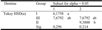
respectively. Table 4 and 5 identified with the Tukey HSD dentine, respectively. For (Retraction Paste %15 AlCl3) significantly lowest values in (P(sig.):0, 00<0,05). It was
(Cord+Viscostat Clear %25
(Cord+Viscostat %20 Ferric
discoloration, Group I ( Cord+Viscostat showed significantly lower ∆E
indexes were created to make the waxes were removed, 3 mm produced by Laboratory Elastic type
A-Zhermack). (Fig. 3).
each specimen, Fig. 3b. Arrangement artificial gingiva
materials and solutions
were divided into three groups for materials. The retraction materials, implementing procedures were shown in were applied for 180 seconds and seconds. Retraction materials were
specimens surfaces.
before and after the application and solutions by Easyshade (Vita) buccal surfaces that were indicated Measurements were used on a white between the tip of Easyshade device the color of artificial gingiva was polymethylmethacrylate block had been used for
Device.
values obtained before and after the recorded for each specimen. Then according to the formula.
dentine and artificial gingiva was analyzed using one-way ANOVA and
difference (HSD) test(a=.05).
ANOVA test performed for the groups,
present the statistical analysis HSD tests for artificial gingiva and artificial gingiva Group III AlCl3) retraction material showed comparison to the other groups was followed by Group I
%25 AlCl3) and Group II
Ferric Sulphate). For dentine
[image:2.595.101.224.602.761.2]II(Cord+Viscostat %20 Ferric Sulphate), whereas no statistically significant differences were observed between Group III(Retraction Paste %15 AlCl3) and the other groups (P(sig.):0,20<0,05).
DISCUSSION
Marginal adaptation is a critical factor for the longevity of fixed prosthetsis. For an adequate marginal fit, gingival retraction provides the clinicans obtain proper impressions and casting models. Inaccurate marginal fit may give rise to periodontal tissue inflammation and risk of seconder caries especially for subgingivally located crown margins (Felton, 1991). In some circumstances like anterior region restorations with esthetic demands, retention requirements of clinically short crowns, restoration of root caries and cervical abrasion and treatments of root sensitivity, subgingival margins should be preferred (Acar, 2014; Padbury, 2003). For acceptable impressions, displacement of gingival tissues, management of hemorrhage and gingival fluid should be proceeded
(Rosenstiel, 2006; Donovan, 2004 and Baharav, 2004).
Mechanical or mechanochemical methods are widely prefered by clinicians where displacement cords are used alone or with hemostatic agents (Johnson, 2010 and Beier, 2009).
Various types of gingival retraction displacement cords and caps, gingival retraction pastes and gels, application methods and impregnation medicaments have been reported. Recently, cordless techniques with retraction pastes or gels have been introduced. Application of the retraction pastes are time saving and more comfortable while being minimally invasive compared to conventinal retraction methods (Albaker, 2010). Retraction pastes commonly include 15% aluminium chloride excipient, mica minerales and kaolin. In the present study, the effect of different retraction materials on discoloration of
dentine and artificial gingiva was evaluated. Wostman et al,
(Wostmann, 2009) reported that animal studies or clinical trials were necessary for evaluation of the gingival retraction phenomenon for not being possible to be simulated by laboratory procedures. Since the animal and human in vivo experiments were rather costly, they decided to work on a semi-clinical model based on the jaws of freshly slaughtered cows. In the present study, we simulated the artificialging ivaaround prepared dentine. One limitation of the current experiment is the lack of blood pressure in the gingival area during impression making. Thus, the results of this study have to be interpreted carefully as the testing design limits its significance compared to the clinical situation. Epinephrine, ferric sulphate and aluminium chloride are widely used
Table 1. Contents of the retraction materials
Retraction Material Contents Product Name Manufacturer Group1 Cord+%25Aluminium chloride Ultrapak+Viscostat Clear Ultradent, ABD Group2 Cord+%20 Ferricsulphade Ultrapak+Viscostat Ultradent, ABD Group3 Retraction Paste (15% 25Aluminium chloride) Astringent Retraction paste 3M Espe, ABD
Table 2. Mean Values and standart deviation of Δ
ArtificialGingiva Groups N Mean Std. dev. Std. Hata Minimum Maximum Group I 12 9,0900 1,20402 0,34757 7,48 11,48 Group II 12 12,2475 2,17404 0,62759 8,52 15,28
GroupIII 12 6,3108 1,69610 0,48962 3,11 9,22
Total 36 9,2161 2,98248 0,49708 3,11 15,28
Dentine Group I 12 6,1758 2,43521 0,70299 2,98 11,22
Group II 12 9,3800 2,64119 0,76245 5,30 14,68
GroupIII 12 7,6792 2,17984 0,62927 4,27 10,96
[image:3.595.118.479.69.320.2]Total 36 7,7450 2,70404 0,45067 2,98 14,68
Table 3. One-way ANOVA results for the groups
Sum of Squares df Mean Square F Sig.
ArtificialGingiva Between Groups 211,750 2 105,875 35,086 0,000 Within Groups 99,582 33 3,018
Total 311,332 35
Dentine Between Groups 61,678 2 30,839 5,239 0,011
Within Groups 194,236 33 5,886
[image:3.595.195.405.361.413.2]Total 255,915 35
Table 4. Mean values of ΔE with statistical comparision using Tukey HSD for artificial gingiva
Artificial Gingiva Group Subset for alpha = 0.05 Tukey HSD(a) III 6,3108 a
[image:3.595.193.407.446.509.2]I 9,0900 b II 12,2475 c Sig. 0,001
Table 5. Mean values of ΔE with statistical comparision using Tukey HSD for dentine
Dentine Group Subset for alpha = 0.05
1 2
Tukey HSD(a) I 6,1758 a
III 7,6792 ab 7,6792 ab
II 9,3800 b
Sig. 0,296 0.214
medicaments to stop haemorrhage in the gingival retraction procedures. Aluminium chloride (AlCl3) and Ferric sulphate (Fe(SO4)3) are metal salts that lead up to proteins and provide contraction in gingival tissues that prevent haemorrhage. Epinephrine causes local vasoconstriction in gingival sulcus; however epinephrine has relatively minimal side effects such as tachycardia, increased respiratory rate, hypertension and anxiety. Hence the use of epinephrine may be contraindicated for the patients with cardiovascular disease (Prasad, 2011). The materials we used in our study are commonly preferred solutions in clinical practice. Materials were applied according to the manufacturers recommendations, 3-5 min. Application periods were recommended we applied 3 min. retraction period the main reason of that is to prevent the irreversible gingival displacement.
Commonly used gingival retraction medicaments have acidic pH values (0.7-3.0) which might dispose the smear layer. Disposal of the smear layer may affect bond strength and longevity of the restoration as well as bacterial invasion into the dentine tubules. Gingival medicaments with acidic iron containing etch dentine unwittingly and that can occured physical resorption of iron into the porous demineralized dentine (Prasad, 2011; Kuphasuck, 2007 and Land, 1994). Due to ferric ions’ affinity to teeth surfaces, ferric sulphate stains the gingival tissue from yellow-brown to black colour within a
few days (Hattab et al., 1999; Strangel et al., 1996; Sulieman
et al., 2005 and Watts, 2001). Aluminium chloride is an agent that acts by precipitation of tissue proteins but causes less vasoconstriction than epinephrine. It is one of the least irritating among all the medicaments used for impregnating
retraction cords (Masek et al., 2005). The null hypothesis that
the retraction methods or materials tested would show no significant difference in respect to discolouration was rejected. In the literature lots of researchers reported that the retraction materials and solutions could affect the colour of the dentine and gingiva. In the present study, we tried to simulate the clinical retraction procedures.
Anterior lower incisors were preferred because of their small anatomical structures, so the discolouration of the dentine
could easily be observed. In the recent years,
spectrophotometers and colorimeters came into use for dental clinicians. On behalf of different colour tabs, a virtual unmistakeable number of tooth colourations could be measured. Hence the colour measurements became more objective and foreseeable; colour data collection became easier and more reliable than naked eye. So that in our study we have used VITA EasyShade Compact electronic colour measuring device which is prevalently preferred in most of the researches (Waltmann, 2012; O’Brien, 2002 and Craig, 2002). CIE l*a*b
values that are defined by CommisionInternationale
d’Eclairage for characterising the colour difference value are attained from the device. Therefore ∆E values which are featured a comparative colour change between replicable
colour measurements are calculated easily. Actually, CIE
l*a*b is a popular and commonly preferred colour system in many studies. In this system, ∆E value of 3.7 is considered clinically noticeable (Ertas, 2006). In the present study, colour change values for all the retraction materials were greater than 3.7 which means that the discolourations tested was visually noticeable. Lighting condition and the background of the area on which the specimens were measured throughout the colour measurement could have effected the metrics (Guler, 2005). In our study, we used a white background and day light during
the measurement. According to our results, aluminium chloride solution showed minimal discolouration in dentine compared to ferricsulphate solution. Discolouration of the ferric sulphate solution might be due to its iron content. As the rate of the aluminium chloride is lower in retraction paste (15%) than Viscostat Clear (% 25), the retraction paste showed minimal change in colour which is in accordance with theliterature. On the other hand, retractionpaste showed higher discolouration then Viscostat Clear (25%) in dentine which may be the result of the presence of mica minerals and kaolin within the retraction paste. Following the preparation of the teeth, dentine tubules could be effected more than the gingival tissues. As a conclusion, it could be reported that using ferric sulphate solutions may cause discolouration both in dentine and gingiva. Hence the clinicians should keep in mind the risk of colour changes in gingival and dentine tissues especially in the anterior regions when choosing the retraction materials and solutions. Despite the results of this study, further in-vivo studies subjecting the discolouration caused by retraction materials should be explored.
Conclusion
Within the limitations of this study following conclusions were drawn; The solution including aluminium chloride caused minimal discolouration in the artificial gingiva. Discolouration of gingiva was in direct proportion to aluminium chloride rate of the solutions. Retraction paste (%15) showed the lowestgingivalcolour change whereas ViscostatClear (%25) showed the least change of colour in dentine. Nevertheless ferric sulphate containing retraction solutions caused discolouration not only in dentine but also in the gingiva.
REFERENCES
Acar, O., Erkut, S., Ozcelik, T.B., Ozdemır, E., Akcil, M., J. 2014. A clinical comparison of cordless and conventional displacement systems regarding clinical performance and
impression quality, Prosthet Dent, 111(5):388–94.
Al-Ani, A., Bennani, V., Chandler, N.P., Lyons, K.M., Thomson, W.M. 2010. New Zealand dentists' use of gingival retraction techniques for fixed prosthodontics and
implants.NZ Dent J, 106:92-6.
Albaker, A.M. 2010. Gingival retraction–techniques and
materials: a review. Pakistan OralDent J, 30(2): 545-551.
Baharav, H., Kupershmidt, I., Laufer, B.Z., Cardash, H.S. 2004. The effect of sulcular width on the linear accuracy of
impression materials in the presence of an undercut. Int J
Prostho- don’t., 17:585-9.
Beier, U.S., Kranewitter, R., Dumfahrt, H. 2009. Quality of impressions after use of the Magic FoamCord gingival
retraction system clinical study of 269 abutment teeth. Int J
Prosthodont, 22:143-7.
Benson, B.W., Bomberg, T.J., Hatch, R.A., Hoffman, W. Jr.
1986. Tissue displacement methods in fixed
prosthodontics. J. Prosthet Dent, 55:175-81.
Bowles, W.H., Tardy, S.J., Vahadi, A. 1991. Evaluation of
new gingival retraction agents. J.Dent Res, 70:1447-9.
Conrad, H.J., Holtan, J.R. 2009. Internalized discoloration of
dentin under porcelain crowns: A clinical report. J Prosthet
Dent., 101:153-7.
Craig, R.G., Powers, J.M. 2002. Restorative dental materials. 11 ed. Mosby, St Louis, pp.39-42.
Donovan, T.E., Winston, W.L. 2004. Current concepts in
Ertas, E., Guler, A.U., Yucel, A.Ç., Koprulu, H., Guler, E. 2006. Color Stability of Resin Composites after Immersion in Different Drinks. Dent Mat J 2006; 2(52):371-376. Felpel, L.P. 1997. A review of pharmacotherapeutics for
prosthetic dentistry: Part I. J. Prosthet Dent, 77:285-92.
Felton, D.A., Kanoy, B.E., Bayne, S.C.,
Wirthman, G.P. 1991. Effect of in vivo crown margin
discrepancies on periodontal health. J Prosthet Dent,
1991;65:357-64.
Guler, A.U., Yilmaz, F., Kulunk, T., Guler, E, Kurt, S. 2005. Effects of different drinks on stainability of composite
resin provisionla restorative materials. J. Prosthet Dent.,
94:118-24.
Hattab, F.N., Qudeimat, M.A., al-Rimawi, H.S. 1999.
Dentldiscoloration:anoverview. J EsthetDent., 11:291-310.
Johnson, G.H., Mancl, L.A., Schwedhelm, E.R., Verhoef, D.R, Lepe, X. 2010. Clinical trial investi- gating success rates for polyether and vinyl polysiloxane impressions made
with full-arch and dual-arch plastic trays. J Prosthet Dent,
103:13-22.
Jokstad, A. 1999. Clinical trial of gingival retraction cords. J.
Prosthet Dent., 81;258-61.
Kuphasuck, W, Harnirattisai, C., Senawongse, P., Tagami, J. Bond strength of two adhesive systems to dentin
contaminated with a hemostatic agent. Oper Dent.,
32:399-405.
Land, M.F., Rosenstiel, S.F., Sandrik, J.L. 1994. Disturbance
of dentinal smear layer by acidic hemostatic agents. J
Prosthet Dent., 72:4-7.
Masek, R. 2005. Marginisolation for opticalim pressions and
adhesion. Int J Comput Dent., 8:69-76.
Nemetz, E.H., Seibly, W. 1990. The use of chemical agents in
gingival retraction. Gen Dent, 38:104-8.
O’Brien, W.J. 2002. Dental materials and their selection, 3rd ed., Quint Pub Inc., Canada, pp.28.
Padbury, A. Jr, Eber, R., Wang, H.L. 2003. Interactions
between the gingiva and the margin of restorations. J
ClinPeriodontol., 30: 379-85.
Perakis, N., Belser, U.C., Magne, P. 2004. Final impressions: a review of material properties and description of a current
technique. Int J Periodontics Restorative Dent;24(2):
109-17.
Prasad, K.D., Hegde, C., Agrawal, G., Shetty, M. 2011. Gingival displacement in prosthodontics: A critical review
of existing methods. J Interdiscip Dentistry, 1:80-6.
Rosenstiel, S.F., Land, M.F., Fujimoto, J. 2006.
Contemporary fixed prosthodontics. 4th ed. St Louis: Mosby, p. 432.
Strangel, I., Valdes, E., Xu, J. 1996. Absorption of iron by
dentin: its role in discoloration. J Biomed Mater Res.,
131:287-92.
Sulieman, M. 2005. An overview of tooth discoloration:
extrinsic, intrinsic and internalized strains. Dent Update,
32:463-4,466-8,471.
Waltmann, D.S. 2012. Contemporary Esthetic Dentistry, 3rd
ed., Mosby, St Louis, p.159.
Wassell, R.W., Barker, D., Walls, A.W. 2002. Crowns and other extracoronal restorations: impression materials and
technique. Br Dent J, 192:679-84, 687-90.
Watts, A., Addy, M. 2001. Tooth discoloration and straining: a
review of the literature. Br Dent J., 190:309-16.
Wostmann, B., Rehmann, P., Trost, D., Balkenhol, M. 2009. Effect of different retraction and impression techniques on
the marginal fit of crowns. Journal of dentistry, 36(7):
508-512.