Co-occurrence of Type 1 Diabetes
and Celiac Disease Autoimmunity
William Hagopian, MD, PhD, a Hye-Seung Lee, PhD, b Edwin Liu, MD, c Marian Rewers, MD, MPH, c Jin-Xiong She, PhD, d Anette-G. Ziegler, PhD, eÅke Lernmark, MD, f Jorma Toppari, MD, PhD, g Stephen S. Rich, PhD, h Jeffrey P. Krischer, PhD, b Henry Erlich, PhD, i Beena Akolkar, PhD, j Daniel Agardh, MD, PhD, f the TEDDY Study Group
BACKGROUND AND OBJECTIVES: Few birth cohorts have prospectively followed development of type
1 diabetes (T1D) and celiac disease (CD) autoimmunities to determine timing, extent of co-occurrence, and associated genetic and demographic factors.
METHODS: In this prospective birth cohort study, 8676 children at high genetic risk of both
diseases were enrolled and 5891 analyzed in median follow-up of 66 months. Along with demographic factors and HLA-DR-DQ, genotypes for HLA-DPB1 and 5 non-HLA loci conferring risk of both T1D and CD were analyzed.
RESULTS: Development of persistent islet autoantibodies (IAs) and tissue transglutaminase
autoantibodies (tTGAs), as well as each clinical disease, was evaluated quarterly from 3 to 48 months of age and semiannually thereafter. IAs alone appeared in 367, tTGAs alone in 808, and both in 90 children. Co-occurrence significantly exceeded the expected rate. IAs usually, but not always, appeared earlier than tTGAs. IAs preceding tTGAs was associated with increasing risk of tTGAs (hazard ratio [HR]: 1.48; 95% confidence interval [CI]: 1.15–
1.91). After adjusting for country, sex, family history, and all other genetic loci, significantly greater co-occurrence was observed in children with a T1D family history (HR: 2.80), HLA-DR3/4 (HR: 1.94) and single-nucleotide polymorphism rs3184504 at SH2B3 (HR: 1.53). However, observed co-occurrence was not fully accounted for by all analyzed factors.
CONCLUSIONS: In early childhood, T1D autoimmunity usually precedes CD autoimmunity.
Preceding IAs significantly increases the risk of subsequent tTGAs. Co-occurrence is greater than explained by demographic factors and extensive genetic risk loci, indicating that shared environmental or pathophysiological mechanisms may contribute to the increased risk.
abstract
NIH
aDiabetes Programs Division, Pacific Northwest Research Institute, Seattle, Washington; bDepartment of Pediatrics, Health Informatics Institute, University of South Florida, Tampa, Florida; cDepartment of Pediatrics, Children’s Hospital Colorado, University of Colorado, Aurora, Colorado; dCenter for Biotechnology and Genomic Medicine, Augusta University, Augusta, Georgia; eInstitute of Diabetes Research, Helmholtz Zentrum München, Oberschleißheim, Germany; fDepartment of Clinical Sciences, Skåne University Hospital, Lund University, Malmö, Sweden; gDepartment of Pediatrics, Turku University Central Hospital, Turku, Finland; hCenter for Public Health Genomics, University of Virginia, Charlottesville, Virginia; iChildren’s Hospital of Oakland Research Institute, Oakland, California; and jDiabetes Branch, National Institute of Diabetes and Digestive and Kidney Diseases, National Institutes of Health, Bethesda, Maryland
Dr Hagopian helped conceptualize and design the study, secured funding, supervised data collection at 1 of 6 sites, helped analyze the data, and drafted and revised the initial manuscript; Dr Lee led the data analysis and statistical testing, formulated the figures, and reviewed and revised the manuscript; Drs Liu and Agardh helped design and supervise the celiac portion of the study, helped with data analysis, and reviewed and revised the manuscript; Drs Rewers, She, Ziegler, and Lernmark helped conceptualize and design the study, secured funding, supervised data collection at 1 of 6 sites, and reviewed and revised the manuscript; Dr Toppari supervised data collection at 1 of 6 sites and reviewed and revised the manuscript; Dr Rich helped design
What’s KnoWn on this subject: Children with type 1 diabetes (T1D) are at greater risk of celiac disease (CD), and screening them for transglutaminase autoantibodies (tTGAs) is useful. T1D and CD share high-risk HLA-DR-DQ genotypes DR3 and DR4, which partly explains disease overlap.
What this study adds: Early markers of T1D and CD autoimmunity coincide more than expected. Overlap associates with familial T1D, HLA-DR3/4, and SH2B3, but is not fully explained by known risk factors. T1D autoantibodies usually precede tTGAs and confer significant risk of subsequent tTGAs.
Celiac disease (CD) and Type 1 diabetes (T1D) involve autoimmune attacks on small bowel mucosa and on pancreatic islet β cells, respectively. Both diseases have a strong genetic predisposition1, 2 and
most often begin in early childhood.3, 4
A subclinical or preclinical phase is characterized by autoantibody positivity with minimal symptoms.3, 5, 6
Later, more severe target organ dysfunction appears, prompting clinical diagnosis and treatment. The frequent coexistence of T1D and CD is widely described, but most researchers measure future risk of CD in children with previous T1D. This ranged from 2.5% to 16.4% (5.7% overall) in 23 studies worldwide, 7 much greater than
background CD rates. Conversely, studies of subsequent T1D in children with previous CD also revealed significantly more T1D than in non-CD controls.8, 9 Presence of either
disease therefore appears to increase the risk of developing the other. Commonly postulated reasons for concurrent CD and T1D include shared HLA genetic risk10–13 and
shared environmental exposures.8, 14
Genetics are indeed important in CD, with ∼75% monozygotic twin concordance and 10% of first- degree relatives (FDRs) affected.2
Genetics are similarly important in T1D, with ∼42% monozygotic twin concordance and 6% to 8% FDRs affected.1 These genetic
effects are greater than for most autoimmune diseases, perhaps because of a remarkably large effect of HLA Class II on both diseases. HLA-DR-DQ accounts for ∼40% to 50% of the overall T1D genetic risk, 15
and 53% of that for CD, using modern prevalence estimates.2 The
2 common haplotypes conferring increased risk in both diseases are DRB1*04-DQA1*03:01-DQB1*03:02 (commonly referred to as the DR4-DQ8 haplotype, abbreviated DR4) and DRB1*03-DQA1*05:01-DQB1*02:01 (commonly referred to
as DR3-DQ2 haplotype, abbreviated DR3). In T1D, DR4 confers greater risk than DR3, but DR3/DR4 heterozygotes are at the greatest risk.15 For CD, DR3 confers more risk
than DR4, and DR3/DR3 confers the greatest risk.2 Studies of the absolute
CD risk conferred by HLA-DR-DQ in unbiased populations (without symptoms, other autoimmunity, or family history [FH]) are sparse, but The Environmental Determinants of Diabetes in the Young (TEDDY) study recently published multicenter risk estimates for several high-risk HLA antigen genotypes that were consistent with most previous literature.4 HLA-DR-DQ strongly
influences both diseases and the risk-conferring haplotypes are shared, so substantial disease overlap is expected solely on that basis. CD and T1D are also concurrently influenced by >10 other genetic loci, 10, 16–18
conferring smaller hazard ratios (HRs) in the 1.14 to 1.35 range but with independent effects that may be cumulative. However, for only 6 loci (HLA-DPB1, RGS1, SH2B3, CTLA4, CCR3/CCR5, and PTPN2) does the relevant polymorphism affect both diseases in the same direction, therefore potentially increasing disease co-occurrence.10
The TEDDY study is an international study of 8676 infants with many years of frequent prospective observation.19 The major inclusion
criterion of elevated HLA antigen risk for T1D20 also confers
elevated CD risk. Frequent blood sampling enabled accurate timing of seroconversion for both islet autoantibodies (IAs) and tissue transglutaminase autoantibodies (tTGAs). The cohort underwent extensive additional genotyping. TEDDY thus represents an unprecedented opportunity to describe the extent and timing of co-occurrence of the 2 diseases. The aim of this study was to investigate if 1 disease might trigger the other, and to test if their co-occurrence can be
explained by common inherited risk genes.
Methods
the teddy cohort and hLa Genotyping
TEDDY is a prospective cohort study designed to identify environmental causes of T1D.19 From 2004 to 2010,
424788 newborns were screened at 6 US and European centers. TEDDY enrolled 8676 infants with high- T1D–risk HLA antigen genotypes by 4.5 months of age, with intent to manage until age 15 years. Eligible HLA-DR-DQ genotypes are abbreviated DR3/4, DR4/4, DR4/8, and DR3/3 (Supplemental Table 2). Of 8676 TEDDY enrollees, 5891 were analyzed herein on the basis of full autoantibody characterization, genotyping on the ImmunoChip, and carrying 1 of the 4 major TEDDY-eligible HLA genotypes. Frequencies of all eligible HLA genotypes for each center are given in Supplemental Table 3. At the time of analysis, median follow-up was 66 months (range 10–111 months, interquartile range [IQR] 50–81 months), covering 32454 person-years of observation. Local institutional review board approval and parental informed consent was obtained for all children. The study is monitored by an
External Evaluation Committee of the US National Institutes of Health.
autoantibody Measurements
Glutamate decarboxylase autoantibodies, insulinoma antigen-2 autoantibodies, and insulin autoantibodies (IAAs) were measured in 2 harmonized core laboratories by using radiobinding assays incorporating extensive quality control.21 Persistent islet
antibodies (IAs) were defined as positive antibodies to the same antigen confirmed by both core laboratories in 2 consecutive samples.19 Tissue tTGAs were
in 2 laboratories with final status determined by a central reference laboratory as previously described.4, 21
Persistent tTGAs were defined as positive tTGAs test results in 2 con secutive samples. Only persistent IAs and tTGAs are considered in the current analyses.
Prospective Follow-up
The TEDDY protocol specifies serum collection every 3 months from ages 3 to 48 months and at least every 6 months thereafter. IAs were measured in every available sample. Concurrent diseases including T1D and CD in the TEDDY child, and immediate family were noted at each visit. T1D was defined by using American Diabetes Association criteria. tTGA testing began at age 24 months and continued annually. If the results of any sample were positive, all previous samples from that child were analyzed to determine when tTGAs first appeared. Celiac symptoms were uncommon at the time of tTGA seroconversion.5 Parents of children
with persistent tTGAs were notified and encouraged to pursue further evaluation under guidance of their physician. This likely led to more rapid ascertainment of CD in study than in general medical practice. In similar fashion, IA surveillance and early T1D ascertainment via glucose tolerance testing was also a feature of TEDDY.22 The decision
on duodenal biopsy was outside of the study protocol, but if a biopsy were performed, a Marsh score of
≥2 defined CD. For individuals who did not receive a biopsy, a mean persistent tTGAs ≥100 U based on 2 consecutive samples was >95% specific for CD and therefore considered diagnostic herein.4
Children developing T1D were no longer on study protocol, but phone interviews and medical records reviews enabled determination of subsequent tTGA status, CD status, biopsy results if any, and use of a
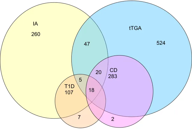
gluten-free diet in 150 out of 167 (90%) of these individuals. At the time of analysis, clinical T1D occurred in 138 (2.3%), CD in 323 (5.5%), and both diseases developed in 18 (0.3%) of the 5891 screened participants, (Fig 1). The low number of subjects concurrently diagnosed with T1D and CD in early childhood limited detailed subanalysis of factors influencing T1D and CD co-occurrence. We therefore focused on IAs and tTGAs as earlier and more prevalent disease markers.
single-nucleotide Polymorphism Genotyping
In addition to HLA-DR-DQ alleles, the cohort was genotyped for single-nucleotide polymorphisms (SNPs) by the Illumina ImmunoChip covering 186 genomic regions associated with 12 autoimmune diseases.23
SNP markers with call rates <90% or allele distributions strongly deviating from Hardy-Weinberg equilibrium in controls (P < 10−6)
were discarded (except within the HLA region). Individuals with call rates <95%, or those discordant with reported sex or previous genotyping
were also discarded. This resulted in data on ∼176586 SNPs in each of 7023 subjects. SNP genotypes were used to determine HLA-DPB1, and 5 other loci reported to affect both T1D and CD in the same direction:
RGS1, SH2B3, CTLA4, CCR3/CCR5, and
PTPN2 (Supplemental Table 4).
statistical Methods
Excess co-occurrence was
calculated by comparison with the expected along with the Wilson 95% confidence interval (CI). Excess proportion was compared by using Fisher’s exact test. To explore the pattern of appearance of each autoantibody (IAs and tTGAs) or disease (T1D and CD), the proportion of event-free survival in which Kaplan-Meier estimates were used was plotted against age of seroconversion or diagnosis. Age was compared between groups by using the Wilcoxon rank sum test.
A Cox proportional hazards model was used to investigate factors associated with IA and tTGA
co-occurrence. Time to co-occurrence was defined as age of seroconversion
FiGuRe 1
for the following autoimmunity. The right-censored time was age at last sample collection. The model considered country, sex, and FH of T1D or CD (defined as an affected FDR), in addition to HLA-DR-DQ, HLA-DPB1, and the 5 other relevant genetic loci. Additionally, we examined the effect of each autoantibody on appearance of the other. For example, the effect of IAs preceding tTGAs on the risk of tTGAs was assessed (by using age of tTGA appearance as time to event) as compared with those without IAs at the time of tTGA appearance. Two sided P values < .05 were considered significant. SAS 9.4 Software (SAS Institute, Cary, NC) was used for all analyses.
ResuLts
The proportion of children who tested positive for IAs that developed T1D was 130 out of 457 (28.5%) compared with 8 out of 5434 (0.15%) of children who tested negative for IAs. The proportion of children who tested positive for tTGAs that developed CD was 321 out of 898 (35.8%) compared with 2 out of 4993 (0.04%) of children who tested
negative for tTGAs. A total of 367 (6.2%) participants had IAs but not tTGAs, 808 (13.7%) had tTGAs but not IAs, and 90 (1.5%) had both (Fig 1). Assuming independence and assuming the observed proportion of tTGAs and IAs in TEDDY are accurate estimates of corresponding population proportions, the product of the observed IA and tTGA
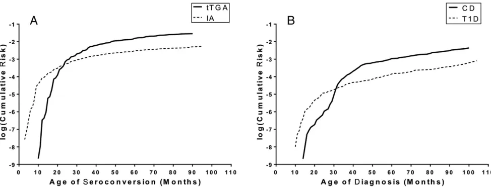
proportions represents the expected proportion of having both antibodies. The observed IA prevalence was 457 out of 5891 (7.8%), whereas that for tTGAs was 898 out of 5891 (15.2%). We therefore expected IAs and tTGAs to occur together in 1.19% (7.8%*15.2%) or 70 children. The observed prevalence of 90 out of 5891 (1.53%) revealed a significant excess of 20 children or 0.34% (95% CI: 0.21%–0.52%). Subgroups by country, sex, FH of T1D and of CD, HLA-DR-DQ, and other genetic loci each retained significant excess co-occurrence of IAs and tTGAs (Supplemental Table 5). In examining the proportion of children with each autoantibody, it can be seen that having IAs increased the proportion with tTGAs from 14.9% to 19.7% (Fig 2A). Conversely, having tTGAs increased the proportion with IAs
from 7.4% to 10.0% (Fig 2B). This represents a 32% greater prevalence of tTGAs in those with IAs, and a 36% greater prevalence of IAs in those with tTGAs. Increases were observed across all individual HLA genotypes but were only significant for DR4/4 (P = .017) and for the combined data set of all HLA genotypes (P = .012). Given the prospective nature of the TEDDY study, it is possible to extend the cross-sectional data of Fig 1 to examine the development of autoimmunity over time. Seroconversion for each of IAs and tTGAs was greatest in the first few years of life (Fig 3). When considered separately, the median age at seroconversion for IAs was 24 (IQR: 14–38.5) months and for tTGAs was 33 (IQR: 24–45) months (P < .0001). There was no significant difference in age at seroconversion when comparing those with 1 type of autoimmunity to those with both. Within the current follow-up, T1D was on average diagnosed earlier than CD (T1D at 36 [23–56] months; CD at 42 [34–57] months; P = .0007). Of the 90 individuals with both IAs and tTGAs, 5 (6%) developed the autoantibodies simultaneously, 61 (68%) developed IAs first at 24
FiGuRe 2
(14–38.5) months, and 24 (27%) developed tTGAs first at 33 (24, 45) months (P < .0001). Comparative timing of IAs versus tTGAs was not different when comparing glutamate decarboxylase autoantibodies or IAAs as the specific IAs (Supplemental Fig 4). Greater HLA-DR3 gene dosage conferred shorter median time interval from development of IAs to tTGAs (median 2 months in DR3/3, 14 months in DR3/4, and 18 months in DR4/4 or DR4/8) (Supplemental Fig 5). The median time from tTGAs to CD was 11.6 (IQR: 7.9–16.1) months, significantly shorter than that from IAs to T1D at 17.2 (7.7–35.7) months (P < .0001).
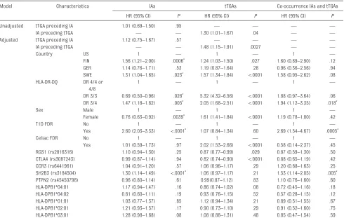
A Cox Proportional Hazards model was used to examine risk factors for each individual type of autoimmunity, including whether each autoantibody influenced the risk for developing the other (Table 1). Known risk factors for each type of autoimmunity showed expected results in the adjusted model. Factors associated separately with increased IAs and with increased tTGAs included the countries of Finland or Sweden and HLA-DR3/4. HLA-DR3/3 increased tTGA risk but decreased IA risk versus the reference genotype. As expected, having an FDR with
CD increased tTGA risk but not IA risk, and having an FDR with T1D increased IA risk but not tTGA risk. RGS1 (rs2816316) and CTLA4 (rs3087243) were associated with increased tTGA risk, whereas SH2B3 (rs3184504) was associated with increased IA risk. Importantly, in unadjusted analysis, IAs preceding tTGAs was associated with greater risk of tTGAs (HR: 1.30; 95% CI: 1.01–1.67), but tTGAs preceding IAs was not associated with risk of IAs (HR: 1.01; 95% CI: 0.69–1.50). Using a similar model adjusted for all other factors did not change this finding; IAs preceding tTGAs was significant (HR: 1.48; 95% CI: 1.15–1.91), whereas tTGAs preceding IAs was not (HR: 1.12; 95% CI: 0.75–1.67). The Cox Proportional Hazards model was then used to analyze demographic and genetic factors conferring risk of co-occurrence of IA and tTGA (Table 1). After adjustment for country, sex and having an FDR with CD, HLA-DR-DQ, HLA-DPB1 and the 5 SNPs at RGS1, SH2B3, CTLA4, CCR3/CCR5, and PTPN2, we found HLA-DR3/4 (HR: 1.94; 95% CI: 1.12–3.35), having an FDR with T1D (HR: 2.69; 95% CI: 1.54–4.67) and rs3184504 at SH2B3
(HR = 1.53, 95% CI: 1.14–2.05) were each independently associated with co-occurrence of IAs and tTGAs.
discussion
The appearance of CD in existing T1D patients is widely appreciated.7 Our
results analyze this relationship in a prospective manner at an earlier stage when autoantibodies to the respective diseases first develop. These autoantibodies are highly predictive of clinical disease and may be encountered more frequently as genetic screening and personalized medicine become more widespread. As with T1D and CD, observation in the first 5 years of life reveals that IAs precede tTGAs more often (67%) than vice versa (27%). Indeed, having IAs significantly increased the risk of developing tTGAs. The reverse relationship was not demonstrated, perhaps because of fewer individuals with the latter pattern detected during limited follow-up.24 Our
results support the notion that children found to have IAs (for example through family screening or population-based prediction) should be screened for tTGAs, as is the practice for T1D children. Having dual T1D and CD
autoimmunity was more common than expected on the basis of the rates of each. It has been widely supposed that HLA antigen explains the overlap7, 12, 13 because similar FiGuRe 3
HLA-DR-DQ genotypes strongly predispose to both diseases. However, this did not appear to be the case. In a detailed analysis of the excess cases, considering HLA antigen loci, non-HLA antigen loci, and demographic features (FH, country) expected to capture additional (although unmeasured) genetic influences on these 2 autoimmune diseases, it is clear that not all the observed overlap between the 2 diseases was accounted for (Table 1). In addition to HLA-DR-DQ, SNP rs3184504 at SH2B3 accounted for significant overlap, but the effect was modest. SH2B3 is an adaptor protein involved in negative regulation of T-cells, consistent with both T1D and CD being T-cell–
mediated diseases.25 Although not
all common minor-effect variants or rare high-effect variants would be captured in the current analysis, 26–28
our finding is in line with a previous study whose authors did not find major contributions by shared genetic risk factors for T1D and CD other than HLA antigen genotype.28 Our results
therefore suggest that nongenetic factors are probably also involved. Prospectively, IAs usually but not always preceded tTGAs, and previous IAs conferred a significantly increased risk of tTGAs. Although previous tTGA did not show a similar effect on subsequent IAs, there may have been insufficient individuals in this category for adequate evaluation. Our significant findings are in contrast to those of Williams et al, 29
who found that tTGA prevalence in adults with an FH of T1D were not different between those with or without IAs, implying that tTGAs were not more likely in the setting
of IAs. However, we prospectively managed individuals from early childhood when both types of autoimmunity first develop, which may explain our findings.
If preexisting T1D autoimmunity triggers CD autoimmunity, a possible mechanism might be coexpression of antigens from both diseases in the same anatomic location. tTG is known to be expressed in islets.30
Cellular endoplasmic reticulum stress leads to tTG overexpression31
including in stressed islets.32 This
raises the intriguing possibility that tTG expressed in the insulitis lesion, in the setting of a susceptible HLA antigen background, might trigger autoimmunity to tTG. Importantly, the duodenum and pancreas are in close proximity and share draining lymph nodes, as shown in elegant
tabLe 1 Cox Proportional Hazards Analysis of Factors Influencing Appearance of IAs and tTGAs, Their Order of Appearance, and Co-occurrence of IA and tTGA
Model Characteristics IAs tTGAs Co-occurrence IAs and tTGAs
HR (95% CI) P HR (95% CI) P HR (95% CI) P
Unadjusted tTGA preceding IA 1.01 (0.69–1.50) .95 — — — —
IA preceding tTGA — — 1.30 (1.01–1.67) .04 — —
Adjusted tTGA preceding IA 1.12 (0.75–1.67) .57 — — — —
IA preceding tTGA — — 1.48 (1.15–1.91) .0027 — —
Country US 1 — 1 — 1 —
FIN 1.56 (1.21–2.00) .0006* 1.24 (1.03–1.50) .027 1.60 (0.89–2.90) .12 GER 1.14 (0.76–1.71) .53 1.19 (0.87–1.64) .28 0.96 (0.36–2.56) .94 SWE 1.31 (1.04–1.65) .023* 1.57 (1.34–1.84) <.0001 1.58 (0.95–2.62) .08 HLA-DR-DQ DR 4/4 or
4/8
1 — 1 — 1 —
DR 3/3 0.69 (0.50–0.96) .028* 5.32 (4.32–6.56) <.0001 1.88 (0.97–3.64) .06 DR 3/4 1.47 (1.18–1.82) .005* 2.05 (1.68–2.51) <.0001 1.94 (1.12–3.35) .018*
Sex Male 1 — 1 1 —
Female 0.76 (0.63–0.92) .0039* 1.61 (1.41–1.84) <.0001 1.19 (0.78–1.80) .42
T1D FDR No 1 — 1 — 1 —
Yes 2.60 (2.03–3.33) <.0001* 1.07 (0.84–1.34) .60 2.69 (1.54–4.67) .0005*
Celiac FDR No 1 — 1 — 1 —
Yes 1.01 (0.59–1.73) .97 2.02 (1.53–2.66) <.0001 0.58 (0.14–2.37) .45 RGS1 (rs2816316) 1.10 (0.94–1.30) .25 0.87 (0.77–0.99) .029 0.87 (0.59–1.30) .50 CTLA4 (rs3087243) 0.99 (0.87–1.14) .94 0.82 (0.74–0.90) <.0001 0.88 (0.65–1.19) .42 CCR3 (rs6441961) 1.04 (0.91–1.20) .57 1.06 (0.96–1.17) .29 1.20 (0.88–1.63) .25 SH2B3 (rs3184504) 1.30 (1.14–1.49) <.0001* 1.06 (0.97–1.17) .21 1.53 (1.14–2.05) .005* PTPN2 (rs45450798) 0.96 (0.80–1.14) .61 0.99(0.87–1.12) .83 1.10 (0.76–1.60) .60 HLA-DPB1*04:01 1.17 (0.94–1.47) .16 0.86 (0.74–1.02) .08 0.72 (0.45–1.16) .18 HLA-DPB1*04:02 0.81 (0.60–1.11) .19 0.93 (0.76–1.15) .52 0.57 (0.28–1.15) .12 HLA-DPB1*01:01 1.03 (0.77–1.37) .85 1.12 (0.94–1.34) .21 0.89 (0.51–1.55) .67 HLA-DPB1*02:01 1.21 (0.93–1.57) .17 0.90 (0.73–1.10) .29 0.91 (0.52–1.60) .75 HLA-DPB1*03:01 1.28 (0.98–1.68) .08 1.08 (0.88–1.31) .48 0.85 (0.47–1.54) .59
Factors analyzed include country, sex, FH of T1D or CD, and HLA-DR-DQ, HLA-DPB1, and 5 non-HLA antigen genetic loci. FIN, Finland; GER, Germany; SWE, Sweden; US, United States; —, not applicable.
cotrafficking studies.33 Inflammation
in islets could thus lead to an anti-tTG immune response in adjacent lymph nodes, thereby promoting CD autoimmunity.
The major identified environmental trigger for CD is ingested gluten. Interestingly, gluten introduction before 4 months or after 7 months of age was shown to increase T1D risk.34, 35
Similarly, gluten exposure before 4 months increased CD risk in some36, 37
but not all35 studies. It has been
proposed that both T1D and CD are favored by an inflammation-mediated increase in gut permeability, 38, 39 which
gluten is known to cause.40 However,
age at introduction of gluten was not found to be significantly associated with tTGA in TEDDY.41
Gut microbes, in direct contact with the intestinal mucosa, are also prime candidates to influence CD risk. Rotavirus infection was implicated in both T1D autoimmunity42 and CD
autoimmunity, 43 but both remain
unconfirmed. These findings are further supported by TEDDY findings that a reported gastrointestinal infection 3 months before was significantly associated with tTGA seroconversion.44 Moreover, rotavirus
vaccination had a protective effect in children exposed to gluten before 6 months of age, indicating a potential interaction between diet and infections on the risk of early autoimmunity.44 Interestingly,
reovirus was recently postulated to be implicated in CD, 45 but its role in T1D
is unknown. Gut bacterial microflora also differ between cases and controls for both CD46 and T1D, 47 with cases
of both diseases having fewer positive firmicutes and more Gram-negative bacteroides, although not all researchers agree and relationships are understandably complicated. The intestinal flora may therefore deviate in a similar way in both diseases. Future metagenomic analyses of TEDDY stool samples will test this possibility.
concLusions
Early T1D and CD autoimmunity, like their clinical diseases, occur together more than expected, with IAs usually preceding tTGAs. In addition to genes and demographics, nongenetic factors appear to contribute to co-occurrence. Elucidation of relevant mechanisms will require additional studies of local immune responses as well as common exposures (eg, gluten and microbial infections) within and outside of the TEDDY Study.
the teddy study GRouP
colorado clinical center
Marian Rewers, MD, PhD, PI, 1, 4–6, 10, 11
Kimberly Bautista, 12 Judith
Baxter, 9, 10, 12, 15 Ruth Bedoy, 2 Daniel
Felipe-Morales, Kimberly Driscoll, PhD, 9 Brigitte I. Frohnert, MD, 2, 14
Patricia Gesualdo, 2, 6, 12, 14, 15 Michelle
Hoffman, 12–14 Rachel Karban, 12
Edwin Liu, MD, 13 Jill Norris, PhD, 2, 3, 12
Adela Samper-Imaz, Andrea Steck, MD, 3, 14 Kathleen Waugh, 6, 7, 12, 15 Hali
Wright.12 University of Colorado,
Anschutz Medical Campus, Barbara Davis Center for Childhood Diabetes.
Finland clinical center
Jorma Toppari, MD, PhD, PI, *, §, 1, 4, 11, 14
Olli G. Simell, MD, PhD, *, §, 1, 4, 11, 13
Annika Adamsson, PhD, §, 12 Suvi
Ahonen, †, ‖, # Heikki Hyöty, MD, PhD, †, ‖, 6
Jorma Ilonen, MD, PhD, *, ††, 3 Sanna
Jokipuu, § Tiina Kallio, § Leena
Karlsson, § Miia Kähönen, ‡, ¶ Mikael
Knip, MD, PhD, †, ‖, 5 Lea Kovanen, †, ‖, #
Mirva Koreasalo, †, ‖, #, 2 Kalle Kurppa,
MD, PhD, †, ‖, 13 Tiina Latva-aho, ‡, ¶
Maria Lönnrot, MD, PhD, †, ‖, 6 Elina
Mäntymäki, § Katja Multasuo, ‡, ¶ Juha
Mykkänen, PhD, *, 3 Tiina Niininen, ‖, †, 12
Sari Niinistö, ‖, # Mia Nyblom, †, ‖ Petra
Rajala, § Jenna Rautanen, ‖, # Anne
Riikonen, †, ‖, # Mika Riikonen, § Jenni
Rouhiainen, § Minna Romo, § Tuula
Simell, PhD, Ville Simell, §, *, 13 Maija
Sjöberg, *, §, 12, 14 Aino Stenius, ‡, ¶, 12
Maria Leppänen, § Sini Vainionpää, §
Eeva Varjonen, *, §, 12 Riitta Veijola,
MD, PhD, ‡, ¶, 14 Suvi M. Virtanen, MD,
PhD, †, ‖, #, 2 Mari Vähä-Mäkilä, § Mari
Åkerlund, †, ‖, # Katri Lindfors, PhD.†, 13
*University of Turku, †University of
Tampere, ‡University of Oulu, §Turku
University Hospital, Hospital District of Southwest Finland, ‖Tampere
University Hospital, ¶Oulu University
Hospital, #National Institute for
Health and Welfare, Finland,
††University of Kuopio.
Georgia/Florida clinical center
Jin-Xiong She, PhD, PI, 1, 3, 4, 11 Desmond
Schatz, MD, *, 4, 5, 7, 8 Diane Hopkins, 12
Leigh Steed, 12–15 Jamie Thomas, *, 6, 12
Janey Adams, *, 12 Katherine Silvis, 2
Michael Haller, MD, *, 14 Melissa
Gardiner, Richard McIndoe, PhD, Ashok Sharma, Joshua Williams, Gabriela Young, Stephen W. Anderson, MD, † Laura Jacobsen, MD, *, 14 Center
for Biotechnology and Genomic Medicine, Augusta University. *University of Florida, †Pediatric
Endocrine Associates, Atlanta.
Germany clinical center:
Anette G. Ziegler, MD, PI, 1, 3, 4, 11
Andreas Beyerlein, PhD, 2 Ezio
Bonifacio PhD, *, 5 Michael Hummel,
MD, 13 Sandra Hummel, PhD, 2 Kristina
Foterek, ‡, 2 Nicole Janz, Mathilde
Kersting, PhD, ‡, 2 Annette Knopff, 7
Sibylle Koletzko, MD, †, 13 Claudia
Peplow, 12 Roswith Roth, PhD, 9
Marlon Scholz, Joanna Stock, 9, 12, 14
Katharina Warncke, MD, 14 Lorena
Wendel, Christiane Winkler, PhD.2, 12, 15
Forschergruppe Diabetes e.V. and Institute of Diabetes Research, Helmholtz Zentrum München, and Klinikum rechts der Isar, Technische Universität München. *Center for Regenerative Therapies, TU Dresden,
†Dr von Hauner Children’s Hospital,
Department of Gastroenterology, Ludwig Maximillians University Munich, ‡Research Institute for Child
Nutrition, Dortmund.
sweden clinical center
Åke Lernmark, PhD, PI, 1, 3–6, 8, 10, 11, 15
Andrén Aronsson, 2, 12, 13 Maria Ask,
Jenny Bremer, Ulla-Marie Carlsson, Corrado Cilio, MD, PhD, 5 Emelie
Ericson-Hallström, Lina Fransson, Thomas Gard, Joanna Gerardsson, Rasmus Bennet, Monica Hansen, Gertie Hansson, Susanne Hyberg, Fredrik Johansen, Berglind Jonsdottir, MD, Helena Elding Larsson, MD, PhD, 6, 14 Marielle Lindström, Markus
Lundgren, MD, 14 Maria Må
nsson-Martinez, Maria Markan, Jessica Melin, 12 Zeliha Mestan, Karin
Ottosson, Kobra Rahmati, Anita Ramelius, Falastin Salami, Sara Sibthorpe, Birgitta Sjöberg, Ulrica Swartling, PhD, 9, 12 Evelyn Tekum
Amboh, Carina Törn, PhD, 3, 15 Anne
Wallin, Åsa Wimar, 12, 14 Sofie Åberg.
Lund University.
Washington clinical center
William A. Hagopian, MD, PhD, PI, 1, 3–7, 11, 13, 14 Michael Killian, 6, 7, 12, 13
Claire Cowen Crouch, 12, 14, 15 Jennifer
Skidmore, 2 Josephine Carson,
Maria Dalzell, Kayleen Dunson, Rachel Hervey, Corbin Johnson, Rachel Lyons, Arlene Meyer, Denise Mulenga, Alexander Tarr, Morgan Uland, John Willis. Pacific Northwest Diabetes Research Institute.
Pennsylvania satellite center
Dorothy Becker, MD, Margaret Franciscus, MaryEllen Dalmagro-Elias Smith, 2 Ashi Daftary, MD, Mary
Beth Klein, Chrystal Yates. Children’s Hospital of Pittsburgh of UPMC.
data coordinating center
Jeffrey P. Krischer, PhD, PI, 1, 4, 5, 10, 11
Michael Abbondondolo, Sarah Austin-Gonzalez, Maryouri Avendano, Sandra Baethke, Rasheedah Brown, 12, 15
Brant Burkhardt, PhD, 5, 6 Martha
Butterworth, 2 Joanna Clasen, David
Cuthbertson, Christopher Eberhard, Steven Fiske, 9 Dena Garcia, Jennifer
Garmeson, Veena Gowda, Kathleen Heyman, Francisco Perez Laras, Hye-Seung Lee, PhD, 1, 2, 13, 15 Shu Liu, Xiang
Liu, PhD, 2, 3, 9, 14 Kristian Lynch,
PhD, 5, 6, 9, 15 Jamie Malloy, Cristina
McCarthy, 12, 15 Steven Meulemans,
Hemang Parikh, PhD, 3 Chris Shaffer,
Laura Smith, PhD, 9, 12 Susan Smith, 12, 15
Noah Sulman, PhD, Roy Tamura, PhD, 1, 2, 13 Ulla Uusitalo, PhD, 2, 15
Kendra Vehik, PhD, 4–6, 14, 15 Ponni
Vijayakandipan, Keith Wood, Jimin Yang, PhD, RD.2, 15 Past staff: Lori
Ballard, David Hadley, PhD, Wendy McLeod. University of South Florida.
Project scientist
Beena Akolkar, PhD, 1, 3–7, 10, 11 National
Institutes of Diabetes and Digestive and Kidney Diseases.
other contributors
Kasia Bourcier, PhD, 5 National
Institutes of Allergy and Infectious Diseases. Thomas Briese, PhD, 6, 15
Columbia University. Suzanne Bennett Johnson, PhD, 9, 12 Florida
State University. Eric Triplett, PhD, 6
University of Florida.
committees
1Ancillary Studies, 2Diet, 3Genetics, 4Human Subjects/Publicity/
Publications, 5Immune Markers, 6Infectious Agents, 7Laboratory
Implementation, 8Maternal Studies, 9Psychosocial, 10Quality Assurance, 11Steering, 12Study Coordinators, 13Celiac Disease, 14Clinical
Implementation, 15Quality Assurance
Subcommittee on Data Quality.
abbReviations
CD: celiac disease CI: confidence interval FDR: first-degree relative FH: family history HR: hazard ratio IA: islet autoantibodies IAA: insulin autoantibodies IQR: interquartile range SNP: single-nucleotide
polymorphism T1D: type 1 diabetes TEDDY: The Environmental
Determinants of Diabetes in the Young tTG: tissue transglutaminase tTGA: tissue transglutaminase
autoantibodies
and supervise the single-nucleotide polymorphism genotyping effort, secured the funding, and reviewed and revised the manuscript; Dr Krischer helped conceptualize and design the study, secured funding, coordinated the study and its central data storage, and reviewed and revised the manuscript; Dr Erlich helped conceptualize, design, and conduct the HLA antigen genotyping effort and reviewed and revised the manuscript; Dr Akolkar helped conceptualize, design, and fund the study, coordinated the genetic typing parts of the study, and reviewed and revised the manuscript; each author has participated sufficiently in the work to take public responsibility for appropriate portions of the content; and all authors approved the final manuscript as submitted and agree to be accountable for all aspects of the work.
doi: https:// doi. org/ 10. 1542/ peds. 2017- 1305 Accepted for publication Aug 11, 2017
Address correspondence to William Hagopian, MD, PhD, Pacific Northwest Research Institute, 720 Broadway, Seattle, WA 98122. E-mail: wah@uw.edu PEDIATRICS (ISSN Numbers: Print, 0031-4005; Online, 1098-4275).
Copyright © 2017 by the American Academy of Pediatrics
FinanciaL discLosuRe: The authors have indicated they have no financial relationships relevant to this article to disclose.
ReFeRences
1. Pociot F, McDermott MF. Genetics of type 1 diabetes mellitus. Genes Immun. 2002;3(5):235–249
2. Sollid LM, Lie BA. Celiac disease genetics: current concepts and practical applications.
Clin Gastroenterol Hepatol. 2005;3(9):843–851
3. Krischer JP, Lynch KF, Schatz DA, et al; TEDDY Study Group. The 6 year incidence of diabetes-associated autoantibodies in genetically at-risk children: the TEDDY study.
Diabetologia. 2015;58(5):980–987
4. Liu E, Lee HS, Aronsson CA, et al; TEDDY Study Group. Risk of pediatric celiac disease according to HLA haplotype and country. N Engl J Med. 2014;371(1):42–49
5. Agardh D, Lee HS, Kurppa K, et al; TEDDY Study Group. Clinical features of celiac disease: a prospective birth cohort. Pediatrics. 2015;135(4): 627–634
6. Dubé C, Rostom A, Sy R, et al. The prevalence of celiac disease in average-risk and at-risk Western European populations: a systematic review. Gastroenterology. 2005; 128(4 suppl 1):S57–S67
7. Szaflarska-Popławska A. Coexistence of coeliac disease and type 1 diabetes.
Prz Gastroenterol. 2014;9(1):11–17
8. Viljamaa M, Kaukinen K, Huhtala H, Kyrönpalo S, Rasmussen M, Collin P. Coeliac disease, autoimmune diseases and gluten exposure. Scand J Gastroenterol. 2005;40(4):437–443
9. Ludvigsson JF, Ludvigsson J, Ekbom A, Montgomery SM. Celiac disease and risk of subsequent type 1 diabetes: a general population cohort study of children and adolescents. Diabetes Care. 2006;29(11):2483–2488 10. Smyth DJ, Plagnol V, Walker NM, et al.
Shared and distinct genetic variants in
type 1 diabetes and celiac disease. N Engl J Med. 2008;359(26):2767–2777 11. Bilbao JR, Calvo B, Aransay AM, et al.
Conserved extended haplotypes discriminate HLA-DR3-homozygous Basque patients with type 1 diabetes mellitus and celiac disease. Genes Immun. 2006;7(7):550–554 12. Bratanic N, Smigoc Schweiger D,
Mendez A, Bratina N, Battelino T, Vidan-Jeras B. An influence of HLA-A, B, DR, DQ, and MICA on the occurrence of celiac disease in patients with type 1 diabetes. Tissue Antigens. 2010;76(3):208–215
13. Viken MK, Flåm ST, Skrivarhaug T, et al. HLA class II alleles in Norwegian patients with coexisting type 1 diabetes and celiac disease. HLA. 2017;89(5):278–284
14. Frisk G, Hansson T, Dahlbom I, Tuvemo T. A unifying hypothesis on the development of type 1 diabetes and celiac disease: gluten consumption may be a shared causative factor. Med Hypotheses. 2008;70(6):1207–1209 15. Erlich H, Valdes AM, Noble J, et al;
Type 1 Diabetes Genetics Consortium. HLA DR-DQ haplotypes and genotypes and type 1 diabetes risk: analysis of the type 1 diabetes genetics consortium families. Diabetes. 2008;57(4):1084–1092
16. Dubois PC, Trynka G, Franke L, et al. Multiple common variants for celiac disease influencing immune gene expression. Nat Genet. 2010;42(4):295–302
17. Hadley D, Hagopian W, Liu E, et al; TEDDY Study Group. HLA-DPB1*04:01 protects genetically susceptible children from celiac disease
autoimmunity in the TEDDY study. Am J Gastroenterol. 2015;110(6):915–920 18. Bugawan TL, Angelini G, Larrick
J, Auricchio S, Ferrara GB, Erlich HA. A combination of a particular
HLA-DP beta allele and an HLA-DQ heterodimer confers susceptibility to coeliac disease. Nature. 1989;339(6224):470–473
19. TEDDY Study Group. The Environmental Determinants of Diabetes in the Young (TEDDY) study: study design. Pediatr Diabetes. 2007;8(5):286–298
20. Hagopian WA, Erlich H, Lernmark A, et al; TEDDY Study Group. The Environmental Determinants of Diabetes in the Young (TEDDY): genetic criteria and international diabetes risk screening of 421 000 infants. Pediatr Diabetes. 2011;12(8):733–743
21. Vehik K, Fiske SW, Logan CA, et al; TEDDY Study Group. Methods, quality control and specimen management in an international multicentre investigation of type 1 diabetes: TEDDY. Diabetes Metab Res Rev. 2013;29(7):557–567
22. Elding Larsson H, Vehik K, Gesualdo P, et al; TEDDY Study Group. Children followed in the TEDDY study are diagnosed with type 1 diabetes at an early stage of disease. Pediatr Diabetes. 2014;15(2):118–126
23. Parkes M, Cortes A, van Heel DA, Brown MA. Genetic insights into common pathways and complex relationships among immune-mediated diseases.
Nat Rev Genet. 2013;14(9):661–673
24. Pham-Short A, Donaghue KC, Ambler G, Chan AK, Craig ME. Coeliac disease in type 1 diabetes from 1990 to 2009: higher incidence in young children after longer diabetes duration. Diabet Med. 2012;29(9):e286–e289
25. Li Y, He X, Schembri-King J, Jakes S, Hayashi J. Cloning and characterization of human Lnk, an adaptor protein with pleckstrin homology and Src homology 2 domains that can inhibit T cell activation. J Immunol. 2000;164(10):5199–5206
DK63829, U01 DK63861, U01 DK63821, U01 DK63865, U01 DK63863, U01 DK63836, U01 DK63790, UC4 DK63829, UC4 DK63861, UC4 DK63821, UC4 DK63865, UC4 DK63863, UC4 DK63836, UC4 DK95300, UC4 DK100238, UC4 DK106955. This work supported in part by the NIH/NCATS Clinical and Translational Science Awards to the University of Florida (UL1 TR000064) and the University of Colorado (UL1 TR001082). Funded by the National Institutes of Health (NIH).
PotentiaL conFLict oF inteRest: The authors have indicated they have no potential conflicts of interest to disclose.
26. Noble JA, Erlich HA. Genetics of type 1 diabetes. Cold Spring Harb Perspect Med. 2012;2(1):a007732
27. Gutierrez-Achury J, Zhernakova A, Pulit SL, et al. Fine mapping in the MHC region accounts for 18% additional genetic risk for celiac disease. Nat Genet. 2015;47(6):577–578
28. Gutierrez-Achury J, Romanos J, Bakker SF, et al; Type 1 Diabetes Genetics Consortium; Diabeter. Contrasting the genetic background of type 1 diabetes and celiac disease autoimmunity.
Diabetes Care. 2015;38(suppl 2):S37–S44
29. Williams AJ, Norcross AJ, Lock RJ, Unsworth DJ, Gale EA, Bingley PJ. The high prevalence of autoantibodies to tissue transglutaminase in first-degree relatives of patients with type 1 diabetes is not associated with islet autoimmunity. Diabetes Care. 2001;24(3):504–509
30. Bungay PJ, Owen RA, Coutts IC, Griffin M. A role for transglutaminase in glucose-stimulated insulin release from the pancreatic beta-cell. Biochem J. 1986;235(1):269–278
31. Lee JH, Jeong J, Jeong EM, et al. Endoplasmic reticulum stress activates transglutaminase 2 leading to protein aggregation. Int J Mol Med. 2014;33(4):849–855
32. Marre M, Profozich J, Coneybeer J, et al. Disruption of tolerance by ER stress in type 1 diabetes (BA8P.128). J Immunol. 2014;192(suppl 1):113.11 33. Turley SJ, Lee JW, Dutton-Swain N,
Mathis D, Benoist C. Endocrine self and gut non-self intersect in the pancreatic
lymph nodes. Proc Natl Acad Sci USA. 2005;102(49):17729–17733
34. Norris JM, Barriga K, Klingensmith G, et al. Timing of initial cereal exposure in infancy and risk of islet autoimmunity.
JAMA. 2003;290(13):1713–1720 35. Ziegler AG, Schmid S, Huber D, Hummel
M, Bonifacio E. Early infant feeding and risk of developing type 1 diabetes-associated autoantibodies. JAMA. 2003;290(13):1721–1728
36. Norris JM, Barriga K, Hoffenberg EJ, et al. Risk of celiac disease autoimmunity and timing of gluten introduction in the diet of infants at increased risk of disease. JAMA. 2005;293(19):2343–2351 37. Lionetti E, Castellaneta S, Francavilla
R, et al; SIGENP (Italian Society of Pediatric Gastroenterology, Hepatology, and Nutrition) Working Group on Weaning and CD Risk. Introduction of gluten, HLA status, and the risk of celiac disease in children. N Engl J Med. 2014;371(14):1295–1303 38. Visser J, Rozing J, Sapone A, Lammers
K, Fasano A. Tight junctions, intestinal permeability, and autoimmunity: celiac disease and type 1 diabetes paradigms. Ann N Y Acad Sci. 2009;1165:195–205
39. Vaarala O, Atkinson MA, Neu J. The
“perfect storm” for type 1 diabetes: the complex interplay between intestinal microbiota, gut permeability, and mucosal immunity. Diabetes. 2008;57(10):2555–2562
40. Vazquez-Roque MI, Camilleri M, Smyrk T, et al. A controlled trial of gluten-free diet in patients with irritable bowel syndrome-diarrhea: effects on bowel frequency and intestinal function.
Gastroenterology. 2013;144(5): 903–911.e3
41. Aronsson CA, Lee HS, Liu E, et al; TEDDY Study Group. Age at gluten introduction and risk of celiac disease. Pediatrics. 2015;135(2):239–245
42. Honeyman MC, Coulson BS, Stone NL, et al. Association between rotavirus infection and pancreatic islet autoimmunity in children at risk of developing type 1 diabetes. Diabetes. 2000;49(8):1319–1324
43. Stene LC, Honeyman MC, Hoffenberg EJ, et al. Rotavirus infection frequency and risk of celiac disease autoimmunity in early childhood: a longitudinal study. Am J Gastroenterol. 2006;101(10):2333–2340
44. Kemppainen KM, Ardissone AN, Davis-Richardson AG, et al; TEDDY Study Group. Early childhood gut microbiomes show strong geographic differences among subjects at high risk for type 1 diabetes. Diabetes Care. 2015;38(2):329–332
45. Bouziat R, Hinterleitner R, Brown JJ, et al. Reovirus infection triggers inflammatory responses to dietary antigens and development of celiac disease. Science. 2017;356(6333):44–50
46. Sanz Y, De Pama G, Laparra M. Unraveling the ties between celiac disease and intestinal microbiota. Int Rev Immunol. 2011;30(4):207–218
DOI: 10.1542/peds.2017-1305 originally published online October 10, 2017;
2017;140;
Pediatrics
Krischer, Henry Erlich, Beena Akolkar, Daniel Agardh and the TEDDY Study Group
Anette-G. Ziegler, Åke Lernmark, Jorma Toppari, Stephen S. Rich, Jeffrey P.
William Hagopian, Hye-Seung Lee, Edwin Liu, Marian Rewers, Jin-Xiong She,
Co-occurrence of Type 1 Diabetes and Celiac Disease Autoimmunity
Services
Updated Information &
http://pediatrics.aappublications.org/content/140/5/e20171305
including high resolution figures, can be found at:
References
http://pediatrics.aappublications.org/content/140/5/e20171305#BIBL
This article cites 47 articles, 14 of which you can access for free at:
Subspecialty Collections
http://www.aappublications.org/cgi/collection/gastroenterology_sub Gastroenterology
http://www.aappublications.org/cgi/collection/diabetes_mellitus_sub Diabetes Mellitus
http://www.aappublications.org/cgi/collection/endocrinology_sub Endocrinology
following collection(s):
This article, along with others on similar topics, appears in the
Permissions & Licensing
http://www.aappublications.org/site/misc/Permissions.xhtml
in its entirety can be found online at:
Information about reproducing this article in parts (figures, tables) or
Reprints
http://www.aappublications.org/site/misc/reprints.xhtml
DOI: 10.1542/peds.2017-1305 originally published online October 10, 2017;
2017;140;
Pediatrics
Krischer, Henry Erlich, Beena Akolkar, Daniel Agardh and the TEDDY Study Group
Anette-G. Ziegler, Åke Lernmark, Jorma Toppari, Stephen S. Rich, Jeffrey P.
William Hagopian, Hye-Seung Lee, Edwin Liu, Marian Rewers, Jin-Xiong She,
Co-occurrence of Type 1 Diabetes and Celiac Disease Autoimmunity
http://pediatrics.aappublications.org/content/140/5/e20171305
located on the World Wide Web at:
The online version of this article, along with updated information and services, is
http://pediatrics.aappublications.org/content/suppl/2017/10/06/peds.2017-1305.DCSupplemental
Data Supplement at:
by the American Academy of Pediatrics. All rights reserved. Print ISSN: 1073-0397.