R E S E A R C H A R T I C L E
Open Access
Evaluation of an optimized metal artifact
reduction algorithm for flat-detector
angiography compared to DSA imaging in
follow-up after neurovascular procedures
Nadine Amelung
1*, Volker Maus
1, Daniel Behme
1, Ismini E. Papageorgiou
2,3, Johanna Rosemarie Leyhe
1,
Michael Knauth
1and Marios Nikos Psychogios
1Abstract
Background:Flat detector CT–angiography (FDCTA) has become a valuable imaging tool in post- and peri-interventional imaging after neurovascular procedures. Metal artifacts produced by radiopaque implants like clips or coils still impair image quality.
Methods:FDCTA was performed in periprocedural or follow-up imaging of 21 patients, who had received
neurovascular treatment. Raw data was sent to a dedicated workstation and subsequently a metal artifact reduction algorithm (MARA) was applied. Two neuroradiologists examined the images.
Results:Application of MARA improved image appearance and led to a significant reduction of metal artifacts. After application of MARA only 8 datasets (34% of the images) were rated as having many or extensive artifacts, before MARA 15 (65%) of the images had extensive or many artifacts. Twenty percent more cases of reperfusion were diagnosed after application of MARA, congruent to the results of digital subtraction angiography (DSA) imaging. Also 3 (13% of datasets) images, which could not be evaluated before application of MARA, could be analyzed after metal artifact reduction and reperfusion could be excluded.
Conclusion:Application of MARA improved image evaluation, reduced the extent of metal artifacts, and more cases of reperfusion could be detected or excluded, congruent to DSA imaging.
Keywords:Angiography, Brain/ brain stem, Flat-detector CT angiography, Coiling, CT metal artifacts reduction
Background
Intravenous Flat-detector computer tomography (FDCT) has become a valuable tool in peri- and postinterven-tional image acquisition after neurovascular procedures [1,2]. It has been shown, that FDCT is a reliable method in follow-up examinations after neurosurgical clipping of aneurysms and aneurysm coiling with or without stent
placement [3]. Metal induced artifacts like streak or
hardening artifacts impaired evaluation of aneurysm re-perfusion, adjacent brain tissue and parent vessels in post-interventional images. Recent improvements in
metal artifact reduction algorithms (MARA) led to a sig-nificant improvement in image appearance and
post-interventional evaluation [2–5]. Previous studies have
shown that application of a dedicated MARA in post-interventional FDCTA improves evaluation of aneurysm reperfusion, parent vessels and small vessels in the level of the implant as well as adjacent brain tissue [6].
Current studies have been performed comparing
ori-ginal and MARA optimized datasets of FDCTA [4, 7].
This study is going to compare original and MARA opti-mized data against the current gold-standard digital sub-traction angiography (DSA).
© The Author(s). 2019Open AccessThis article is distributed under the terms of the Creative Commons Attribution 4.0 International License (http://creativecommons.org/licenses/by/4.0/), which permits unrestricted use, distribution, and reproduction in any medium, provided you give appropriate credit to the original author(s) and the source, provide a link to the Creative Commons license, and indicate if changes were made. The Creative Commons Public Domain Dedication waiver (http://creativecommons.org/publicdomain/zero/1.0/) applies to the data made available in this article, unless otherwise stated. * Correspondence:[email protected]
Methods Patients
Over a period of 3 months, 21 patients have been exam-ined via flat detector computer tomography angiography (FDCTA). Twenty-one patients had follow-up examina-tions via DSA as well as FDCTA with and without MARA. Of those, 13 patients received follow-up examinations after endovascular coiling, 2 patients had intracranial clips, 3 patients had intracranial clips and coils, 1 patient under-went intracranial clipping and stent assisted coiling, 1 pa-tient had a flow diverter and a clip, and 1 papa-tient received FDCTA after stent-assisted coiling.
According to the guidelines of the local ethics com-mittees, ethic approval was given for the retrospective evaluation of patient data, which was conducted in ac-cordance to the Declaration of Helsinki.
Image acquisition
Images were acquired on an angiography suite (Artis Q Angiography System; Siemens Healthcare GmbH, Forchheim, Germany) immediately after patients under-went DSA for follow-up after endovascular or neurosurgi-cal therapy. If DSA and rotational angiography were performed simultaneously, the 5-s protocol was per-formed. If FDCTA was performed as a separate examin-ation and DSA was performed later, we applied a 10 s or 20 s protocol. The 10-s protocol was used only for clips or coils, the 20-s protocol was applied to patients after stent-assisted coiling.
Each protocol covered a rotational angle of 220°. The 5-s rotation protocol, resulting in 133 projections was performed with arterial injection of contrast agent. The 10 or 20 s protocol resulted in 248 and 496 projections, respectively, and was performed with intravenous injec-tion of contrast agent. After injecinjec-tion of contrast agent the rotation was started manually after bolus tracking in the carotid siphon. Sixteen patients received a 5 s rota-tion protocol with intra-arterial injecrota-tion of 16 ml of contrast agent (Imeron 400, Bracco Altana Pharma, Konstanz, Germany) at a flow rate of 2.4 ml/s via a 5F -diagnostic catheter in the anterior circulation. Two pa-tients were treated in the posterior circulation. They re-ceived an intra-arterial injection of 12 ml contrast agent at a flow rate of 1.8 ml/s.
Five patients received a 10s rotation protocol and 2 pa-tients received a 20s rotation protocol. Those protocols in-cluded application of 40 ml contrast agent (Imeron 400, Bracco Altana Pharma, Konstanz, Germany) followed by 40 ml of saline flush injected into a cubital vein at a flow rate of 5 ml/s. Fluoroscopy imaging was used for bolus tracking before starting the 10s and 20s protocol. The 3D dataset was processed on a designated Siemens Worksta-tion (Syngo X Workplace, Siemens, Forchheim, Germany).
For reconstruction a “sharp” reconstruction kernel was
applied, images had a 512 × 512 pixel matrix and an iso-tropic voxel size of about 0,3 × 0,3 × 0,3 mm.
Metal artifact reduction
The metal artifact reduction algorithm is a prototype based on the normalized metal artifact reduction
algorithm, which has been described before [1–3, 6].
Except for minimal changes in the optimization process, the algorithm remained unchanged, compared to earlier studies [1–3,6].
Raw data was processed on an offline research work-station by Siemens. The metal artifact reduction algo-rithm consists of several steps. At first, an uncorrected volume is calculated. In this volume the metal contain-ing implants, either clips or coils, are identified and the correction process is started. After segmentation a bin-ary metal volume is created.
Forward projection of the binary metal volume yields a projection image of metal regions on the detector in each position. These metal bearing regions are respon-sible for the artifacts, thus the data itself and on the boundary line must be replaced. Then normalized jection images are computed by relating (dividing pro-jection images by uncorrected volume) projected and uncorrected images to each other. The masked metal in the normalized projections is removed by interpolation and the corrected projections are denormalized and
re-constructed as described before [2]. Finally, data along
the boundary line is smoothened and a procedure min-imizing total variation is applied in order to reduce streak artifacts [2].
Multiplanar reconstructions (MPR) in standard projec-tions and maximum intensity projecprojec-tions (MIP) were created using the datasets of original and MARA opti-mized data. Additionally to reconstructions using the standard field of view (FOV) of the whole head, a smaller FOV (256 × 256 pixel) was created containing the implant and the parent vessel as well as adjacent brain parenchyma.
Image analysis
Corrected and original images were randomized. Two raters (D.B. and N.A.) evaluated the images on a
desig-nated Syngo X Workplace (Siemens, Forchheim,
adjacent brain parenchyma, adjacent small vessels, bleeding, and infarction could be evaluated on a scale from 1 to 3 (1 = yes, 2 = no, 3 = no evaluation possible). Adjacent brain parenchyma was defined as brain paren-chyma within a radius of 2 cm around the metal implant. They had to evaluate how well the parent vessel, the ip-silateral adjacent brain parenchyma, the contralateral brain parenchyma as well as stent struts could be identi-fied, on a scale from 1 to 4 (1 = excellent, 2 = good, 3 = limited, 4 = not possible). Ipsilateral brain parenchyma was defined as parenchyma of the same hemisphere as the metal implant. Contralateral brain parenchyma was defined as brain parenchyma of the contralateral hemisphere.
The corresponding DSA images were evaluated by the same criteria except for those cases, where this was im-possible (i.e. infarction, evaluation of brain parenchyma). Raters could choose to use a smaller FOV, which might add some information regarding smaller structures like stent struts.
To avoid recall bias, data was divided into two groups, evaluated separately after an interval of 4 weeks.
Statistical analysis
Interrater agreement was calculated with Cohen’s Kappa.
In cases of disagreement a third experienced rater (M.P.) evaluated the datasets again. Original and MARA-opti-mized data were compared to DSA data. Statistical
ana-lysis included McNemar’s testing and chi-squared test
for testing the results without MARA against he results with MARA concerning reperfusion at the aneurysm base, and Wilcoxon signed ranking test of data con-cerning evaluation of adjacent structures, ipsi- and contralateral brain parenchyma, stent struts, parent-vessels, and the extent of artifacts. Results of both raters regarding reperfusion of the aneurysm without and with MARA were visualized on an ROC curve per-formed with MedCalc Version 12,1, MedCalc Software, Ostend, Belgium.
Results
Twenty-one patients were examined via FDCTA. One of these patients received a 5 s rotational angiography and a 10s FDCTA, one patient received a 10s FDCTA as well as two 5 s rotational angiographies, one patient received a 5 s rotational angiography and a 20s FDTCA, and one patient received a 10s and a 20s FDCTA thus the
exami-nations add up to more than 21. In Table 1 indication
and type of examination are listed for each patient. Application of MARA significantly changed the detec-tion or exclusion of reperfusion compared to data before application of MARA (chi-squared testing: 0.0369). Application of MARA improved sensitivity of detection or exclusion of reperfusion, AUC without MARA 0,713,
95%CI 0,534 –0,847, AUC with MARA 0,837, 95%CI 0,
682–0,937 (see Fig.1). In all FDCTA images it was
pos-sible to identify the implants correctly. The parent vessel could be named correctly in all cases. Application of MARA improved evaluation of the implants concerning identification of reperfusion significantly as opposed to evaluation before application of MARA (8/9 cases with
MARA, 2/9 cases without MARA; p< 0.05). Nine cases
of reperfusion could be identified with DSA. After appli-cation of MARA to FDCTA imaging, 8 cases of reperfu-sion could be identified (88%), whereas without MARA only 2 cases (22%) could be identified in FDCTA im-aging (Fig.2).
Only 2 (8%) datasets could not be evaluated con-cerning reperfusion after application of MARA as op-posed to 6 (24%) datasets before. Among those FDCT images not possible to evaluate, were 4 (16%) cases without MARA and 2 (8%) cases with MARA where reperfusion should have been excluded according to DSA imaging.
The extent of artifacts was rated “extensive” in 3 out
of 25 (12%) cases and as “many” in 12 out of 25 cases
(48%) before application of MARA. After application of MARA only one case was rated having extensive artifacts (4%), and 7 were rated having“many”artifacts (28%).
Application of MARA to FDCTA imaging reduced the cases where no statement concerning parent vessel sten-osis, 8 (32%) with MARA instead of 9 (36%) without MARA, stenosis near stent borders, 1 (4%) with MARA instead of 2 (8%) without MARA, or adjacent small ves-sels, 1 (4%) with MARA instead of 2 (8%) without MARA could be made. Bleeding detection as well as identification of infarction or small vessels nearby was not changed significantly.
In general, assessment of parent vessel, adjacent brain parenchyma, as well as ipsi- and contralateral brain par-enchyma was rated superior in MARA-corrected images compared to original data (see Fig.3).
Concerning the evaluation of parent vessels, 13 (52%) of the MARA optimized images were rated superior to the non-optimized images, resulting in a rating of“good” or“excellent”as compared to“limited” or“not possible” before MARA. In 11 cases (44%) application of MARA led to a superior rating of adjacent brain parenchyma. Eight cases (32%), which were rated as“limited”or “not
possible” before application of MARA were rated as
“good”after application of MARA. Two cases (8%) were
rated as “good” before application of MARA, after
ap-plication of MARA these cases were rated as “
excel-lent”. In one case (4%) application of MARA improved
evaluation of adjacent brain parenchyma from “not
application of MARA ipsilateral brain parenchyma was rated as“good”in those cases.
Wilcoxon signed-rank test showed significantly im-proved assessment of contralateral brain parenchyma in 14 cases (56%). Without application of MARA evalu-ation of contralateral brain parenchyma was rated as
“limited” in 9 cases (36%), after application of MARA
contralateral brain parenchyma was rated “good” on
those cases (p< 0.05). In 5 cases (20%) assessment of
contralateral brain parenchyma was not possible before application of MARA, after application of MARA, lim-ited evaluation was possible.
The extent of artifacts was rated significantly less severe in 16 cases (64%) after application of MARA
compared to original data (p≤0.001). There was no
statistically significant difference in assessment of par-ent vessel and brain parenchyma of the ipsilateral hemisphere.
Inter rater agreement was 100% calculated by Cohen’s
Kappa, concerning identification of implants, parent ves-sels and reperfusion in DSA images.
Discussion
In this study, FDCTA images were evaluated before and after application of an optimized metal artifact reduction
algorithm. Images were also compared to DSA – the
current gold standard in follow-up of endovascular or surgical treated aneurysms.
As shown before [1, 3, 4, 6, 7], application of the im-proved MARA imim-proved evaluation of FDCTA images in all cases. More images could be evaluated after application of MARA. After application of MARA, image quality was rated superior, the artifact load was rated less severe, and evaluation of adjacent as well as contralateral brain paren-chyma improved. Soft tissue resolution could not be im-proved after application of MARA. Detection of infarction or bleeding did not improve significantly after metal artifact reduction compared to raw images.
It has been shown, that application of MARA im-proved evaluation of FDCTA images after endovascular or surgical intervention as opposed to unmodified data
[6], the correlation with DSA images has not been made
before. MARA improved evaluation of FDCTA images Table 1Indication and type of examination are listed for each patient
Patient Target Vessels Metal Implants Protocol
1 ACom clip, coiling iv FDCTA 10s
2 ACom coiling 5 s rotational angiography
3 ICA flow diverter, clip iv FDCTA 10s
flow diverter, clip iv FDCTA 20s
4 ICA clip, stent-assisted coiling iv FDCTA 10s
5 ACom coiling 5 s rotational angiography
6 ACom coiling 5 s rotational angiography
7 VA coiling 5 s rotational angiography
8 ICA coiling 5 s rotational angiography
9 MCA clip 5 s rotational angiography
10 MCA 3 clips, coiling iv FDCTA 10s
11 ACom coiling 5 s rotational angiography
12 ACom coiling 5 s rotational angiography
13 ICA coiling 5 s rotational angiography
14 ACom coiling 5 s rotational angiography
15 ACom stent-assisted coiling 5 s rotational angiography
stent assisted coiling iv FDCTA 20s
16 ICA 2 clips, coiling 5 s rotational angiography
17 ACom coiling 5 s rotational angiography
18 ACom coiling 5 s rotational angiography
19 ACom coiling 5 s rotational angiography
20 ACom coiling 5 s rotational angiography
21 ICA and PCA 3 clips 5 s rotational angiography
3 clips iv FDCTA 10s
3 clips 5 s rotational angiography
concerning reperfusion of aneurysms. A higher rate of reperfusion could be detected. In 6 cases, where reperfu-sion could not be diagnosed due to artifacts in FDCTA imaging without MARA, application of MARA showed reperfusion of the aneurysm, according to DSA images (see Fig.4).
Without application of MARA, there were 4 cases (16%), where according to DSA imaging reperfusion should be excluded but due to extensive metal artifacts evaluation of FDCTA imaging concerning reperfusion was not possible. After application of MARA the raters were able to exclude reperfusion in 2 of those cases. Only 2 cases (8%) remained, where reperfusion could not be determined after application of MARA.
In other metal artifact reduction algorithms, it was reported, that application of MARA led to an alter-ation of data in the level of implant, primarily not
af-fected by metal artifacts [2]. Due to these artifacts
information regarding brain parenchyma and vessels in the level of implants is lost. Additional diagnostic might be necessary to gather this information. As
shown before [4], the application of the improved
MARA did not alter data not affected by metal arti-facts. No information regarding brain parenchyma and vessels is lost after application of this improved MARA.
In 5 cases (20%), where patients had been treated with multiple clips or clips and coils, MARA could not be
Fig. 2In those cases, where reperfusion was diagnosed via DSA, application of MARA increased the detection of reperfusion. Eight out of ten cases of reperfusion were diagnosed correctly after application of MARA, whereas only 6 cases were diagnosed in the images without MARA. Additionally there were 2 cases, in which reperfusion should have been detected but images without MARA were not diagnostic
applied to all implants simultaneously. This led to per-sisting metal artifacts in the level of implants. A future improvement of a metal artifact algorithm might be the application to multiple implants simultaneously.
Additionally to the standard FOV of 512 × 512 pixel, a smaller FOV, containing only the implant and the adjacent parent vessel and brain parenchyma was cal-culated. This additional FOV was rated helpful in 10 out of 25 cases (40%) by both raters, regarding evalu-ation of stent struts or helping in the final decision, to exclude or diagnose reperfusion at aneurysm base.
This study was limited by its small sample size. Only 21 patients were included in this study. Depend-ing on the implants used in each patient, the protocol differed. Three different protocols were used depend-ing on the implants used in each patient and the time of the examination. Rotational angiographies were
acquired during diagnostic catheter angiography, and intravenous FDCTA was acquired during follow-up examinations. A 5 s rotational angiography protocol with intraarterial application of contrast agent, a 10 s or a 20 s protocol with intravenous application of contrast agent were applied. Expectedly soft tissue resolution was best in the 20-s protocol and metal artifact load was least but also radiation exposure in-creased due to the inin-creased number of projections. Application of MARA improved image evaluation in all applied imaging protocols. Further limitations were the variety of used metal implants and the different metals.
In cases of patient’s movement, high volume coils
or multiple implants, the algorithm is still limited be-cause residual artifacts may persist in the level of implants.
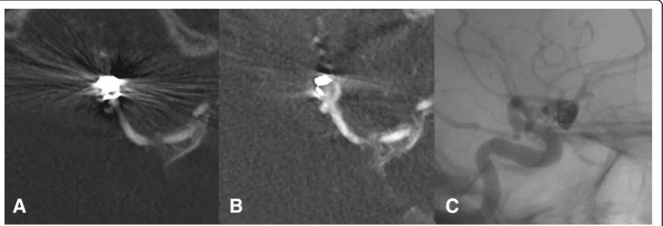
Fig. 4In imageaa statement concerning residual perfusion of the aneurysm was not possible due to streak artifacts and image hardening caused by the coil material. After application of MARA the residual perfusion could be detected as seen in imageb, congruent to the result of the digital subtraction angiography as seen in imagec
Conclusion
Application of the optimized MARA improved image analysis after endovascular and surgical therapy of aneu-rysms. In cases, where metal artifacts made it impossible to evaluate reperfusion at aneurysm base, application of MARA improved images and quality of evaluation. In 6 cases, reperfusion of aneurysms could be depicted only after application of MARA, corresponding to DSA im-aging. Extent of artifacts was reduced, whereas no sig-nificant information was lost i.e. concerning parent vessels or adjacent brain parenchyma as well as brain parenchyma in level of implant. FDCTA images added additional information, regarding implants located in other vessels, compared to DSA. Application of MARA leads to better evaluation of FDCTA images and can re-duce the necessity to examine patients via DSA in fol-low-up examinations after neurovascular procedures.
Abbreviations
CI:Confidence interval; DSA: Digital subtraction angiography; FDCT: Flat-detector CT; FDCTA: Flat–detector CT angiography; FOV: Field of view; MARA: Metal artifact reduction algorithm; MIP: Maximum intensity projection; MPR: Multiplanar reconstruction
Acknowledgements Not applicable.
Authors’contributions
The study was designed and conducted by NA. NA developed the study design, evaluated the images, processed the images on the workstation, applied MARA and processed the data statistically. The article was written by NA. VM read and corrected the article. DB and MNP evaluated the images. MNP supervised the study and helped with the study design. IEP evaluated the results and made the statistical analysis. JRL identified the patients. MK supervised the study and developed the study design.
Availability of data and materials
The datasets used and analysed during the current study are available from the corresponding author on reasonable request.
Ethics approval and consent to participate
According to the guidelines of the local ethics committees, ethic approval was given for the retrospective evaluation of patient data, which was conducted in accordance to the Declaration of Helsinki.
Consent for publication Not applicable
Competing interests
The department of neuroradiology of university clinic Goettingen has a research agreement with Siemens healthcare GmbH, Forchheim, Germany.
Author details
1Institute for Diagnostic and Interventional Neuroradiology, University Medicine Göttingen, Robert Koch Straße 40, 37075 Göttingen, Germany. 2
Institute for Diagnostic and Interventional Radiology, University Hospital of Jena, Am Klinikum 1, 07747 Jena, Germany.3Institute for Radiology, Südharz Klinikum Nordhausen, Dr. Robert Koch Straße 39, 99734 Nordhausen, Germany.
Received: 2 April 2019 Accepted: 23 June 2019
References
1. Stidd DA, Theessen H, Deng Y, et al. Evaluation of a metal artifacts reduction algorithm applied to postinterventional flat panel detector CT imaging. AJNR Am J Neuroradiol. 2014;35:2164–9.
2. Psychogios MN, Scholz B, Rohkohl C, et al. Impact of a new metal artefact reduction algorithm in the noninvasive follow-up of intracranial clips, coils, and stents with flat-panel angiographic CTA: initial results. Neuroradiology. 2013;55:813–8.
3. Prell D, Kyriakou T, Struffert T, et al. Metal artifact reduction for clipping and coiling in interventional C-arm CT. AJNR Am J Neuroradiol. 2010;31:634–9. 4. Mennecke A, Svergun S, Scholz B, et al. Evaluation of a metal artifact
reduction algorithm applied to post-interventional flat detector CT in comparison to pre-treatment CT in patients with acute subarachnoid haemorrhage. Eur Radiol. 2017;27:88–96.
5. Buhk JH, Groth M, Sehner S, et al. Application of a novel metal artifact correction algorithm in flat-panel CT after coil embolization of brain aneurysms: intraindividual comparison. Rofo. 2013;185:824–9. 6. Pjontek R, Onenkoprulu B, Scholz B, et al. Metal artifact reduction for flat
panel detector intravenous CT angiography in patients with intracranial metallic implants after endovascular and surgical treatment. J Neurointerv Surg. 2016;8:824–9.
7. Yuki I, Kambayashi Y, Ikemura A, et al. High-resolution C-arm CT and metal artifact reduction software: a novel imaging modality for analyzing aneurysms treated with stent-assisted coil embolization. AJNR Am J Neuroradiol. 2016;37:317–23.
Publisher’s Note