Open Access
Research
Fbw7/hCDC4 dimerization regulates its substrate interactions
Markus Welcker
1and Bruce E Clurman*
1,2,3Address: 1Division of Human Biology, Fred Hutchinson Cancer Research Center, 1100 Fairview Ave N, Seattle, Washington, 98109, USA, 2Clinical
Research Division, Fred Hutchinson Cancer Research Center, 1100 Fairview Ave N, Seattle, Washington, 98109, USA and 3Department of
Medicine, University of Washington School of Medicine, Seattle, Washington, 98104, USA
Email: Markus Welcker - [email protected]; Bruce E Clurman* - [email protected] * Corresponding author
Abstract
Background: The Fbw7 ubiquitin ligase promotes the rapid degradation of several important oncogenes, such as cyclin E, c-Myc, c-Jun, and Notch. The two fission yeast homologs of Fbw7, pop1 and pop2, have previously been shown to dimerize. In this study, we asked whether Fbw7 can also dimerize and how dimerization affects Fbw7 function.
Results: We found that Fbw7 binds efficiently to itself through a domain just upstream of its F-box. We further show that dimerization is essential for the stable interaction of Fbw7 with the cyclin E T380 phospho-degron. Surprisingly, the requirement for dimerization can be suppressed by an additional phosphorylation of this phospho-degron at the +4 position (S384), which creates a binding site with higher affinity for monomeric Fbw7.
Conclusion: Degradation of cyclin E by the Fbw7 pathway can, thus, be conditionally regulated either by Fbw7 dimerization or by hyperphosphorylation of the T380 phospho-degron. Other substrates, which cannot accommodate an extra phosphate in their phospho-degrons, or which don't provide a negatively charged amino acid in the +4 position, may be absolutely dependent on Fbw7 dimerization for their turnover. Our results point to an additional level of regulation for substrate interaction and turnover by Fbw7.
Background
Fbw7 is the mammalian homolog of budding yeast CDC4 and mediates the degradation of several proteins involved in cell growth and division, including cyclin E, Myc, c-Jun, Notch, Presenilin, and SREBP [1-10]. Fbw7 recog-nizes a phospho-epitope, termed CPD (for Cdc4 Phos-pho-Degron), contained within these substrates. Via its F-box, Fbw7 recruits the remainder of an SCF ubiquitin ligase complex, thus promoting substrate ubiquitination and rapid degradation by the proteasome [11]. Mamma-lian cells contain three Fbw7 isoforms (Fbw7α, Fbw7β, and Fbw7γ) that are produced by alternative splicing and
that localize to the nucleoplasm, cytoplasm, and nucleo-lus, respectively [12-14].
Numerous cancer-associated mutations have been identi-fied within CPDs of Fbw7 substrates that render them insensitive to Fbw7 regulation. Accordingly, the Fbw7 gene is deleted in a large number of tumors. Moreover, many somatic point mutations have been found that eliminate Fbw7's function either by terminating the pro-tein prematurely or disabling its substrate recognition domain, the C-terminal WD40 repeats [15].
Published: 13 February 2007
Cell Division 2007, 2:7 doi:10.1186/1747-1028-2-7
Received: 29 November 2006 Accepted: 13 February 2007
This article is available from: http://www.celldiv.com/content/2/1/7 © 2007 Welcker and Clurman; licensee BioMed Central Ltd.
Cell Division 2007, 2:7 http://www.celldiv.com/content/2/1/7
The eight WD40 repeats form a beta-propeller structure creating a phospho-epitope binding pocket that can rec-ognize phosphorylated CPDs [16]. All currently known mammalian CPDs consist of a central phosphorylated threonine immediately followed by proline (pT-P). For substrates like c-Myc, c-Jun, SREBP, and possibly cyclin E, a phosphorylated serine in the +4 position serves as prim-ing phosphate for GSK3 in order to phosphorylate the central threonine. However, beyond simply priming for GSK3, there appear to be additional requirements for this negative charge in the +4 position for substrate turnover by Fbw7.
The first evidence for this role of the +4 negative charge came from studies of cyclin E degradation. Cyclin E's turn-over by Fbw7 depends on the phosphorylation of T380, the central phospho-threonine [2,3,17,18]. We previously identified S384 as a second phosphorylation site that plays a key role in the formation of this CPD [19,20]. However, we noticed that degradation of cyclin E by either Fbw7β or Fbw7γ strictly required S384 phosphorylation, whereas the requirement for this phosphorylation by Fbw7α was depended on the stoichiometry of Fbw7α and cyclin E. That is, high Fbw7α abundance relative to cyclin E can overcome the need for S384 phosphorylation. Moreover, an S384 glutamate mutant (S384E) is degraded like wild-type cyclin E, emphasizing the importance of a negative charge rather than a (priming) phosphate in this position. Further evidence for a role of the +4 negative charge was provided by the interaction between Fbw7 and the SV40 Large T oncoprotein, which also binds to Fbw7 via a CPD and interferes with Fbw7 function [21]. Large T
contains a glutamate in the +4 position, and this residue is crucial for its interaction with Fbw7. In fact, almost every substrate of Fbw7 and its homologs potentially pro-vides a negative charge in the +4 position in form of either phosphoserine/threonine or a negatively charged gluta-mate (Table 1) [3-5,10,17,18,21-27]. Thus, the +4 nega-tive charge is highly conserved and likely plays a central role in substrate regulation. Ironically, one exception to this rule is the best-studied CDC4 substrate, Sic1, which contains a number of imperfect CPDs that interact allova-lently with CDC4 [16]. However, for CDC4 binding to CPDs, the -1 and -2 positions have also been shown to be important instead of +4, generating a slightly different consensus: I/L-I/L/P-pTP <K/R>4 where <K/R> are disfa-vored residues in positions +2 to +5 [11]. Although cyclin E's T380 degron completely conforms with this consensus we observed the additional requirement of S384 phos-phorylation for its turnover by Fbw7.
Because the fission yeast Fbw7 homologs, pop1 and pop2, have previously been shown to dimerize [28], we tested whether Fbw7 can also form dimers and how dimerization affects its functions. We identified a dimeri-zation domain in the region common to all Fbw7 iso-forms just upstream of the F-box. Fbw7 mutants that cannot dimerize are fully active toward cyclin E and c-Myc in turnover assays in cells, indicating that dimerization is not strictly required for Fbw7 function. However, we found that stable interactions between dimerization-defi-cient Fbw7 and cyclin E are largely impaired and that cyc-lin E turnover by monomeric Fbw7 completely depends on phosphorylated S384 – even by the Fbw7α isoform.
Table 1: Alignment of CPDs.
Conservation of the negative charge in the +4 position of CPDs
0 +4 references
cyclin E: T380 LLTPPQSGK mammalian (1–3)
T62 IPTPDKEDD (3)
c-Myc : T58 LPTPPLSPS (4,5)
c-Jun: T239 GETPPLSPI (22)
SREBP 1: T456* TLTPPPSDA (10)
SV40/Large T: T701 PPTPPPEPE (21)
Notch 1: T2512* FLTPSPESP
Presenilin 1: T116 IYTPFTEDT
Gcn4: T165 LPTPVLEDA yeast (23)
Far1 : S87 PISPPPSLK (24)
Cdc6: T39 DVTPESSPE (25)
T368 PLTPTTSPV (25)
Clb6: S6 IPSPISERK (26)
T39 NLTPHSTNE (26)
Rum1 : T58 PPTPAKTPK (27)
Our results link Fbw7 dimerization to the negative charge in a CPD's +4 position and point to an additional level of complexity in substrate turnover by Fbw7.
Results
Identification of an Fbw7 dimerization domain
In order to determine whether Fbw7 can form dimers, we generated Flag-tagged or Myc-tagged versions of Fbw7α. Both versions were transiently expressed in 293A cells and immunoprecipitates from cell lysates were analyzed for their interaction. The results demonstrated that Myc-tagged Fbw7α efficiently co-precipitated with Flag-tagged Fbw7α (Fig. 1A), while a truncated version of Fbw7 that only consists of the WD40 repeats did not interact with full length Fbw7α. Identical results were achieved in a reciprocal immunoprecipitation (anti Myc tag) from the same lysates (not shown). Therefore, like pop1 and pop2, Fbw7α can dimerize. In fact, dimerization may be a more common feature of F-box proteins, since β-TrCP has also been shown to form dimers [29].
We then attempted to define the region on Fbw7α neces-sary for dimerization. A panel of N-terminal truncation mutants was constructed and tested for interaction with full-length Fbw7α as above. This analysis revealed that a region common to all three Fbw7 isoforms (but lacking the isoform-specific N-terminus) was required to bind to full-length Fbw7 (Fig. 1B). However, the M73 truncation mutant (starting at methionine 73 of the common region) was incapable of binding to Fbw7α, suggesting that resi-dues upstream of M73 are essential for dimerization. Sub-strate interaction is not required for Fbw7 dimerization, since a cancer-associated Fbw7 point mutant that no longer recognizes CPDs, still bound to wild-type Fbw7α (Fig. 1B, last lane).
In order to more precisely define the dimerization domain, we generated several additional truncation mutants beginning just upstream of M73 and tested them in co-immunoprecipitation assays as above. This demon-strated that the M67 mutant (beginning at amino acid 67 of the common region) was still capable of binding to Fbw7α, suggesting that dimerization occurs via a domain immediately downstream of amino acid 67 (Fig. 1C). We next generated a panel of C-terminal truncation mutants to determine the extent of the dimerization domain. As expected from the data shown above truncating Fbw7α at amino acid 70 of the common region eliminated dimeri-zation with full-length Fbw7α (Fig. 2A). Termination at amino acid 80 also failed to enable dimerization. How-ever, terminating Fbw7α at amino acid 90 restored stable binding to full-length Fbw7α by about 50%, and was completely restored at amino acid 100 of the common region. Based on the analyses of the truncation mutants we then generated two adjacent deletion mutants in
full-length Fbw7α just downstream of amino acid 67 (∆69–72 and ∆74–78) and tested them for dimerization defects. Figure 2B clearly demonstrates that both mutants are severely impaired with regards to dimerization. The dimerization of an additional deletion mutant lacking amino acids 82 to 87 was also largely compromised (not shown). We conclude that Fbw7 dimerizes via an extended domain between amino acids 67 and at least 90 of the common region (see model, Fig. 2C).
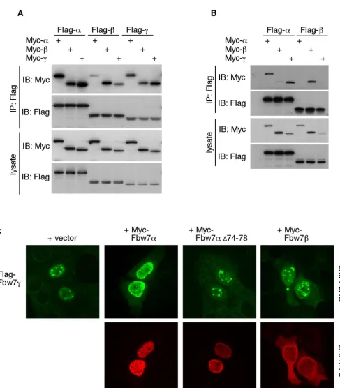
Because we mapped the dimerization domain to the com-mon region of Fbw7, we asked whether Fbw7β and Fbw7γ also homodimerize, and whether the different isoforms heterodimerize. Myc- and Flag-tagged versions of all three Fbw7 isoforms were analyzed for their interactions by co-immunoprecipitation. As expected both the Fbw7β and Fbw7γ isoforms could homodimerize (Fig. 3A). Interest-ingly, there was a clear preference for homo- over het-erodimerization, most likely reflecting the differing sub-cellular localization of each isoform. Furthermore, some isoforms were able to form heterodimers, at least when overexpressed in 293A cells. Since vast overexpression can lead to leaking of the Fbw7 isoforms into different subcel-lular compartments, we repeated this experiment using 5-fold lower amounts of plasmid DNA. This demonstrated a more pronounced tendency for homo- over het-erodimerization, in particular for cytoplasmic Fbw7β (Fig. 3B). However, nuclear Fbw7α and nucleolar Fbw7γ were still able to interact efficiently under these conditions.
Fbw7 dimerization in cells
In order to determine whether heterodimerization may be a post-lysis artifact or actually occurs in cells, we analyzed these interactions by immunofluorescence of fixed cells. U2OS cells were grown on coverslips and transfected with each individual Flag-tagged Fbw7 isoform either alone or together with Myc-tagged Fbw7 isoforms in every het-erodimeric combination. The localization of Flag-tagged Fbw7 was then assayed by immunofluorescence. As shown in figure 3C, co-expression of the nucleoplasmic Fbw7α protein caused the normally nucleolar Fbw7γ pro-tein to become mislocalized to the nucleoplasm suggest-ing that they interact in cells, and this was dependent on an intact dimerization domain of Fbw7α. In contrast, co-expression of cytoplasmic Fbw7β did not relocalize Fbw7γ. Consistent with the preference for homodimeriza-tion observed in figures 3A and 3B, Fbw7α and Fbw7β were largely unaffected by co-expressing any other iso-form (not shown).
co-Cell Division 2007, 2:7 http://www.celldiv.com/content/2/1/7
Mapping of the Fbw7 dimerization domain (1)
Figure 1
Mapping of the Fbw7 dimerization domain (2)
Figure 2
Cell Division 2007, 2:7 http://www.celldiv.com/content/2/1/7
Fbw7 isoforms interact in cells (1)
Figure 3
expression of Fbw7α relocalized both NLS mutants into the nucleoplasm in a dimerization-dependent fashion, clearly demonstrating their ability to interact with one another in situ (Fig. 4). These experiments were also per-formed in 293A cells with similar results (not shown). Taken together, all three Fbw7 isoforms can homo- and partially heterodimerize in intact cells, at least when expressed ectopically. This observation may have implica-tions for the general compartmentalization of the endog-enous Fbw7 isoforms (see discussion).
Dimerization is not strictly required for Fbw7 function
We next tested whether Fbw7 dimerization mutants are capable of promoting cyclin E and c-Myc turnover. Cyclin E and CDK2 were co-expressed with either wild-type Fbw7α or dimerization mutants in 293A cells and steady-state levels of cyclin E were analyzed by western blotting. As shown in figure 5A, both the non-dimerizing deletion mutant (∆74–78) and the M73 truncation mutant were able to degrade cyclin E similar to wild-type Fbw7. We did not observe any significant impairment of the dimeriza-tion mutant compared to wild-type Fbw7α, even upon titration of either cyclin E or Fbw7 (Fig. 5B and 5C). Dimerization-defective Fbw7 also promoted c-Myc turno-ver (Fig. 5D) and bound well to SV40 Large T (not shown and [21]). Therefore, at least in these types of assays, Fbw7 dimerization is not strictly required for substrate degrada-tion.
Conditional requirement for Fbw7 dimers
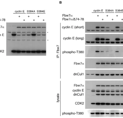
The requirement for S384 phosphorylation in cyclin E turnover by Fbw7α is conditional and depends upon Fbw7 stoichiometry [20]. We therefore tested whether S384 manipulation would affect cyclin E's binding and turnover by Fbw7 dimerization mutants. Wild-type cyclin E or S384 mutants changed to alanine or glutamate were expressed in 293A cells together with either Fbw7α or the dimerization mutant. Steady-state abundance of cyclin E was then analyzed by western blotting. Figure 6A demon-strates that wild-type cyclin E and the S384E mutant are both efficiently degraded by Fbw7α and by the dimeriza-tion mutant. As expected, Fbw7α was also able to elimi-nate the S384A mutant at this stoichiometry. However, the S384A mutant was completely resistant to non-dimer-izing Fbw7α (Fig. 6A), and this was independent of the stoichiometry between the S384A and Fbw7 mutants (not shown).
In addition to its potential priming role for GSK3, S384 phosphorylation participates in the formation of the T380 CPD and is likely to mediate direct contacts with Fbw7 [19]. We therefore directly tested the role of Fbw7 dimer-ization in binding to cyclin E and the S384 mutants by co-immunoprecipitation. In order to capture this transient interaction we co-expressed a dominant-negative version
of Cul1, an essential core component of SCF complexes, in an otherwise identical experiment as in figure 6A. This prevents cyclin E ubiquitination and degradation and allows stable binding of cyclin E to Fbw7 [20]. Lysates were immunoprecipitated against Fbw7α or the dimeriza-tion mutant and western blotted for co-immunoprecipi-tated cyclin E (Fig. 6B). The results confirmed that the stable interaction between the S384A mutant and Fbw7α is largely impaired compared with wild-type cyclin E. This is not simply due to decreased T380 phosphorylation of this mutant (see lysate). In fact, the extent of T380 phos-phorylation is comparable between the S384A and S384E mutants, yet only S384E is efficiently bound to Fbw7. This supports the notion that a negative charge in +4 directly contributes to Fbw7 binding.
Dimerization-deficient Fbw7α, on the other hand, was partly compromised in binding to wild-type cyclin E and completely lost its ability to interact with the S384A mutant (Fig. 6B), even on very long exposures (not shown). Surprisingly, S384E failed to restore efficient binding to the Fbw7α dimerization mutant despite the finding that the S384E mutant is degraded by monomeric Fbw7 (compare Figs. 6A and 6B). The reason for this dis-crepancy is unclear, but probably reflects differences in contact formations of phospho-serine versus glutamate with the WD40 repeats of Fbw7. The implications of our findings are summarized in figure 7.
Discussion
Cell Division 2007, 2:7 http://www.celldiv.com/content/2/1/7
Fbw7 isoforms interact in cells (2)
Figure 4
Monomeric Fbw7 is active
Figure 5
Cell Division 2007, 2:7 http://www.celldiv.com/content/2/1/7
For instance, the extent of heterodimerization between Fbw7α and Fbw7γ may regulate the amount of nucleolar Fbw7γ and, hence, the activities of nucleolar substrates.
Besides potentially affecting isoform localization, dimeri-zation may directly regulate other functional aspects of Fbw7. For instance, since the dimerization domain is adjacent to the F-box, we initially tested the idea that dimerization may prevent the interaction with Skp1 and, hence, inactivate Fbw7. However, we found no evidence
meric and dimerized Fbw7 (not shown). Instead, we uncovered a requirement of Fbw7 dimerization for the regulation of certain substrates. Our results establish a link between Fbw7 dimerization and the presence of a negative charge in the +4 position of cyclin E's T380-CPD. The implications of our findings suggest differential modes of action by Fbw7 and sub-divide Fbw7 targets into three groups. First are substrates that always provide the negative charge in +4 via glutamates, aspartates, or constitutive (priming) phosphorylation. This subgroup A role for Fbw7 dimerization
Figure 6
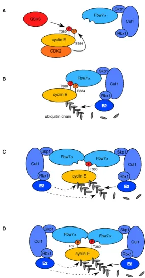
Model for role of Fbw7α dimerization
Figure 7
Cell Division 2007, 2:7 http://www.celldiv.com/content/2/1/7
ond are substrates that sometimes, perhaps conditionally, provide this charge and may switch their CPD affinity in response to environmental cues. Cyclin E is an example of this group and can be degraded independent of Fbw7 dimerization when S384 is phosphorylated, but becomes dependent on S384 phosphorylation for monomeric Fbw7 (see model in figure 7). Also c-Myc may belong to this subgroup, since it has been suggested that S62 became de-phosphorylated after having served as primer for T58 phosphorylation [30]. However, in our assays c-Myc regulation by Fbw7 is not dependent on Fbw7 dimers (Fig. 5D). Finally, some substrates may never provide this charge and therefore are completely dependent on Fbw7 dimerization for their degradation. Such CPDs probably mediate weaker interactions with Fbw7 and may entirely rely on cooperative effects together with other (weak) CPDs. The activities of these substrates may in part be con-trolled by the state of Fbw7 dimerization and are possibly subjected to an additional level of regulation, if dimeriza-tion itself was controlled.
Sic1 requires at least six phosphorylated CPDs for optimal destruction by CDC4, each of which are flawed by the absence of the +4 negative charge, the presence of non-favorable basic amino acids, or both [11,16]. Therefore, the low-affinity CPDs of Sic1, like cyclin E that is not phosphorylated on S384, may entirely depend on CDC4 dimerization for efficient turnover. Indeed, recent studies have shown that Sic1 degradation requires CDC4 dimeri-zation (Mike Tyers personal communication). Why low affinity CPDs require F-box protein dimerization remains unclear, but perhaps their affinity is so low that the recruitment of several SCF complexes is required for effi-cient substrate degradation. Dimerized Fbw7 (or CDC4) may simply be twice as efficient in substrate ubiquitina-tion, because two SCF complexes can recruit two E2 ubiq-uitin ligases, and this may be necessary for the turnover of low-affinity substrates.
Several mechanistic ideas arise from our observations. One possibility is that dimerized Fbw7 binds to two dif-ferent CPDs concurrently to increase substrate affinity and facilitate more efficient ubiquitination of its substrate. This model would be a plausible explanation to account for the more transient interactions of Fbw7/CDC4 with low-affinity CPDs and is depicted in figure 7D. Alterna-tively, a dimer may bind to a single CPD via one of the dimer moieties while the other one simply recruits a sec-ond E2 enzyme, perhaps for more efficient ubiquitination (Figure 7C). Moreover, a dimerized SCF might elongate the same ubiquitin chain, act on two different chains, or perhaps several dimers elongate one single ubiquitin chain when bound to different CPDs. All of these models may apply in a substrate-specific fashion or even for
dif-Conclusion
Our results suggest that Fbw7's affinity for certain sub-strates underlies yet another level of regulation that links the negatively charged +4 position of CPDs and Fbw7's ability to form dimers. We propose that weak CPDs that do not contain the + 4 charge must either cooperate with another (weak?) CPD or bring a second SCF to the same low-affinity CPD for efficient substrate destruction via Fbw7 dimerization.
Methods
Cell culture, transfections, plasmids
U2OS (human osteosarcoma) and HEK-293A (human embryonic kidney) cell lines were cultured under stand-ard conditions in DMEM containing 10% FCS. For tran-sient transfections experiments, cells were seeded into 6 cm dishes and transfected the next day by Ca-precipitation overnight at 30 to 40% confluence. 24 hrs after washing and replacing the media cells were harvested either for protein extracts or immunostaining. Typically, we express 1/10 of the plasmid amount of pFlag-Fbw7α (0.3 µg) compared to Fbw7β and Fbw7γ (3 µg each). The in vivo
Fbw7-driven cyclin E turnover assay was performed as described [19]. All point mutants and deletions were introduced by the Quick Change method (Stratagene) and confirmed by sequencing. The deletions ∆69–72 and ∆74–78 correspond to amino acids EWLK and FQSWS, respectively. Truncation mutants were generated by PCR and sequenced. All amino acid numbers indicate their positions within the common region of Fbw7.
Immunofluorescence, immunoprecipitation, immunoblotting
For immunofluorescence, cells were seeded and trans-fected on glass coverslips. Slips were fixed with ice-cold Methanol/Acetone (1:1) for 5 min, air-dried and immu-nostained with primary antibody. After washing with PBS, slips were incubated with secondary FITC-coupled anti-mouse antibody, washed, dried and mounted. For double stainings mouse Flag-tag and rabbit Myc-tag antibodies were mixed and visualized with FITC- and dopamine-cou-pled secondary antibodies. All protein extracts were made with Tween 20 lysis buffer [19]. Protein expression was determined by standard procedures (SDS-PAGE, western blot, immunoblot). Protein complexes were analyzed by immunoprecipitation. The following primary antibodies were used in this study: anti-Flag for Fbw7 (Sigma, M2), anti-HA for CDK2 and dnCul1 (12CA-5), anti-c-Myc (Santa Cruz, sc-N262), anti-Myc-tag for cyclin E and Fbw7 (9E-10 and Cell Signaling #2278), anti-pT380 for phos-pho-cyclin E (Santa Cruz, sc-12917-R).
Competing interests
inter-Publish with BioMed Central and every scientist can read your work free of charge
"BioMed Central will be the most significant development for disseminating the results of biomedical researc h in our lifetime."
Sir Paul Nurse, Cancer Research UK
Your research papers will be:
available free of charge to the entire biomedical community
peer reviewed and published immediately upon acceptance
cited in PubMed and archived on PubMed Central
yours — you keep the copyright
Submit your manuscript here:
http://www.biomedcentral.com/info/publishing_adv.asp
BioMedcentral
Authors' contributions
MW performed all experiments and wrote this manu-script. BEC edited the manumanu-script. Both authors have approved the final version of this manuscript.
Note added in proof
While this manuscript was under revision a similar study was published by Zhang and Koepp (Mol Cancer Res December 2006).
Acknowledgements
This work was supported by grants from the National Cancer Institute (NCI) RO1CA84069 and NCI 1 RO1CA102742-01 (B.E.C.) and the Leuke-mia and Lymphoma Society (M.W.). We like to thank Mike Tyers and Wade Harper for prepublication information, and Steve Reed for 293A cells.
References
1. Moberg KH, Bell DW, Wahrer DC, Haber DA, Hariharan IK: Archi-pelago regulates Cyclin E levels in Drosophila and is mutated in human cancer cell lines. Nature 2001, 413(6853):311-316. 2. Koepp DM, Schaefer LK, Ye X, Keyomarsi K, Chu C, Harper JW,
Elledge SJ: Phosphorylation-dependent ubiquitination of cyclin E by the SCFFbw7 ubiquitin ligase. Science 2001, 294(5540):173-177.
3. Strohmaier H, Spruck CH, Kaiser P, Won KA, Sangfelt O, Reed SI: Human F-box protein hCdc4 targets cyclin E for proteolysis and is mutated in a breast cancer cell line. Nature 2001, 413(6853):316-322.
4. Welcker M, Orian A, Jin J, Grim JE, Harper JW, Eisenman RN, Clur-man BE: The Fbw7 tumor suppressor regulates glycogen syn-thase kinase 3 phosphorylation-dependent c-Myc protein degradation. Proc Natl Acad Sci U S A 2004, 101(24):9085-9090. 5. Yada M, Hatakeyama S, Kamura T, Nishiyama M, Tsunematsu R, Imaki
H, Ishida N, Okumura F, Nakayama K, Nakayama KI: Phosphoryla-tion-dependent degradation of c-Myc is mediated by the F-box protein Fbw7. Embo J 2004, 23(10):2116-2125.
6. Moberg KH, Mukherjee A, Veraksa A, Artavanis-Tsakonas S, Hariha-ran IK: The Drosophila F box protein archipelago regulates dMyc protein levels in vivo. Curr Biol 2004, 14(11):965-974. 7. Nateri AS, Riera-Sans L, Da Costa C, Behrens A: The ubiquitin
ligase SCFFbw7 antagonizes apoptotic JNK signaling. Science
2004, 303(5662):1374-1378.
8. Hubbard EJ, Wu G, Kitajewski J, Greenwald I: sel-10, a negative regulator of lin-12 activity in Caenorhabditis elegans, encodes a member of the CDC4 family of proteins. Genes Dev
1997, 11(23):3182-3193.
9. Wu G, Hubbard EJ, Kitajewski JK, Greenwald I: Evidence for func-tional and physical association between Caenorhabditis ele-gans SEL-10, a Cdc4p-related protein, and SEL-12 presenilin.
Proc Natl Acad Sci U S A 1998, 95(26):15787-15791.
10. Sundqvist A, Bengoechea-Alonso MT, Ye X, Lukiyanchuk V, Jin J, Harper JW, Ericsson J: Control of lipid metabolism by phospho-rylation-dependent degradation of the SREBP family of tran-scription factors by SCF(Fbw7). Cell Metab 2005, 1(6):379-391. 11. Nash P, Tang X, Orlicky S, Chen Q, Gertler FB, Mendenhall MD, Sicheri F, Pawson T, Tyers M: Multisite phosphorylation of a CDK inhibitor sets a threshold for the onset of DNA replica-tion. Nature 2001, 414(6863):514-521.
12. Kimura T, Gotoh M, Nakamura Y, Arakawa H: hCDC4b, a regula-tor of cyclin E, as a direct transcriptional target of p53. Cancer Sci 2003, 94(5):431-436.
13. Welcker M, Orian A, Grim JE, Eisenman RN, Clurman BE: A nucle-olar isoform of the Fbw7 ubiquitin ligase regulates c-Myc and cell size. Curr Biol 2004, 14(20):1852-1857.
14. Spruck CH, Strohmaier H, Sangfelt O, Muller HM, Hubalek M, Muller-Holzner E, Marth C, Widschwendter M, Reed SI: hCDC4 gene mutations in endometrial cancer. Cancer Res 2002, 62(16):4535-4539.
15. Minella AC, Clurman BE: Mechanisms of tumor suppression by the SCF(Fbw7). Cell Cycle 2005, 4(10):1356-1359.
16. Orlicky S, Tang X, Willems A, Tyers M, Sicheri F: Structural basis for phosphodependent substrate selection and orientation by the SCFCdc4 ubiquitin ligase. Cell 2003, 112(2):243-256. 17. Won KA, Reed SI: Activation of cyclin E/CDK2 is coupled to
site-specific autophosphorylation and ubiquitin-dependent degradation of cyclin E. Embo J 1996, 15(16):4182-4193. 18. Clurman BE, Sheaff RJ, Thress K, Groudine M, Roberts JM: Turnover
of cyclin E by the ubiquitin-proteasome pathway is regulated by cdk2 binding and cyclin phosphorylation. Genes Dev 1996, 10(16):1979-1990.
19. Welcker M, Singer J, Loeb KR, Grim J, Bloecher A, Gurien-West M, Clurman BE, Roberts JM: Multisite phosphorylation by Cdk2 and GSK3 controls cyclin E degradation. Mol Cell 2003, 12(2):381-392.
20. Ye X, Nalepa G, Welcker M, Kessler BM, Spooner E, Qin J, Elledge SJ, Clurman BE, Harper JW: Recognition of phosphodegron motifs in human cyclin E by the SCF(Fbw7) ubiquitin ligase. J Biol Chem 2004, 279(48):50110-50119.
21. Welcker M, Clurman BE: The SV40 large T antigen contains a decoy phosphodegron that mediates its interactions with Fbw7/hCdc4. J Biol Chem 2005, 280(9):7654-7658.
22. Wei W, Jin J, Schlisio S, Harper JW, Kaelin WG Jr.: The v-Jun point mutation allows c-Jun to escape GSK3-dependent recogni-tion and destrucrecogni-tion by the Fbw7 ubiquitin ligase. Cancer Cell
2005, 8(1):25-33.
23. Meimoun A, Holtzman T, Weissman Z, McBride HJ, Stillman DJ, Fink GR, Kornitzer D: Degradation of the transcription factor Gcn4 requires the kinase Pho85 and the SCF(CDC4) ubiquitin-ligase complex. Mol Biol Cell 2000, 11(3):915-927.
24. Henchoz S, Chi Y, Catarin B, Herskowitz I, Deshaies RJ, Peter M: Phosphorylation- and ubiquitin-dependent degradation of the cyclin-dependent kinase inhibitor Far1p in budding yeast.
Genes Dev 1997, 11(22):3046-3060.
25. Perkins G, Drury LS, Diffley JF: Separate SCF(CDC4) recogni-tion elements target Cdc6 for proteolysis in S phase and mitosis. Embo J 2001, 20(17):4836-4845.
26. Jackson LP, Reed SI, Haase SB: Distinct mechanisms control the stability of the related S-phase cyclins Clb5 and Clb6. Mol Cell Biol 2006, 26(6):2456-2466.
27. Benito J, Martin-Castellanos C, Moreno S: Regulation of the G1 phase of the cell cycle by periodic stabilization and degrada-tion of the p25rum1 CDK inhibitor. Embo J 1998, 17(2):482-497.
28. Kominami K, Ochotorena I, Toda T: Two F-box/WD-repeat pro-teins Pop1 and Pop2 form hetero- and homo-complexes together with cullin-1 in the fission yeast SCF (Skp1-Cullin-1-F-box) ubiquitin ligase. Genes Cells 1998, 3(11):721-735. 29. Suzuki H, Chiba T, Suzuki T, Fujita T, Ikenoue T, Omata M, Furuichi
K, Shikama H, Tanaka K: Homodimer of two F-box proteins betaTrCP1 or betaTrCP2 binds to IkappaBalpha for signal-dependent ubiquitination. J Biol Chem 2000, 275(4):2877-2884. 30. Yeh E, Cunningham M, Arnold H, Chasse D, Monteith T, Ivaldi G,