R E S E A R C H
Open Access
Impact of simulation-based training in
surgical chest tube insertion on a model
of traumatic pneumothorax
Alexandre Léger
1, Aiham Ghazali
2,3, Franck Petitpas
2,4, Youcef Guéchi
5, Amélie Boureau-Voultoury
6and Denis Oriot
2,6*Abstract
Background:Chest tube insertion is required for most cases of traumatic pneumothorax. However, this procedure entails risks of potentially life-threatening complications. A“surgical”approach is widely recommended to minimize these risks. Simulation-based education has previously been used in surgical chest tube insertion, but not been subjected to rigorous evaluation.
Methods:The primary objective was to evaluate the success rate of surgical chest tube insertion in a task trainer (previously published). Secondary objectives were to assess performance with a performance assessment scale (previously designed), to measure the time of insertion, and to seek out a correlation between the learner’s status, experience, and performance and success rate. Participants were surveyed for realism of the model and satisfaction; 65 participants (18 residents, 47 senior physicians) were randomized into SIM+ or SIM−groups. Both groups received didactic lessons. The SIM+ group was assigned deliberate practice on the model under supervision. Both groups were assessed on the model 1 month later.
Results:There was no difference between the SIM+ (n= 34) and SIM−(n= 31) groups regarding status (p= 0.44) or previous surgical insertion (p= 0.12). Success rate was 97 % (SIM+) and 58 % (SIM−),p= 0.0002. Performance score was 16.29 ± 1.82 (SIM+) and 11.39 ± 3.67 (SIM−),p= 3.13 × 10⁻⁸. SIM+ presented shorter dissection time than SIM−(p= 0.047), but procedure time was similar (p= 0.71). Status or experience was not correlated with success rate, performance score, procedure time, or dissection time. SIM+ gained more self-confidence, judged the model more realistic, and were more satisfied than SIM−.
Conclusions:Simulation-based education significantly improved the success rate and performance of surgical chest tube insertion on a traumatic pneumothorax model.
Keywords:Simulation-based education, Trauma, Pneumothorax, Chest tube, Assessment, Success rate, Performance, Task trainer
Background
Insertion of a chest tube is a mandatory procedure for treatment of a significant traumatic pneumothorax [1, 2]. However, this procedure remains somewhat difficult, stressful, and at risk of potentially life-threatening complications [3–10]. Numerous physicians acknow-ledge their lack of training and feel uncomfortable carrying out a chest tube insertion, particularly in a
young patient [11, 12]. Furthermore, many physicians may have difficulty supervising novices while inserting a chest tube with a surgical approach [13]. For three decades, simulation-based education has been developed, allowing learners to gain competence and develop their skills through performance in a safe environment, prior to practice in a clinical setting [14–20]. Nevertheless, no study to date has reported a rigorous evaluation of the benefit of simulation on surgical approach for chest tube insertion.
The aim of this study was to evaluate the benefit of simulation-based education on the performance and * Correspondence:[email protected]
2Emergency Department, Pitié-Salpétrière University Hospital, Paris, France 6Pediatric Emergency Department, University Hospital, 2 rue de la Milétrie,
86000 Poitiers, France
Full list of author information is available at the end of the article
success rate of surgical insertion of a chest tube on a model of traumatic pneumothorax.
Methods
Study
This was a prospective, randomized, controlled, bicentric study, carried out from May 2013 to October 2013 in the Simulation Laboratory of the Faculty of Medicine of Poitiers and in the Medical Center of Cayenne, French Guyana, after approval by the Institutional Research Board (Scientific Committee of the Faculty of Medicine of Poitiers, file number 11–37). All participants signed an informed consent form. Results were kept anonymous.
Objectives
The primary objective was to evaluate the success rate of surgical insertion of a chest tube in a task trainer simulator of traumatic pneumothorax.
The secondary objectives were (1) to assess the performance of insertion procedure according to a performance assessment scale, (2) to measure the global procedure time and dissection time during insertion, (3) to seek out a correlation between the learner’s status, experience, and performance and success rate, and (4) to survey the learners for their evaluation of the realism of the model, self-confidence, and satisfaction after simula-tion experience.
Population
Sixty-five healthcare providers (18 residents and 47 senior emergency physicians), representing two groups of registered participants (Poitiers and Cayenne) for the pediatric emergency procedure course at the University of Poitiers (carried out in Poitiers and in Cayenne), were given a chance to participate.
Intervention
Since the surgical approach is rarely taught and practiced in France, all participants received a 1-h academic lesson, prior to the simulation session, on current international recommendations for surgical chest tube insertion for traumatic pneumothorax [1, 21–24]. This didactic lesson was performed by three Advanced Trauma Life Support (ATLS)-certified supervisors. The session consisted in a
presentation of the currently recommended safest
approach to chest tube insertion in case of traumatic pneumothorax: landmarks on the medio-axillary line, in the fourth or fifth intercostal space [21, 22, 25], use of a tube without chuck or handle, dissection of chest wall muscular layers with a Kelly clamp, followed by insertion of a gloved finger probing into the chest cavity to confirm pleural placement, and strip any adhesions facing the insertion site [21]. In small children, it is recommended to tunnel the chest tube by a skin incision at the underlying
intercostal space. This provides a subcutaneous path for securing the tube and avoids its falling out due to a thin chest wall [22].
Comparison
Randomization was based on a list of 65 random
numbers—even numbers for SIM+ and odd ones for
SIM− group allocation. The SIM+ group participants (n= 34) were assigned for deliberate practice on the simulator just after the didactic lesson, whereas the SIM− group participants (n= 31) were not assigned any simulation practice. The SIM+ group participants had the opportunity to practice several times (mean = four times) on the simulator with immediate feedback from the supervisor. The SIM− group participants were exposed 5 min to the simulator and its environment (webcam, laptop, chest tube equipment) but did not perform chest tube insertions. One month later, all participants (SIM+ and SIM−) were evaluated during an assessment session on the simulator.
Every participant received a questionnaire on clinical experience in chest tube insertion—especially surgical approach—before the didactic lesson and during the month that followed prior to the assessment on the simulator. We arbitrarily distinguished experienced participants who had inserted at least five chest tubes (with a surgical approach) during the last 5 years from novices who had inserted less than five or none during the same span time.
At the end of the study, all participants were offered a chance for deliberate practice on the simulator with supervision.
Outcomes
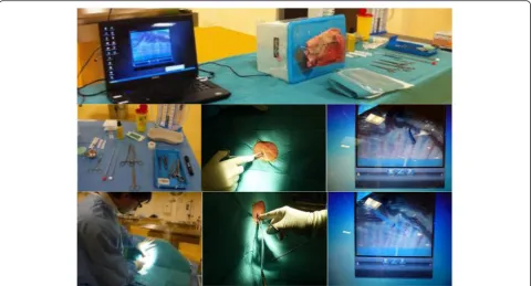
The scenario consisted in an emergency surgical chest tube insertion for a traumatic pneumothorax in a teenager. The model was the one we had previously de-veloped and published [26]. Briefly, the model assembled a lamb half chest tightly fixed on a box cover (Fig. 1). A spread-out two-layer plastic film simulated the pleural membranes. Inside the box, a webcam connected to a laptop made it possible to assess the intrathoracic steps of the procedure. All the required disposable equipment was set on a table: a 24-Fr-wide Joly drain (diameter 5.4 mm, length 40 cm, with two lateral holes, without chuck or handle) (Fig. 1). The landmark of the fourth rib was indicated to the participant. All simulation sessions were videotaped.
Assessment of performance was carried out by an independent observer expert in surgical approach for chest tube insertion (ATLS-certified or ATLS instructor), who was neither a supervisor nor a research investigator. He was unaware of the breakdown into two groups. Four observers participated in the assessment (two emergency physicians, one intensivist, and one pediatric intensivist). All were trained for surgical chest tube insertion assessment according to a specific scale that we had previously designed and validated [27]. It included eight steps, detailing the procedure: asepsis, local anesthesia, cutaneous incision and dissection of the thoracic wall, confirmation of insertion, introduction of the chest tube with a Kelly clamp, securing the water seal tubing, securing the chest tube, and location of the incision site (which was examined by the observer after the procedure had been completed). Each item was ranked 1 (correctly performed) or 0 (not done or incorrectly performed). The maximum total score was over 20 points. This scale had acceptable internal coherence (Cronbach’s alpha coefficient = 0.747) and high inter-observer reproducibility (intra-class correl-ation coefficient = 0.966). Because of the webcam connected to a laptop inside the model, the observer could assess the performance of the extra- and intra-thoracic steps of the procedure.
Timing of the different steps of the procedure was recorded. A stopwatch was started at the beginning of the procedure (t0) to record different times: beginning of the cutaneous incision (t1), chest tube passing through the skin (t2), connection of chest tube to water seal tubing (t3), and end of the procedure after securing the chest tube on the skin (t4). Dissection time was consequently defined ast2−t1.
After each chest tube insertion, a debriefing was performed by the observer, and all the observers were trained in debriefing by good judgment [28]. Videotape replay during debriefing could render participants aware of their gaps in performance [29].
After each assessment, the participant was asked to fill out a questionnaire on realism of the model, self-confidence, and overall satisfaction with simulation-based education using 10-point Likert scales.
Statistics
Analysis was carried out on Biostat TGV software and Excel 2010. Descriptive analysis included percentage, mean, and standard deviation (SD) of every variable. Comparative analysis used paired Student’s t test, with an ANOVA for repeated measures when necessary. Correlation between training and performance or success rate used Spearman’s test. Correlation between success
and performance used Pearson’s test. Ap value of <0.05 was considered as significant.
Results
Population studied
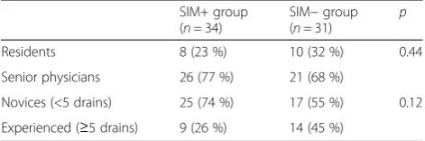
Among the 65 included participants, 47 were senior physicians (72.3 %) and 18 were residents (27.7 %). Twenty-three participants (only 49 % of senior physicians) were considered as experienced as they had previously surgically inserted more than five chest tubes during the previous 2 years. Thirty-four participants were random-ized in the SIM+ group and 31 in the SIM− group, independently from their status and experience in chest tube insertion. There were no differences between the groups regarding status (p= 0.44) or previous experience in surgical chest tube insertion (p= 0.12) (Table 1). None of the participants had inserted chest tubes during the month having elapsed between the didactic lesson and the assessment.
Primary objective
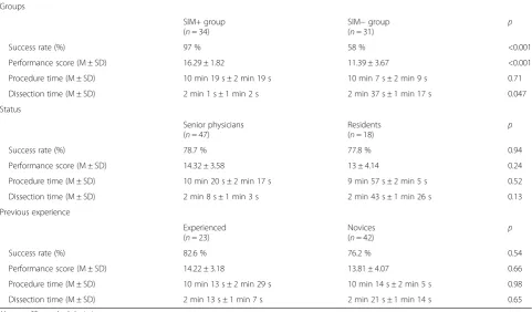
Mean success rate for surgical chest tube insertion was 78.5 % for the whole population. It was 97 % (33 drains/34) in the SIM+ group vs. 58 % (18 drains/31) in the SIM− group, p< 0.001 (Table 2).
Secondary objectives
The mean performance assessment score was 13.95 ± 3.76 for the whole population. It was significantly higher in the SIM+ group (16.29 ± 1.82) than in the SIM− group (11.39 ± 3.67), p< 0.001 (Table 2). Details of scores per item of the scale showed that the typically surgical steps of the procedure were those with a significant difference between groups, i.e., location of the insertion site, incision and dissection, confirm-ation of locconfirm-ation by probing a gloved finger into the pleural space, introduction of the chest tube with a Kelly clamp, and securing the tubing and chest tube (Table 3). Success was found to be correlated to perform-ance with Pearson’s correlation coefficient of 0.85.
There was no difference in the procedure time between SIM+ and SIM−, p= 0.71. By contrast, the SIM+ group had a shorter dissection time than the SIM− group, p= 0.047. There were no correlations
between status and procedure time (p= 0.52) or
dissection time (p= 0.16), nor between a participant’s previous experience and procedure time (p= 0.98) or dissection time (p= 0.65) (Table 2). Status was not correlated with either performance score (p= 0.24) or success rate (p= 0.94).
There was no difference between experienced partici-pants and novices regarding performance score (p= 0.66) or success rate (p= 0.54) (Table 2).
The SIM+ group participants gained more self-confidence than the SIM−ones (p= 0.036). The former judged the surgical chest tube insertion model with more realism (p= 0.0039) and were more satisfied with the simulation experience than the SIM− participants (8.21 ± 0.88 vs. 7.71 ± 0.74,p= 0.016) (Table 4).
Discussion
Main results
On a traumatic pneumothorax model, simulation-based education was associated with higher success rate and performance score. These results were directly due to the simulation training, with no influence of status or previous clinical experience in surgical chest tube inser-tion. To our knowledge, this is the first study reporting the benefit of simulation-based education on success rate and performance for a surgical chest tube insertion.
Limitations
This study nevertheless presented some limitations. All participants were registered for the pediatric emergency procedure university course, which could have repre-sented a confounding factor. The SIM− participants were exposed to the model only 5 min prior to the evaluation. Therefore, on evaluation day, they might have been surprised and experienced a lack in confi-dence, higher stress, and poorer performance due to ignorance of how the model would respond to their approach [17]. Nevertheless, the fact that assessment was performed 1 month after initial didactic and training reduced this déjà-vu phenomenon in the SIM+ group and might have counterbalanced the lack of familiarity with the task trainer of the SIM−group.
Discussion about the primary objective
The results of the present study on the benefit of simulation-based education for surgical chest tube insertion are similar to those reporting the increase of success rate for non-surgical chest tube insertion after simulation training [15–17, 19]. One distinctive feature of the present study is to have reported that simulation increased not only the success rate but also the perform-ance assessment score [27]. This approach could reveal that, as expected, the specific“surgical”steps of the pro-cedure were those that benefit the most from simulation
Table 1Comparison of SIM+ and SIM−groups according to
status and previous clinical experience of participants
SIM+ group (n= 34)
SIM−group
(n= 31) p
Residents 8 (23 %) 10 (32 %) 0.44
Senior physicians 26 (77 %) 21 (68 %)
Novices (<5 drains) 25 (74 %) 17 (55 %) 0.12
training. In fact, there was a high correlation be-tween success and performance score. During the validation process of the performance assessment scale, a score ≥14 was always found to be associated with a functional chest tube [27].
Discussion about secondary objectives
Simulation-based education was not associated with a decrease in procedure time for surgical chest tube insertion, as one would have expected. However,
dissection time—the crucial step of the surgical pro-cedure—significantly decreased in the SIM+ group. This implied that failure of surgical chest tube inser-tion was associated with poor mastery of this major step of the procedure [20–22].
Surprisingly, there was no effect of status or previ-ous experience on the performance or success rate of surgical chest tube insertion [5, 30]. This fact might be related to the rarity of this technique (surgical approach) in France and a consequent need for adequate training, according to the recommendations [18]. Besides that, it underlines the fact that only simulation-based training was responsible for the gain in performance and success rate.
Compared with SIM−participants, SIM+ group partic-ipants judged the training model more realistic (with more anatomic features), probably because they had
Table 2Comparison of success rate, performance score, procedure time, and dissection time according to group allocation, status,
and previous clinical experience Groups
SIM+ group (n= 34)
SIM−group (n= 31)
p
Success rate (%) 97 % 58 % <0.001
Performance score (M ± SD) 16.29 ± 1.82 11.39 ± 3.67 <0.001
Procedure time (M ± SD) 10 min 19 s ± 2 min 19 s 10 min 7 s ± 2 min 9 s 0.71
Dissection time (M ± SD) 2 min 1 s ± 1 min 2 s 2 min 37 s ± 1 min 17 s 0.047
Status
Senior physicians (n= 47)
Residents
(n= 18) p
Success rate (%) 78.7 % 77.8 % 0.94
Performance score (M ± SD) 14.32 ± 3.58 13 ± 4.14 0.24
Procedure time (M ± SD) 10 min 20 s ± 2 min 17 s 9 min 57 s ± 2 min 5 s 0.52
Dissection time (M ± SD) 2 min 8 s ± 1 min 3 s 2 min 43 s ± 1 min 26 s 0.13
Previous experience
Experienced (n= 23)
Novices (n= 42)
p
Success rate (%) 82.6 % 76.2 % 0.54
Performance score (M ± SD) 14.22 ± 3.18 13.81 ± 4.07 0.66
Procedure time (M ± SD) 10 min 13 s ± 2 min 29 s 10 min 14 s ± 2 min 5 s 0.98
Dissection time (M ± SD) 2 min 13 s ± 1 min 7 s 2 min 21 s ± 1 min 14 s 0.65
Mmean,SDstandard deviation
Table 3Details of the scores (mean ± standard deviation)
obtained at the different steps of the performance assessment scale for the SIM+ and SIM−groups
Steps of the scale (maximum score) SIM+
n= 34
SIM−
n= 31
p
Antiseptic procedure (3) 2.61 ± 0.65 2.36 ± 0.66 0.11
Location of incision site (1) 0.94 ± 0.23 0.69 ± 0.47 0.005
Local anesthesia (1) 0.88 ± 0.33 0.85 ± 0.37 0.61
Incision and dissection (6) 4.41 ± 0.70 2.43 ± 1.31 <0.001
Confirmation of location (2) 1.62 ± 0.69 0.98 ± 0.91 0.001
Introduction of chest tube with a Kelly clamp (4)
3.28 ± 1.03 2.46 ± 1.09 0.002
Securing water seal tubing (1) 0.81 ± 0.38 0.23 ± 0.42 <0.001
Securing chest tube (2) 1.74 ± 0.44 1.39 ± 0.61 0.01
Total (20) 16.29 ± 1.82 11.39 ± 3.67 <0.001
Table 4Comparison of the assessment of realism of the model,
gain in self-confidence, and global satisfaction according to the group (on a 0–10 scale). Mean ± standard deviation
SIM+ group (n= 34)
SIM−group
(n= 31) p
Realism of the model 7.65 ± 1.01 6.97 ± 0.83 0.0039
Gain in self-confidence 7.94 ± 0.69 7.55 ± 0.93 0.0364
been trained on it repeatedly a month earlier. Similarly, there was a higher gain in self-confidence in the SIM+ group. But these differences were minimal. Indeed, self-confidence is a major issue in the performance of a stressful procedure or when the practitioner is uncom-fortable with the procedure because of his/her scarce experience [11, 12]. The satisfaction rate was high in both groups—more pronouncedly in SIM+—thereby implying that participants are particularly receptive and attracted to simulation-based education as a means of improving technical skills [14, 26, 30, 31].
External validity
We think that simulation-based education for surgi-cal chest tube insertion can be spread to other groups of learners with the same results, in initial learning (residents) as well as continuous education (emergency physicians, intensivists) [5, 6, 13–15]. We observed that one simulation attempt did not suffice to achieve 100 % success for the whole group. This observation underscores the great interest of repeated simulation training for low-volume/high-stake procedures [20–22]. Finally, the assessment session was scheduled 1 month after the initial di-dactic session in both groups and simulation training in the SIM+ group. This delay might have been short as regards the occurrence of clinical surgical chest tube insertion (in our occupational field) and performance scores may have been better in simula-tion than in reality [16, 20, 30, 32]. Future studies should investigate if practicing on a task trainer improves the performance of the technique when performed on human subjects.
Conclusions
Simulation-based education significantly improved the success rate and performance of surgical chest tube insertion on a traumatic pneumothorax model. This benefit was explained by a better mastery of the chest wall dissection step. It was accompanied by an in-crease in self-confidence contributing to performance and success. Such training appears well suited to an infrequent, difficult procedure, responsible for poten-tial severe complications if poorly performed.
Future studies should investigate the frequency of repetition of simulation sessions as a way of sustaining the benefit of simulation-based education.
Acknowledgements
We are grateful to our colleague—Dr Paul Contal—who consented for the publication of his picture while using our model for chest tube insertion (Fig. 1). Furthermore, we would like to thank Jeffrey Arsham, an American medical translator, for having reviewed the English-language text.
Authors’contributions
DO is responsible for the content of the manuscript, including the data analysis. DO had the idea of the project and supervised the whole process of research and writing. AL and AG contributed substantially to the study design, analyzed the literature, and wrote the text. AG supervised the data management. FP, YG, and ABV contributed substantially to the assessment phase. All authors read and approved the final manuscript.
Competing interests
The authors declare that they have no competing interests.
Author details
1Pediatric Department, Basse-Terre Medical Center, Guadeloupe, France. 2
Emergency Department, Pitié-Salpétrière University Hospital, Paris, France.
3Simulation Laboratory, Faculty of Medicine, University of Poitiers, Poitiers,
France.4Surgical Intensive Care Unit, University Hospital, Poitiers, France.
5Emergency Department, University Hospital, Poitiers, France.6Pediatric
Emergency Department, University Hospital, 2 rue de la Milétrie, 86000 Poitiers, France.
Received: 8 February 2016 Accepted: 31 May 2016
References
1. Alrahbi R, Easton R, Bendinelli C, Enninghorst N, Sisak K, Balogh ZJ. Intercostal catheter insertion: are we really doing well? ANZ J Surg. 2012;82:392–4. 2. Cullinane DC, Morris JA JR, Bass JG, Rutherford EJ. Needle thoracostomy
may not be indicated in the trauma patient. Injury. 2001;32:749–52. 3. Millikan JS, Moore EE, Ateiner E, Aragon GE, Van Way III CW. Complications
of tube thoracostomy for acute trauma. Am J Surg. 1980;140:738–41. 4. Bailey R. Complications of tube thoracostomy in trauma. J Accid Emerg
Med. 2000;17:111–4.
5. Sethuraman KN, Duong D, Mehta S, Director T, Crawford D, ST George J, et al. Complications of tube thoracostomy placement in the emergency department. J Emerg Med. 2011;40:14–20.
6. Chan I, Reilly KM, Henderson C, Kahn F, Salluzzo RF. Complication rates of tube thoracostomy. Am J Emerg Med. 1997;15:368–70.
7. Meisel S, Ram Z, Priel I, Nass D, Lieberman P. Another complication of thoracostomy—perforation of the right atrium. Chest. 1990;98:772–3. 8. Fraser RS. Lung perforation complicating tube thoracostomy: pathologic
description of three cases. Hum Pathol. 1988;19:518–23.
9. Osinowo O, Softah A, Eid Zahrani M. Ectopic chest tube insertions: diagnosis and strategies for prevention. Afr J Med Med Sci. 2002;31:67–70.
10. Remerand F, Luce V, Badachi Y, Lu Q, Bouhemad B, Rouby JJ. Incidence of chest tube malposition in the critically ill: a prospective computed tomography study. Anesthesiology. 2007;106:1112–9.
11. Elsayed H, Rebecca R, Emadi M, Whittle I, Shackcloth M. Chest drain insertion is not a harmless procedure—are we doing it safely? Interact Cardiovasc Thorac Surg. 2010;11:745–8.
12. Maritz D, Wallis L, Hardcastle T. Complications of tube thoracostomy for chest trauma. South Afr med J. 2009;99:114–7.
13. Ball CG, Lord J, Laupland KB, Gmora S, Mulloy RH, Ng AK, et al. Chest tube complications: how well are we training our residents? Can J Surg. 2007;50:450–8.
14. Park CS, Rochlen LR, Yaghmour E, Higgins N, Bauchat JR, Wojciechowski KG, et al. Acquisition of critical intraoperative event management skills in novice anesthesiology residents by using high-fidelity simulation-based training. Anesthesiology. 2010;112:202–11.
15. Yoshimura A, Kosaihira S, Morimoto T, Kim C, Tsueshita T, Adachi K, et al. An effective training program for chest tube drainage for medical interns in a clinical simulation laboratory. J Nippon Med Sch. 2012;79:403–8. 16. Homan CS, Viccellio P, Thode HC, Fisher W. Evaluation of an
emergency-procedure teaching laboratory for the development of proficiency in tube thoracostomy. Acad Emerg Med. 1994;1:382–7.
17. Hishikawa S, Kawano M, Tanaka H, Konno K, Yasuda Y, Kawano R, et al. Mannequin simulation improves the confidence of medical students performing tube thoracostomy: a prospective, controlled trial. Am Surg. 2010;76:73–8.
it improve clinical team performance when added to an existing didactic teamwork curriculum? Qual Saf Health Care. 2004;13:417–21.
19. Hutton IA, Kenealy H, Wong C. Using simulation models to teach junior doctors how to insert chest tubes: a brief and effective teaching module. Intern Med J. 2008;38:887–91.
20. Cook DA, Hatala R, Brydges R, Zendejas B, Szostek JH, Wang AT, et al. Technology-enhanced simulation for health professions education: a systematic review and meta-analysis. Jama. 2011;306:978–88.
21. American College of Surgeons. Chapter 4 Thoracic trauma, skill station VII: chest trauma management, skill VII-b chest tube insertion. In: advanced trauma life support, student course manual. 9th ed. Chicago: American College of Surgeons; 2012. p. 119–20.
22. Kathleen M, Terndrup C, Terndrup T. Tube thoracostomy and needle decompression of the chest. In: King C, Henretig FM, editors. Textbook of pediatric emergency procedures. Baltimore: Williams & Wilkins; 2007. p. 389–407. 23. Kirmani B, Zacharias J. Insertion of a chest drain for pneumothorax. Anaesth
Intensive Care Med. 2013;14:163–5.
24. Durai R, Ng PCH. How to insert a perfect chest drain. Acta Chir Belg. 2009;109:652–4.
25. Laws D, Neville E, Duffy J. BTS guidelines for the insertion of a chest drain. Thorax. 2003;58 suppl 2:ii53–9.
26. Ghazali A, Brèque C, Léger A, Scépi M, Oriot D. Testing of a complete training model for chest tube insertion in traumatic pneumothorax. Sim Healthcare. 2015;10:239–44.
27. Ghazali A, Léger A, Petitpas F, Guéchi Y, Boureau-voultoury A, Oriot D. Development and validation of a performance assessment scale for chest tube insertion in traumatic pneumothorax. J Pulm Respir Med. 2016;6:346. doi:10.4172/2161-105x.1000346.
28. Rudolph JW, Simon R, Raemer DB, Eppich WJ. Debriefing as formative assessment: closing performance gaps in medical education. Acad Emerg Med. 2008;15:1010–6.
29. Seagull FJ, Mackenzie CF, Xiao Y, Bochicchio GV. Video-based ergonomic analysis to evaluate thoracostomy tube placement techniques. J Trauma. 2006;60:227–32.
30. Jiang G, Chen H, Wang S, Zhou Q, Li X, Chen K, et al. Learning curves and long-term outcome of simulation-based thoracentesis training for medical students. Bmj Med Educ. 2011;11:39–42.
31. Carter YM, Wilson BM, Hall E, Marshall MB. Multipurpose simulator for technical skill development in thoracic surgery. J Surg Res. 2010;163:186–91. 32. Cumin D, Weller JM, Henderson K, Merry AF. Standards for simulation in
anaesthesia: creating confidence in the tools. Br J Anaesth. 2010;105:45–51.
• We accept pre-submission inquiries
• Our selector tool helps you to find the most relevant journal • We provide round the clock customer support
• Convenient online submission • Thorough peer review
• Inclusion in PubMed and all major indexing services • Maximum visibility for your research
Submit your manuscript at www.biomedcentral.com/submit