R E S E A R C H
Open Access
Expression of genes involved in
progesterone receptor paracrine signaling
and their effect on litter size in pigs
Xiao Chen
1,2, Jinluan Fu
1*and Aiguo Wang
1*Abstract
Background:Embryonic mortality during the period of implantation strongly affects litter size in pigs. Progesterone receptor (PGR) paracrine signaling has been recognized to play a significant role in embryonic implantation.IHH,
NR2F2,BMP2,FKBP4andHAND2were proved to involve inPGRparacrine signaling. The objective of this study was to evaluate the expression ofIHH,NR2F2,BMP2,FKBP4andHAND2in endometrium of pregnant sows and to further investigate these genes’effect on litter size in pigs. Real-time PCR, western blot and immunostaining were used to study target genes/proteins expression in endometrium in pigs. RFLP-PCR was used to detect single nucleotide polymorphisms (SNPs) of target genes.
Results:The results showed that the mRNA and protein expression levels ofIHH,NR2F2andBMP2were up-regulated during implantation period (P< 0.05 orP< 0.01). All target proteins were mainly observed in luminal epithelium and glandular epithelium. Interestingly, the staining of NR2F2 and HAND2 was also strong in stroma. SNPs detection revealed that there was a -204C > A mutation in promoter region ofNR2F2gene. Three genotypes were found in Large White, Landrace and Duroc sows. A total of 1847 litter records from 625 sows genotyped atNR2F2gene were used to analyze the total number born (TNB) and number born alive (NBA). The study of the effect on litter size suggested that sows with genotype CC tend to have higher litter size.
Conclusions:These results showed the expression patterns of genes/proteins involved inPGRparacrine signaling over implantation time. And the candidate gene for litter size was identified from genes involved in this signaling. This study could be a resource for further studies to identify the roles of these genes for embryonic implantation in pigs.
Keywords:Expression, Implantation, Litter size, Pigs, SNPs
Background
Most reproductive traits are complex in terms of their genetic architecture [1]. Litter size is one of the most important economical traits in pig production. But as a quantitative trait, the heritability of litter size is low (0.1–0.15) [2]. Also litter size cannot be measured until the age of sexual maturity. However, these biological constraints can be potentially ameliorated by a better knowledge of the genetic regulation of litter size, which
will lead to new tools to implement gene and/or marker assisted selection [3].
Implantation process is one of the important factors that affect litter size in pigs, owing to the high embry-onic mortality during this stage. Due to the significant role that progesterone receptor (PGR) plays in preg-nancy [4–7], paracrine signaling initiated byPGRwithin the uterine microenvironment during implantation period promotes implantation of conceptus and also promotes the development and maintenance of gestation [8, 9]. It has been proved that during early stage of preg-nancy the function ofPGRcan be successfully transmit-ted through HH–NR2F2signaling axis. Indian hedgehog (IHH), which was identified as an acutePGRtarget gene [10], is a known member of the hedgehog (HH) signaling pathway. The HH signaling pathway has been demonstrated * Correspondence:fujinlian@126.com;agwang@cau.edu.cn
1College of Animal Sciences and Technology, National Engineering Laboratory for Animal Breeding & Key Laboratory of Animal Genetics, Breeding and Reproduction, Ministry of Agriculture, China Agricultural University, Beijing 100193, People’s Republic of China
Full list of author information is available at the end of the article
to be critical for embryonic development, which operates in an epithelial to mesenchymal manner within the uterus (reviewed in [11]). NR2F2 (nuclear receptor subfamily 2, group F, member 2) has been identified to be a critical regu-lator in cell differentiation and tissue development as well as angiogenesis and metabolism (reviewed in [12]). IHH
and NR2F2 interaction works as HH–NR2F2 axis, which plays a role in transducing an epithelial to stromal signal that initiates embryonic implantation and subsequently decidualization.BMP2(bone morphogenetic protein 2) and
FKBP4(FK506 binding protein 4) worked as down-stream target genes ofHH-NR2F2axis, which were necessary and sufficient for implantation and decidualization.BMP2acts via a paracrine mechanism to initiate decidualization after embryonic implantation, and also plays a fundamental role in preparing the epithelium for implantation through the regulation of Fkbps and Wnt ligands. HAND2 is a basic helix-loop-helix (bHLH) transcription factor and a known downstream target ofPGR. HAND2is a critical mediator between active paracrine signaling by PGR signaling and the inhibition of estrogen-induced proliferation within the epithelium, which is critical for embryonic implantation.
Therefore, PGR paracrine signaling is critical for em-bryonic implantation. Porcine embryos begin to attach to the uterus on pregnancy day 13 and 14, and implant-ation completes from pregnancy day 18 to day 24 [13]. In this research, we detected the expression level of the genes/proteins involved in PGR paracrine signaling, in-cludingIHH,NR2F2,BMP2, FKBP4andHAND2, in the endometrium on d 13, 18 and 24 of gestation in pigs. SNPs of these genes were detected and the association between the polymorphism and litter size in Large White, Landrace and Duroc pigs was analyzed. The re-sults will provide information towards a better under-standing of PGR paracrine signaling, which regulates implantation and subsequently affect litter size in pigs.
Methods
Animal materials
The Animal Care and Use Committee of China Agricul-tural University reviewed and approved the experimental protocol used in this study (Code: SYXK (Jing) 2009-0030). Multiparous Large White sows (5th parity) were observed daily for standing heat in the presence of a boar. The sows of the pregnant groups (three groups, three sows each group) were inseminated twice, 12 h and 24 h after heat detection, respectively [14]. The sows of the non-pregnant group (three sows) were treated with inactivated sperm from the same boar [14]. Preg-nant sows were slaughtered by electrocution on d 13, 18 and 24 after insemination. Samples of the endometrium attachment sites and inter-sites were taken. Samples were taken from three locations of each uterine horn:
proximal (the end, close to the ovaries), medial, and dis-tal (next to the corpus uteri) [14]. Non-pregnant sows were slaughtered on d 13 after insemination. Samples were taken from the comparable locations. Endometrial tissue sampling was carried out according to the proced-ure of Lord, with minor modifications [15]. The samples used for real time PCR and western-blot were collected immediately, snap frozen in liquid nitrogen and stored at −80 °C. The samples used for immunohistochemistry were collected and placed in a tube containing pre-cooling paraformaldehyde solution (4 %, pH = 7.4) and placed on a rocker overnight for fixation of the tissue. Once the period of fixation was finished, the tissue was rinsed in PBS, and then processed through a series of ethanol washes to displace the water. Then the tissue was infiltrated with and embedded in paraffin. Paraffin-embedded tissues were sliced at 5μm thickness using a microtome (Leica2016, Germany).
Animals used to identify candidate genes for litter size were from Beijing Huadu Swine Breeding Company LTD. All sows were reared and feed in the same condi-tion. Ear tissue samples of 625 Large White, Landrace and Duroc sows were collected in centrifuge tubes (1.5 mL) with 70 % ethanol and stored at 4 °C until DNA extraction. DNA was extracted by phenol and chloroforms (1:1) extraction. There are eight sire fam-ilies in Large White, eight sire famfam-ilies in Landrace, and seven sire families in Duroc sows. 1847 litters’ records were used for statistical analysis. Litter size records such as total number born (TNB) and number born alive (NBA) were recorded by parity.
RNA isolation and real time quantitative PCR
the kit and 0.02μmol/L of both forward and reverse gene specific primers. Glyceraldehyde-3-phosphate dehydrogen-ase (GAPDH) served as the internal reference gene. Cyc-ling conditions were 95 °C for 10 min, followed by 45 cycles of 95 °C (10 s) and 60 °C (10 s) where the fluores-cence was acquired. Finally, a dissociation curve to test PCR specificity was generated by one cycle at 95 °C (10s) followed by 60 °C (1 min) and ramp up to 95 °C with ac-quired fluorescence during the ramp to 0.2 °C/s. PCR effi-ciency of each gene was estimated by standard curve calculation using four points of cDNA serial dilutions. Ct values were transformed to quantities using the compara-tive Ct method, setting the relacompara-tive quantities of non-pregnant group for each gene to 1 (Qty= 10-ΔCt/slope). Data normalization was carried out usingGAPDH as the reference gene. Comparisons of genes expression levels were done using at-test.
Western-blot
Frozen sections of endometrial samples were prepared and western blotting was performed as previously de-scribed with minor modification [16]. Tissues protein was extracted (0.05 mol/L Tris–HCl, NaCl 8.76 mg/mL, 1 % TritonX-100 and 100μg/mL PMSF) (Sunbio, China) by vortex meter (Kylinbell, China). Total protein con-centrations were detected using the BCA Protein Assay Kit (Sunbio, China) according to the manufacturer’s recommendations.
Sample 80–120 μg was separated in a 10 % Tris–HCl polyacrylamide gel in electrophoresis system (Liuyi, China), and protein from the gel was transferred onto a single PVDF membrane (BioRad, USA). After rinsed in TBST for 5 min at room temperature (RT), the membrane was soaked in 5 % skim milk (in TBST) for 1 h. Next, the membrane was immerged into specific dilution (IHH, Santa Cruz Biotechnology, Inc., sc-13088, 1:100; NR2F2, Abcam (Hong Kong) Ltd., ab50487, 1:100; BMP2, Abcam (Hong Kong) Ltd., ab14933, 1:100; FKBP4, Abcam (Hong Kong) Ltd., ab97306, 1:150; HAND2, Biobyt, orb36304, 1:100;β-Actin 1:200) of the primary anti-body at 4 °C overnight. After rinsed in TBST for 5 min three times at RT, the membrane was immerged into
1:1000 dilution of the secondary antibody (HRP) (Santa Cruz, USA) for 1 h, and then rinsed in TBST for 5 min three times at RT. Finally, the membrane was colored using the DAB kit (Invitrogen, USA) and exposed using Chemiluminescence Detection Kit for HRP (Sunbio, China). Scanned images were quantified using Image J analysis software.
Immunohistochemistry
Sows endometrial slides were subjected to immunohisto-chemical analysis with immunostaining kit, Histostain-Plus Mouse Primary (Invitrogen, USA) according to the manu-facturer’s recommendations. After being washed in PBS, the sections were incubated with 10 % horse serum (Invi-trogen, USA) at RT for 30 min. The washed sections were then reacted with primary antibodies (rabbit polyclonal to IHH, Santa Cruz Biotechnology, Inc., sc-13088; rabbit polyclonal to NR2F2, Abcam (Hong Kong) Ltd., ab50487; rabbit polyclonal to BMP2, Abcam (Hong Kong) Ltd., ab14933; rabbit polyclonal to FKBP4, Abcam (Hong Kong) Ltd., ab97306; rabbit polyclonal to HAND2, Biobyt, orb36304; mouse monoclonal toβ-Actin, Santa Cruz Bio-technology, Inc., sc-81178) at 4 °C overnight. Followed by incubation with biotinylated second antibody (Invitrogen, USA) at 37 °C for 25 min, and after being washed in PBS for 15 min three times, the sections were incubated with streptavidin-peroxidase (HRP) (Invitrogen, USA) at 37 °C for 25 min. Finally, the slides were washed with PBS and stained with DAB kit (Invitrogen, USA). After being washed fully with water for 5 min, the slides were stained with hematoxylin and eosin, and then examined by micro-scope (BH2, Olympus). Instead of primary antibodies, PBS was used as a negative control. Endometrial tissues of non-pregnant sows were used as positive control [17]. ImagePro Plus software was used to measure the level of staining. The gray value of the portion of the picture without tissue was set as 0 to correct the background. Scoring of staining was carried out according to the procedure of Constantine A. Axiotis (1991), with minor modifications [18]. Expres-sion of target protein was determined by assessing the staining intensity and the percentage of stained cells. The staining intensity was rated as follows: weak staining
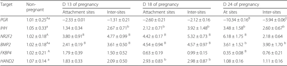
Table 1The mRNA level of target genes in the endometrium during implantation (M ± S.D.)
Target Non-pregnant
D 13 of pregnancy D 18 of pregnancy D 24 of pregnancy
Attachment sites Inter-sites Attachment sites Inter-sites At sites Inter-sites
PGR 1.01 ± 0.25Aa −2.33 ± 0.01 −1.31 ± 0.21 −2.60 ± 0.21 −2.12 ± 0.16 −10.34 ± 0.16B −3.94 ± 0.06b
IHH 1.05 ± 0.33a 1.34 ± 0.34 2.67 ± 0.71b 2.12 ± 0.71b 3.92 ± 1.48b 3.48 ± 1.58b 2.60 ± 0.67b
NR2F2 1.02 ± 0.18A 3.80 ± 0.91B 4.77 ± 0.99B 4.42 ± 0.17B 5.32 ± 0.73B 6.18 ± 1.75B 2.18 ± 0.64
BMP2 1.02 ± 0.18Aa 2.41 ± 0.19B 3.61 ± 0.50B 4.54 ± 0.94B 4.57 ± 0.97B 3.61 ± 1.52b 3.90 ± 1.70b
FKBP4 1.02 ± 0.21A 1.79 ± 0.39 1.50 ± 0.52 0.63 ± 0.19 0.99 ± 0.15 0.35 ± 0.08B 0.76 ± 0.21
HAND2 1.07 ± 0.14a 1.83 ± 0.33 2.09 ± 0.50 2.93 ± 0.83b 2.98 ± 0.87b 1.08 ± 0.16 1.11 ± 0.16
(score = 1), moderate staining (score = 2), strong staining (score = 3). The percentages of positive cells was calcu-lated using ImagePro plus. This formula was used to cal-culated the final score: ∑(percentage of positive cells)* (score of positive staining). Average of five different areas per picture was recorded. According to the final score, the protein expressed as follows: <1.0, weak, 1.0–1.5, moder-ate; >1.5, strong.
Detection of SNPs and litter size association analysis DNA was extracted by phenol and chloroforms (1:1) stand-ard techniques. 18 PCR primer pairs (see Additional file 2: Table S2) were designed to detect SNPs of target genes. PCR amplifications were carried out on an Eppendorf Mastercycler gradient 5331 PCR System (Eppendorf, Germany). The polymerase chain reaction amplification was performed using 50–100 ng of genomic DNA, 25μL Taq PCR MasterMix (Taq DNA Polymerase: 0.05 units/μL; MgCl2: 4 mM/μL; dNTPs: 0.4 mM/μL), 10 pM of each primers in a 50 μL final volume. All reagents
Fig. 1The protein relative abundance of target proteins in endometrium of sows. Note: NP, endometrium of non-pregnant sows; D13a, endometrial attachment sites on d 13 of gestation; D13b, the endometrial inter-sites on d 13 of gestation; D18a, endo-metrial attachment sites on d 18 of gestation; D13b, the endoendo-metrial inter-sites on d 18 of gestation; D24a, endometrial
attachment sites on d 24 of gestation; D24b, the endometrial inter-sites on d 24 of gestation
Table 2The protein relative abundance of target proteins in endometrium of sows
Target
Non-pregnant
D 13 of pregnancy D 18 of pregnancy D 24 of pregnancy
Attachment sites Inter-sites Attachment sites Inter-sites Attachment sites Inter-sites IHH 0.28 ± 0.10Aa 0.33 ± 0.15 0.48 ± 0.11 0.48 ± 0.11b 1.00 ± 0.02B 1.03 ± 0.21B 0.72 ± 0.03B NR2F2 0.89 ± 0.08 0.71 ± 0.05 0.99 ± 0.10 1.09 ± 0.02 1.15 ± 0.06 1.16 ± 0.07 1.11 ± 0.06 BMP2 0.37 ± 0.14a 0.77 ± 0.13b 0.58 ± 0.01b 0.38 ± 0.10 0.44 ± 0.11 0.33 ± 0.19 0.40 ± 0.21 FKBP4 0.57 ± 0.14 0.66 ± 0.16 1.00 ± 0.20 0.45 ± 0.19 0.66 ± 0.21 0.63 ± 0.18 0.84 ± 0.16 HAND2 0.61 ± 0.03A 0.73 ± 0.03B 0.71 ± 0.01B 0.57 ± 0.06 0.78 ± 0.01B 0.82 ± 0.03B 0.87 ± 0.05B
a, bP< 0.05, A, BP< 0.01
Fig. 2Immunhistochemical localization of IHH in pig uterus. GE = glandular epithelium; LE = luminal epithelium; S = stroma.
aNegative control;bImmunohistochemical staining of non-pregnanct sows uterus with IHH antibody;cImmunohistochemical staining of porcine uterus attachment site with IHH antibody on d 13 of pregnancy;dImmunohistochemical staining of porcine uterus inter-site with IHH antibody on d 13 of pregnancy;
eImmunohistochemical staining of porcine uterus attachment site with IHH antibody on d 18 of pregnancy;fImmunohistochemical staining of porcine uterus inter-site with IHH antibody on d 18 of pregnancy;gImmunohistochemical staining of porcine uterus attachment site with IHH antibody on d 24 of pregnancy;hImmunohistochemical staining of porcine uterus inter-site with IHH antibody on d 24 of pregnancy
were collected from the National Laboratories for Agrobiotechnology, China Agricultural University. The following conditions of PCR amplification were used: a denaturation step at 95 °C for 4 min, 30 cycles at 95 °C for 30 s, 52 °C ~ 55 °C for 30 s, and 72 °C for 30 s ~ 1 min 30 s, a final extension step of 72 °C for 10 min. Amplified fragments were separated by 1.5 % agarose gel electrophoresis (AGE).
Using pooled DNA amplification and sequencing, several mutations were found. Mutation −204C > A in promoter region of NR2F2 gene caused the deletion of transcription factor binding sites (TFBS) CREB (cAMP-response-element-binding protein).
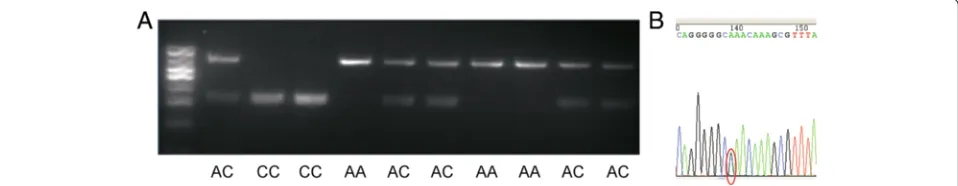
NR2F2 was selected to be the candidate gene for litter size based on its mRNA/protein expression level during embryonic implantation period and the muta-tion found in promoter region. PCR- Restricmuta-tion frag-ment length polymorphism (PCR-RFLP) was used to detect different genotypes. HaeIII (NEB R0108L, BioLabs Inc.) was used. The PCR products of three genotypes were random selected and sequenced to validate the results.
Alleles and genotypes frequencies of NR2F2 were calculated from the 625 sows, respectively. GLM proced-ure of SAS 8.02 software was used to compute the least square means of TNB and NBA. According to the Fig. 3Immunhistochemical localization of NR2F2 in pig uterus.
GE = glandular epithelium; LE = luminal epithelium; S = stroma.
aNegative control;bImmunohistochemical staining of non-pregnanct sows uterus with NR2F2 antibody;cImmunohistochemical staining of porcine uterus attachment site with NR2F2 antibody on d 13 of pregnancy;dImmunohistochemical staining of porcine uterus inter-site with NR2F2 antibody on d 13 of pregnancy;eImmunohistochemical staining of porcine uterus attachment site with NR2F2 antibody on d 18 of pregnancy;fImmunohistochemical staining of porcine uterus inter-site with NR2F2 antibody on d 18 of pregnancy;g
Immunohistochemical staining of porcine uterus attachment site with NR2F2 antibody on d 24 of pregnancy;hImmunohistochemical staining of porcine uterus inter-site with NR2F2 antibody on d 24 of pregnancy
Fig. 4Immunhistochemical localization of BMP2 in pig uterus. GE = glandular epithelium; LE = luminal epithelium; S = stroma.aNegative control;bImmunohistochemical staining of non-pregnanct sows uterus with BMP2 antibody;cImmunohistochemical staining of porcine uterus attachment site with BMP2 antibody on d 13 of pregnancy;
analysis, the effect of sire and dam on litter size was not significant, so the following linear model was used to analyze the genotype effect ofNR2F2.
Yijkl¼μþHYSiþPjþGkþeijkl
Where Yijkl is the traits of TNB and NBA, μ is the overall mean, HYSiis the effect of herd-year-season (i= 1 to 52), Pjis the effect of parity (j= 1, 2,≥3 and all par-ities), Gkis the effect of genotype (k= 1 to 3) and eijklis the random residual. The data was analyzed separately for the first parity, the second parity, the third and fol-lowing parities, and all parities. The additive effect and
the dominant effect were calculated according to the methods of Rothschild et al. [19].
Results
mRNA expression in porcine endometrium
The effect of the day of pregnancy on mRNA expression
of IHH, NR2F2, BMP2, FKBP4 and HAND2 in sows’
endometrium during implantation period was shown in Table 1. In pregnant sows, the expression of IHH was significantly higher than that of non-pregnant sows on d 18 and d 24 of pregnancy (P< 0.05) (Table 1). The ex-pression ofIHH in attachment sites showed an uptrend. Fig. 5Immunhistochemical localization of FKBP4 in pig uterus. GE =
glandular epithelium; LE = luminal epithelium; S = stroma.aNegative control;bImmunohistochemical staining of non-pregnanct sows uterus with FKBP4 antibody;cImmunohistochemical staining of porcine uterus attachment site with FKBP4 antibody on d 13 of pregnancy;
dImmunohistochemical staining of porcine uterus inter-site with FKBP4 antibody on d 13 of pregnancy;eImmunohistochemical staining of porcine uterus attachment site with FKBP4 antibody on d 18 of pregnancy;
fImmunohistochemical staining of porcine uterus inter-site with FKBP4 antibody on d 18 of pregnancy;gImmunohistochemical staining of porcine uterus attachment site with FKBP4 antibody on d 24 of pregnancy;hImmunohistochemical staining of porcine uterus inter-site with FKBP4 antibody on d 24 of pregnancy
Fig. 6Immunhistochemical localization of HAND2 in pig uterus. GE = glandular epithelium; LE = luminal epithelium; S = stroma.aNegative control;bImmunohistochemical staining of non-pregnanct sows uterus with HAND2 antibody;cImmunohistochemical staining of porcine uterus attachment site with HAND2 antibody on d 13 of pregnancy;d
Immunohistochemical staining of porcine uterus inter-site with HAND2 antibody on d 13 of pregnancy;eImmunohistochemical staining of porcine uterus attachment site with HAND2 antibody on d 18 of pregnancy;fImmunohistochemical staining of porcine uterus inter-site with HAND2 antibody on d 18 of pregnancy;g
Immunohistochemical staining of porcine uterus attachment site with HAND2 antibody on d 24 of pregnancy;hImmunohistochemical staining of porcine uterus inter-site with HAND2 antibody on d 24 of pregnancy
This was consistent with the expression ofNR2F2which was significantly up-regulated during implantation time.
The expression ofBMP2was significantly up-regulated (P< 0.05 or P< 0.01) during implantation time (Table 1), which was consistent withIHHandNR2F2. ForFKBP4, at attachment sites, the expression of FKBP4 was signifi-cantly down-regulated on d 24 of pregnancy (P< 0.01) (Table 1). The expression of HAND2 was the highest on d 18 of pregnancy (P< 0.05) (Table 1).
Protein expression in porcine endometrium
The protein expressions of IHH, NR2F2, BMP2, FKBP4 and HAND2 in the porcine endometrium during the em-bryonic implantation period were shown in Fig. 1 and Table 2. The protein expression of IHH was significantly up-regulated on d 18 and d 24 of pregnancy (P< 0.05 orP
< 0.01) (Fig. 1 and Table 2), which was similar to its mRNA expression. The protein expression of BMP2 was higher on d 13 of pregnancy (P< 0.05) (Fig. 1 and Table 2). For the protein expression of FKBP4, there was not significantly difference between pregnant groups and non-pregnant group (Fig. 1 and Table 2), which was not consistent with its mRNA expression pattern. The protein expression of HAND2 was higher in pregnant sows (P< 0.01) (Fig. 1 and Table 2), except at attachment sites on d 18 of pregnancy.
Protein localization in porcine endometrium
During implantation period, IHH, NR2F2, BMP2, FKBP4 and HAND2 were observed in luminal epithelium and glandular epithelium (Figs. 2, 3, 4, 5, 6). In stroma, the staining of BMP2 and FKBP4 were weak, but the staining of NR2F2 and HAND2 was strong (Figs. 2, 3, 4, 5, 6). The result was summarized in Table 3.
Detection of SNPs of target genes and association analysis
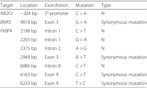
After analysis samples of 625 sows, several mutations were found (Table 4). Mutation -204C > A in promoter region of NR2F2 gene was found, and this mutation caused the deletion of TFBS CREB (Fig. 7). Synonymous mutation 9619G > A in exon 3 ofBMP2gene was found (Table 4). Seven mutations in FKBP4 gene were found, but no one is missense mutation (Table 4).
NR2F2was selected to be the candidate gene for litter size based on its mRNA/protein expression level during embryonic implantation period and the mutation found in promoter region. PCR-RFLP was used to detect differ-ent genotypes. The represdiffer-entative SNPs sequencing out-put for genotypes were shown in Fig. 8. The genotype frequencies and allele frequencies at each polymorphic locus in Large White, Landrace and Duroc sows were shown in Table 5. The genotype frequencies of AA, AC and CC in large white were 0.388, 0.414, and 0.198. In Landrace, the genotype frequencies were 0.088, 0.366, and 0.546. In Duroc, the genotype frequencies were 0.358, 0.433, and 0.208. None of the three breeds was found to be in Hardy-Weinberg equilibrium (HWE).
The data for TNB and NBA were observed for the first parity, the second parity, the third and the following par-ities and all parpar-ities. The least square means in Large White, Landrace and Duroc were shown in Tables 6, 7 and 8. In Large White, in the first parity, the sows with AA genotype had an advantage of 0.81 (P< 0.05) NBA per litter over the sows with CC genotype. In the second par-ity, the sows with CC genotype had an advantage of 1.76 (P< 0.01) and 1.56 (P< 0.01) TNB per litter over the sows with AA and AC, respectively. NBA of CC genotype were Table 3The expression of different position of target proteins in endometrium of sows
Target Non-pregnant D 13 of pregnancy D 18 of pregnancy D 24 of pregnancy
Attachment sites Inter-sites Attachment sites Inter-sites Attachment sites Inter-sites
GE LE S GE LE S GE LE S GE LE S GE LE S GE LE S GE LE S
IHH ++ ++ ± ++ ++ + ++ ++ ± ++ ++ + ++ ++ + ++ ++ + ++ ++ ±
NR2F2 ++ ++ ± ++ ++ + ++ ++ + ++ ++ ++ ++ ++ ++ ++ ++ ++ ++ ++ ++
BMP2 ++ ++ ± ++ ++ ± ++ ++ ± ++ ++ + ++ ++ ± ++ ++ + ++ ++ ±
FKBP4 ++ ++ ± ++ ++ ± ++ ++ ± ++ ++ ± ++ ++ ± ++ ++ ± ++ ++ ±
HAND2 ++ ++ ± ++ ++ + ++ ++ + ++ ++ + ++ ++ + ++ ++ + ++ ++ +
GEglandular epithelium;LEluminal epithelium;Sstroma ± weak; +moderate; ++strong
Table 4Location and type of nucleotide mutation of target genes
Target Location Exon/Intron Mutation Type
NR2F2 −204 bp 5′-promoter C > A N
BMP2 9619 bp Exon 3 G > A Synonymous mutation
FKBP4 2198 bp Intron 1 C > T N 2203 bp Intron 1 G > A N 2375 bp Intron 2 A > G N
2949 bp Exon 3 A > T Synonymous mutation 6086 bp Intron 8 C > T N
of 0.99 (P< 0.05) more piglets per litter than that of the AA genotype. In the third and following parities, NBA sig-nificantly increased for the CC genotype with 0.60 (P< 0.05) and 0.85 (P< 0.01) more piglets in comparison with the AA and AC genotype, respectively. In all parities, the sows with CC genotype had an advantage (P< 0.05) of 0.89 and 0.64 for TNB per litter over the AA and AC genotype sows, respectively. And NBA of CC genotype were of 0.97 (P< 0.01) and 0.88 (P< 0.01) more piglets per litter than that of the AA and AC genotype, respectively.
In Landrace, in the third and following parities, the sows with CC genotype had an advantage of 0.53 for TNB and 0.61 for NBA per litter over the sows with AA genotype, and 0.53 for TNB over the sows with AC genotype, but not significantly. In all parities, TNB of genotype CC was 1.05 (P< 0.05) piglets higher than that of the AA genotype. And the sows with the CC genotype had an advantage of 0.53 and 0.22 for NBA per litter over the sows with AA and AC, but not significantly.
In Duroc, in the second parity, the sows with the CC genotype had an advantage of 0.66 piglets (P< 0.05) for TNB and 1.34 (P< 0.05) piglets for NBA per litter over the sows with AA genotype. In the third and following parities, the sows with the CC genotype had
an advantage of 1.35 piglets (P< 0.01) for TNB and 1.34 (P< 0.05) for NBA per litter over the sows with AA genotype.
Discussion
Expression of genes participated in paracrine signaling in sows endometrium
The embryonic peri-implantation time of pigs is espe-cially longer. During the peri-implantation period of pregnancy, uterine LE and conceptus trophectoderm de-velop adhesion competency in synchrony to initiate the adhesion cascade within a restricted period of the uter-ine cycle termed the“window of receptivity” [20–22]. In pigs, this window is orchestrated through the actions of progesterone and estrogen to regulate locally produced cytokines, growth factors, cell surface glycoproteins, cell surface adhesion molecules, and extracellular matrix (ECM) proteins [23]. A fundamental paradox of early pregnancy is that cessation of expression of PGR and
ESR1by uterine epithelia is a prerequisite for uterine re-ceptivity to implantation, expression of genes by uterine epithelia and selective transport of molecules into the uterine lumen that support conceptus development. Thus, effects of P4 are mediated via PGR expressed in uterine stromal and myometrial cells by stromal cell Fig. 7Change of transcription factor caused by mutation.aC at 204 bp;bA at 204 bp
Fig. 8PCR-RFLP results of swineNR2F2gene and sequence image of the different genotypes.aGenotypes of the RFLP marker of PCR products;
bSequence image of mutation -204C > A
derived growth factors known as “progestamedins” [24, 25]. As previous indicated, progesterone down regulated the expression of PGR in the uterine epithelia of pigs after d 10 of pregnancy, immediately prior to the time when the endometrium becomes receptive to implant-ation [26–28]. In pigs, down-regulimplant-ation of PGRin uter-ine epithelia is a prerequisite for the expression of genes for uterine secretions and transport of molecules into the uterine lumen that support conceptus development. Down-regulation of PGR is associated with down-regulation of mucin1 (MUC1), as well as up-regulation of the expression of secreted phosphoprotein 1 (SPP1) and insulin-like growth factor binding protein 1 (IGFBP1). During conceptus elongation and the early peri-implantation period, the endometrium increases the release of a number of growth factors and cytokines such as epidermal growth factor (EGF), insulin-like growth factor-1 (IGF-1), fibroblast growth factor 7 (FGF7), vascular endothelial growth factor (VEGF), interleukin 6 (IL-6), transforming growth factor beta (TGFβ), and leukemia inhibitory factor (LIF) [29, 30]. Some of these genes had been reported to have signifi-cant effect on litter size in pigs, such as SPP1, VEGF, MUC1, LIFet al [1, 31–33].
PGRparacrine signaling has been recognized to play a significant role in pregnancy in human and mouse, which have not been studied in pigs [5].IHHis a proges-terone receptor target activated within the epithelium which signals downstream to NR2F2 in the stroma es-tablishing the HH–NR2F2 axis within the dual uterine compartments. Strong evidence exists to propose a role of a HH–NR2F2 axis in the regulation of reproduction in human and mice [12, 34]. Identification of the signal-ing pathway from stroma to epithelium would aid in the understanding of how the stroma contributes to embryo implantation. Changes in endometrial transcriptome during early stages of conceptus attachment to uterine LE in previous study showed thatIHH regulated signifi-cantly during pregnancy period in the pigs. In the present study, compared with non-pregnant sows, the mRNA and protein expression of IHH were up-regulated during implantation. The expression ofIHHin bovine uterus had been studied. The result showedIHH
is modulated by progesterone in bovine uterus, and may be required to be down-regulated to allow expression of genes that drive conceptus elongation in cattle [35]. In
pigs, the conceptus elongated rapidly before d 13 of ges-tation, and the filamentous conceptus continue to elong-ate but slowly after d 13 of gestation. The expression of
IHH did not show significantly changed at d 13 of preg-nancy in our result. It may be because the conceptus elongate slowly after d 13 of pregnancy in pigs [36]. The expression ofNR2F2 was significantly up-regulated dur-ing implantation time and the expression in attachment sites showed an upward trend. This was consistent with previous study, which foundNR2F2up-regulated in d 12 of gestation in Yorkshire pigs [37].NR2F2was shown to activate hypoxia-inducible factor 1 alpha (HIF-1α) and
HIF-1 is an important mediator of estrogen-induced
VEGF expression in the uterus [38, 39]. They thought that the expression of NR2F2 is associated with greater activation of angiogenesis at the stage of implantation in the Yorkshire breed [37]. The expression of IHH and
NR2F2were consistent with their functional role in em-bryonic implantation and also consistent with previous studies [40–44]. It was reported that HH-NR2F2 axis can transmit the paracrine signaling by PGR from epi-thelium to stroma [42]. The protein localization of IHH in porcine endometrium showed that IHH mainly ob-served strongly in luminal epithelium and glandular epi-thelium. NR2F2 was especially observed strongly in stroma. This confirmed that HH–NR2F2 axis was im-portant in mediating the signal from epithelial to other effect or genes in the stroma.
BMP2, as a downstream gene of HH-NR2F2 axis, has demonstrated to be a critical effector for decidualization and the maintenance of pregnancy during post-implantation. BMP2 likely acts as a paracrine signaling factor for the initiation of the proliferative response after embryonic implantation within the uterine stroma. In the present study, the mRNA expression of BMP2 was significantly up-regulated during implantation time, which was consistent with the expression of IHH and
NR2F2. In previous study, researchers found that BMP2 and BMP6 can significantly suppress progesterone pro-duction in pigs in vitro [45]. So this was consistent with our result, which showedBMP2up-regulated along with
PGR down-regulated during implantation period. The protein expression of BMP2 was significantly up-regulated on d 13 of pregnancy, which demonstrated that BMP2 promotes implantation cooperated with IHH and NR2F2. But on d 18 and 24, the expression did not regulate Table 5Number of alleles (n), allele and genotype frequencies ofNR2F2, observed heterozygosity (h)
Breed Sows Genotype distribution Genotype frequencies Allele frequencies h
AA AC CC AA AC CC A C
Large White 232 90 96 46 0.388 0.414 0.198 0.595 0.405 4.648
Landrace 273 24 100 149 0.088 0.366 0.546 0.271 0.729 1.458
Table 7Effects of theNR2F2polymorphism on total number born (TNB) and number born alive (NBA) in Landrace (LS means ± S.E.)
Breed Genotype First parity Second parity Third to ninth parity All parities
Litters TNB NBA Litters TNB NBA Litters TNB NBA Litters TNB NBA
Landrace AA 24 11.02 ± 0.32 10.44 ± 0.31 11 10.33 ± 0.53 10.06 ± 0.47 15 10.68 ± 0.30 10.24 ± 0.33 50 10.62 ± 0.19a 10.13 ± 0.23
AC 100 11.15 ± 0.25 10.54 ± 0.25 52 10.85 ± 0.39 10.25 ± 0.34 78 11.19 ± 0.23 10.79 ± 0.30 230 10.98 ± 0.15 10.44 ± 0.20
CC 149 11.36 ± 0.27 10.75 ± 0.27 97 11.26 ± 0.45 10.76 ± 0.40 151 11.21 ± 0.26 10.85 ± 0.30 397 11.67 ± 0.16b 10.66 ± 0.21
Values with different superscripts show significant levels within columns: a, bP< 0.05, A, BP< 0.01
Table 6Effects of theNR2F2polymorphism on total number born (TNB) and number born alive (NBA) in Large White (LS means ± S.E.)
Breed Genotype First parity Second parity Third to ninth parity All parities
Litters TNB NBA Litters TNB NBA Litters TNB NBA Litters TNB NBA
Large White AA 90 11.12 ± 0.25 10.33 ± 0.23a 60 11.10 ± 0.40A 10.21 ± 0.35a 144 11.078 ± 0.40 10.09 ± 0.35a 294 10.99 ± 0.32A 10.23 ± 0.28A
AC 96 11.43 ± 0.25 10.59 ± 0.24 66 11.30 ± 0.41A 10.01 ± 0.37 190 10.96 ± 0.40 9.84 ± 0.36A 352 11.24 ± 0.32A 10.32 ± 0.29A
CC 46 11.72 ± 0.32 11.14 ± 0.31b 26 12.86 ± 0.55B 11.20 ± 0.49b 93 10.69 ± 0.46 10.69 ± 0.41Bb 165 11.88 ± 0.35B 11.20 ± 0.32B
Values with different superscripts show significant levels within columns: a, bP< 0.05, A, BP< 0.01
Table 8Effects of theNR2F2polymorphism on total number born (TNB) and number born alive (NBA) in Duroc (LS means ± S.E.)
Breed Genotype First parity Second parity Third to ninth parity All parities
Litters TNB NBA Litters TNB NBA Litters TNB NBA Litters TNB NBA
Duroc AA 43 10.43 ± 0.35 9.53 ± 0.37 21 10.88 ± 0.39a 9.87 ± 0.35A 58 9.68 ± 0.34A 9.26 ± 0.39a 122 10.02 ± 0.20 9.30 ± 0.24
AC 52 10.54 ± 0.30 10.07 ± 0.32 36 11.12 ± 0.39 9.98 ± 0.35A 103 10.48 ± 0.25 9.97 ± 0.31 161 10.21 ± 0.16 9.63 ± 0.21
CC 25 10.60 ± 0.43 10.10 ± 0.46 11 11.54 ± 0.45b 10.81 ± 0.40B 40 11.03 ± 0.46B 10.60 ± 0.4b 76 10.17 ± 0.26 9.47 ± 0.30
Values with different superscripts show significant levels within columns: a, bP< 0.05, A, BP< 0.01
Chen
et
al.
Journal
of
Animal
Science
and
Biotechnolo
gy
(2016) 7:31
Page
10
of
significantly. It may be because decidualization did not happen in pigs.
HAND2was another downstream target ofPGR[8]. In the stroma, HAND2 plays an important role in the hibition of the FGF pathway, a pathway known to be in-volved in the promotion of epithelial proliferation by estrogen signaling [8]. Therefore, HAND2 is important to inhibit the estrogen-induced epithelial proliferation in the uterus [8]. The inhibition of epithelial proliferation byPGRsignaling was possibly viaHH–NR2F2axis.HH–
NR2F2axis then activatedHAND2, which caused the in-hibition of estrogen signaling and subsequent allowance for proper embryonic implantation. In the present study, the expression of mRNA and protein of HAND2 were both up-regulated on d 13 of pregnancy. This may re-lated with its inhibition of estrogen signaling, and fur-ther more promoted the positive role of PGR in implantation. In previous studies, HAND2had been de-tected up-regulated at implantation period and late ges-tation period in pigs [31, 46]. The researchers find
HAND2 related with receptivity of uterus and vascular development of placenta [31, 46]. The mRNA ofHAND2
was up-regulated on d 18 of pregnancy, but the protein expression was not. Maybe there is regulation mechan-ism at translation level, which needs further research. The protein localization in porcine endometrium showed that HAND2 observed strongly in luminal epi-thelium, glandular epiepi-thelium, and stroma. This indi-cated thatHAND2played an important role in transmit thePGRsignaling from epithelium to stroma.
The variations ofNR2F2and its association with litter size Marker-assisted selection (MAS) in conjunction with traditional selection methods is most effective for the traits such as litter size, which are either expressed later in life, are sex-dependent, or are of low heritability [47]. The candidate gene approach has led to notable success in demonstrating reproduction-related genetic markers or major genes, such as ESR, PRLR, the erythropoietin receptor (EPOR) and so on [19, 48–50].
In the present study, we selectedNR2F2as the candidate gene for litter size in pigs, due to its biological function and the interesting mutation. Three genotypes were found: AA, AC and CC. The association with litter size revealed that CC genotype is the favorable genotype. Through ana-lysis using Consite database (http://consite.genereg.net/cgi-bin/consite?rm=t_input_single), the C→A mutation caused deletion of TFBS CREB (Fig. 7). CREB has been proved played an important role in activation of transcrip-tion and regulatranscrip-tion of gene transcriptranscrip-tion [51, 52]. The dele-tion of CREB may affect the expression of NR2F2 in porcine endometrium and stroma. The effect ofNR2F2on litter size possibly associated with its expression in endo-metrium during embryonic implantation. This certainly
will affect the signal ofPGRfrom endometrium to stroma, in consideration of the PGR-IHH-NR2F2 axis. Subse-quently, the embryonic implantation process and litter size was affected.
Conclusions
In current research, the expression patterns of genes/ proteins involved in PGR paracrine signaling over im-plantation time were studied. And candidate gene for litter size was identified from genes involved in this signaling. The present study could be a resource for further studies to identify the roles of these genes for embryonic implantation in pigs.
Additional files
Additional file 1: Table S1.Primers used for Real-time PCR (RT-PCR). (DOCX 19 kb)
Additional file 2: Table S2.Primer pairs and PCR conditions used for SNPs detection. (DOCX 20 kb)
Abbreviations
AGE:agarose gel electrophoresis; CREB: cAMP-response-element-binding protein; D13a: endometrial attachment sites on day 13 of gestation; D13b: the endometrial inter-sites on day 13 of gestation; D13b: the endometrial inter-sites on day 18 of gestation; D18a: endometrial attachment sites on day 18 of gestation; D24a: endometrial attachment sites on day 24 of gestation; D24b: the endometrial inter-sites on day 24 of gestation; HWE: Hardy-Weinberg equilibrium; MAS: Marker-assisted selection; NBA: number born alive; NP: endometrium of non-pregnant sows; PCR-RFLP: PCR-Restriction fragment length polymorphism; PGR: progesterone receptor; TFBS: transcription factor binding sites; TNB: total number born.
Acknowledgements
This study was supported by National Natural Science Foundation of China (No.31172176), China Agriculture Research System (No. CARS-36), Program for Changjiang Scholar and Innovation Research Team in University (IRT1191).
Authors’contributions
The contributions of the authors are as follows: XC conducted the research, analysis the results and wrote the paper. XC and JLF participated in the animal experiment. AGW was in charge of the whole trail. All authors read and approved the final manuscript.
Competing interests
The authors declare that they have no competing interests.
Author details
1
College of Animal Sciences and Technology, National Engineering Laboratory for Animal Breeding & Key Laboratory of Animal Genetics, Breeding and Reproduction, Ministry of Agriculture, China Agricultural University, Beijing 100193, People’s Republic of China.2Institute of Apicultural Research, Chinese Academy of Agricultural Sciences, Beijing 100093, People’s Republic of China.
Received: 27 November 2015 Accepted: 5 May 2016
References
1. Spotter A, Muller S, Hamann H, Distl O. Effect of polymorphisms in the genes for LIF and RBP4 on litter size in two German pig lines. Reprod Domest Anim. 2009;44:100–5.
3. Fernandez-Rodriguez A, Munoz M, Fernandez A, Pena RN, Tomas A, Noguera JL, et al. Differential gene expression in ovaries of pregnant pigs with high and low prolificacy levels and identification of candidate genes for litter size. Biol Reprod. 2010;84:299–307.
4. Brayman MJ, Julian J, Mulac-Jericevic B, Conneely OM, Edwards DP, Carson DD. Progesterone receptor isoforms A and B differentially regulate MUC1 expression in uterine epithelial cells. Mol Endocrinol. 2006;20:2278–91.
5. Lydon JP, DeMayo FJ, Funk CR, Mani SK, Hughes AR, Montgomery CA, et al. Mice lacking progesterone receptor exhibit pleiotropic reproductive abnormalities. Genes Dev. 1995;9:2266–78.
6. Tibbetts TA, Conneely OM, O’Malley BW. Progesterone via its receptor antagonizes the pro-inflammatory activity of estrogen in the mouse uterus. Biol Reprod. 1999;60:1158–65.
7. Mote PA, Arnett-Mansfield RL, Gava N, deFazio A, Mulac-Jericevic B, Conneely OM, et al. Overlapping and distinct expression of progesterone receptors A and B in mouse uterus and mammary gland during the estrous cycle. Endocrinology. 2006;147:5503–12.
8. Wetendorf M, Demayo FJ. The progesterone receptor regulates implantation, decidualization, and glandular development via a complex paracrine signaling network. Mol Cell Endocrinol. 2012;357(1–2):108-18. 9. Bazer FW, Spencer TE, Johnson GA, Burghardt RC, Wu G. Comparative
aspects of implantation. Reproduction. 2009;138:195–209.
10. Takamoto N, Zhao B, Tsai SY, DeMayo FJ. Identification of Indian hedgehog as a progesterone-responsive gene in the murine uterus. Mol Endocrinol. 2002;16:2338–48.
11. Varjosalo M, Taipale J. Hedgehog: functions and mechanisms. Genes Dev. 2008;22:2454–72.
12. Lin FJ, Qin J, Tang K, Tsai SY, Tsai MJ. Coup d’Etat: an orphan takes control. Endocr Rev. 2011;32:404–21.
13. Kyriazakis I, Whittemore C. Whittemore’s science and practice of pig production. 3rd ed. Oxford: Blackwell; 2006. p. 105–47.
14. Samborski A, Graf A, Krebs S, Kessler B, Bauersachs S. Deep sequencing of the porcine endometrial transcriptome on day 14 of pregnancy. Biol Reprod. 2013;88:84.
15. Lord E, Murphy BD, Desmarais JA, Ledoux S, Beaudry D, Palin MF. Modulation of peroxisome proliferator-activated receptor delta and gamma transcripts in swine endometrial tissue during early gestation. Reproduction. 2006;131:929–42.
16. Patel V, Ramesh A, Traicoff JL, Baibakov G, Emmert-Buck MR, Gutkind JS, et al. Profiling EGFR activity in head and neck squamous cell carcinoma by using a novel layered membrane Western blot technology. Oral Oncol. 2005;41:503–8.
17. Hewitt SM, Baskin DG, Frevert CW, Stahl WL, Rosa-Molinar E. Controls for immunohistochemistry: the Histochemical Society’s standards of practice for validation of immunohistochemical assays. J Histochem Cytochem. 2014;62:693–7.
18. Axiotis CA, Monteagudo C, Merino MJ, LaPorte N, Neumann RD. Immunohistochemical detection of P-glycoprotein in endometrial adenocarcinoma. Am J Pathol. 1991;138:799–806.
19. Rothschild M, Jacobson C, Vaske D, Tuggle C, Wang L, Short T, et al. The estrogen receptor locus is associated with a major gene influencing litter size in pigs. Proc Natl Acad Sci U S A. 1996;93:201–5.
20. Bazer FW, Spencer TE, Johnson GA, Burghardt RC. Uterine receptivity to implantation of blastocysts in mammals. Front Biosci (Schol Ed). 2011;3:745–67.
21. Fazleabas AT, Kim JJ, Strakova Z. Implantation: embryonic signals and the modulation of the uterine environment–a review. Placenta. 2004;25 Suppl A:S26–31.
22. Spencer TE, Johnson GA, Bazer FW, Burghardt RC. Fetal-maternal interactions during the establishment of pregnancy in ruminants. Soc Reprod Fertil Suppl. 2007;64:379–96.
23. Johnson GA, Bazer FW, Burghardt RC, Spencer TE, Wu G, Bayless KJ. Conceptus-uterus interactions in pigs: endometrial gene expression in response to estrogens and interferons from conceptuses. Soc Reprod Fertil Suppl. 2009;66:321–32.
24. Cunha GR, Cooke PS, Kurita T. Role of stromal-epithelial interactions in hormonal responses. Arch Histol Cytol. 2004;67:417–34.
25. Spencer TE, Bazer FW. Biology of progesterone action during pregnancy recognition and maintenance of pregnancy. Front Biosci. 2002;7:d1879–1898.
26. Bailey DW, Dunlap KA, Erikson DW, Patel AK, Bazer FW, Burghardt RC, et al. Effects of long-term progesterone exposure on porcine uterine gene expression: progesterone alone does not induce secreted phosphoprotein 1 (osteopontin) in glandular epithelium. Reproduction. 2010;140:595–604. 27. Bazer FW, Burghardt RC, Johnson GA, Spencer TE, Wu G. Interferons and
progesterone for establishment and maintenance of pregnancy: interactions among novel cell signaling pathways. Reprod Biol. 2008;8:179–211. 28. Geisert RD, Pratt TN, Bazer FW, Mayes JS, Watson GH.
Immunocytochemical localization and changes in endometrial progestin receptor protein during the porcine oestrous cycle and early pregnancy. Reprod Fertil Dev. 1994;6:749–60.
29. Bazer FW, Wu G, Spencer TE, Johnson GA, Burghardt RC, Bayless K. Novel pathways for implantation and establishment and maintenance of pregnancy in mammals. Mol Hum Reprod. 2010;16:135–52.
30. Geisert RD, Lucy MC, Whyte JJ, Ross JW, Mathew DJ. Cytokines from the pig conceptus: roles in conceptus development in pigs. J Anim Sci Biotechnol. 2014;5:51.
31. Chen X, Li A, Chen W, Wei J, Fu J, Wang A. Differential gene expression in uterine endometrium during implantation in pigs. Biol Reprod. 2015;92:52.
32. Xiao C, Jinluan F, Aiguo W. Effect of VNTR polymorphism of the Muc1 gene on litter size of pigs. Mol Biol Rep. 2012;39:6251–8.
33. Putnova L, Kolarikova O, Knoll A, Dvorák J. Association study of osteopontin (SPP1) and estrogen receptor (ESR) genes with reproduction traits in pigs. Acta Universitatis Agriculturae et Silviculturae Mendelianae Brunensis (Czech Republic); 2001.
34. Pereira FA, Qiu Y, Zhou G, Tsai MJ, Tsai SY. The orphan nuclear receptor COUP-TFII is required for angiogenesis and heart development. Genes Dev. 1999;13:1037–49.
35. Forde N, Mehta JP, Minten M, Crowe MA, Roche JF, Spencer TE, et al. Effects of low progesterone on the endometrial transcriptome in cattle. Biol Reprod. 2012;87:124.
36. Blomberg LA, Long EL, Sonstegard TS, Van Tassell CP, Dobrinsky JR, Zuelke KA. Serial analysis of gene expression during elongation of the peri-implantation porcine trophectoderm (conceptus). Physiol Genomics. 2005;20:188–94.
37. Gu T, Zhu MJ, Schroyen M, Qu L, Nettleton D, Kuhar D, et al. Endometrial gene expression profiling in pregnant Meishan and Yorkshire pigs on day 12 of gestation. BMC Genomics. 2014;15:156.
38. Kim EJ, Yoo YG, Yang WK, Lim YS, Na TY, Lee IK, et al. Transcriptional activation of HIF-1 by RORalpha and its role in hypoxia signaling. Arterioscler Thromb Vasc Biol. 2008;28:1796–802.
39. Koos RD, Kazi AA, Roberson MS, Jones JM. New insight into the transcriptional regulation of vascular endothelial growth factor expression in the endometrium by estrogen and relaxin. Ann N Y Acad Sci. 2005;1041:233–47.
40. Ingham PW, McMahon AP. Hedgehog signaling in animal development: paradigms and principles. Genes Dev. 2001;15:3059–87.
41. Johnson RL, Scott MP. New players and puzzles in the Hedgehog signaling pathway. Curr Opin Genet Dev. 1998;8:450–6.
42. Kurihara I, Lee DK, Petit FG, Jeong J, Lee K, Lydon JP, et al. COUP-TFII mediates progesterone regulation of uterine implantation by controlling ER activity. PLoS Genet. 2007;3:e102.
43. Matsumoto H, Zhao X, Das SK, Hogan BL, Dey SK. Indian hedgehog as a progesterone-responsive factor mediating epithelial-mesenchymal interactions in the mouse uterus. Dev Biol. 2002;245:280–90. 44. McMahon AP. More surprises in the Hedgehog signaling pathway. Cell.
2000;100:185–8.
45. Webb R, Garnsworthy PC, Campbell BK, Hunter MG. Intra-ovarian regulation of follicular development and oocyte competence in farm animals. Theriogenology. 2007;68 Suppl 1:S22–29.
46. Zhou Q-Y, Fang M-D, Huang T-H, Li C-C, Yu M, Zhao S-H. Detection of differentially expressed genes between Erhualian and Large White placentas on day 75 and 90 of gestation. BMC Genomics. 2009;10:337.
47. Soller M. Marker assisted selection - overvier. Anim Biotech. 1994;5:193–207. 48. Li N, Zhao Y F, Xiao L, Zhang FJ, Chen YZ, Dai RJ, Zhang JS, et al. Candidate
gene approach for identification of genetic loci controlling litter size in swine[C]//Proc. 6th World Congress on Genetics Applied to Livestock Production. Armidale, Australia, vol. 26. 1998. p. 183–86.
49. van Rens BT, Evans GJ, van der Lende T. Components of litter size in gilts with different prolactin receptor genotypes. Theriogenology. 2003;59:915–26.
50. Vincent V, Goffin V, Rozakis-Adcock M, Mornon JP, Kelly PA. Identification of cytoplasmic motifs required for short prolactin receptor internalization. J Biol Chem. 1997;272:7062–8.
51. Nichols M, Weih F, Schmid W, DeVack C, Kowenz-Leutz E, Luckow B, et al. Phosphorylation of CREB affects its binding to high and low affinity sites: implications for cAMP induced gene transcription. EMBO J. 1992;11:3337–46. 52. Lee KA, Masson N. Transcriptional regulation by CREB and its relatives.
Biochim Biophys Acta. 1993;1174:221–33.
• We accept pre-submission inquiries
• Our selector tool helps you to find the most relevant journal
• We provide round the clock customer support
• Convenient online submission
• Thorough peer review
• Inclusion in PubMed and all major indexing services
• Maximum visibility for your research
Submit your manuscript at www.biomedcentral.com/submit