Validity And Reliability Of Tri-Goniometric
Method For Measuring Pelvic Tilt And Pelvic
Range Of Motion In Standing Posture
Thiruvarangan S, Dassanayake T.D.M.S.B, Samaranayake D.B.D.L
Department of Allied Health Sciences, Faculty of Medicine, University of Colombo Sri Lanka, +94 778470824
Department of Allied Health Sciences, Faculty of Medicine, University of Colombo Sri Lanka, +94 713009859
Department of Community Medicine, Faculty of Medicine, University of Colombo Sri Lanka, +94 716613296
Abstract: The purpose of this study was to examine the validity and reliability of a test designed to measure the pelvic-tilt angle, active posterior and anterior pelvic-tilt angles and ranges of motion, and the total pelvic-tilt range of motion (ROM) in standing position. After an instruction session, the pelvic-tilt angles of the both right and left side of 49 subjects were calculated using trigonometric functions. Ranges of motion were determined from the pelvic-tilt angles. Validity coefficients (Pearson r) for trigonometric measurements were .86; .90 for the standing pelvic-tilt angle, .91; .93 for the anterior pelvic-tilt angle, .86; .89 for the posterior pelvic-tilt angle and .89; .98 for the total ROM right and left side respectively. Reliability coefficients (Pearson r) for trigonometric measurements were .73; .82 for the standing pelvic-tilt angle, .74; .68 for the anterior pelvic-tilt angle, .76; .73 for the posterior pelvic-tilt angle and .88; .95 for the total ROM right and left side respectively. The factors may have influenced the validity and reliability of the measurements, the clinical implications and limitations of the test.
Key Words:Pelvic motion, Pelvic tilt angle, Trigonometric method
1.
Introduction
The pelvic tilt is the angle between the horizontal plan and a line drawn from the anterior superior iliac spine (ASIS) to the posterior superior iliac spine (PSIS) in quiet standing. It is determined by the muscular and ligamentous forces that act between the pelvis and adjacent segments (Alviso et al., 1988). A forward rotation of the pelvis, referred to as anterior pelvic tilt, is accompanied by an increase in lumbar lordosis and is believed to be associated with a number of common musculoskeletal conditions, including low back pain (Day J. W et al., 1984). In addition, anterior pelvic tilt has been associated with a loss of core stability, and therefore the degree of pelvic tilt has been used to assess routinely therapeutic procedures that either directly or indirectly affects the standing position of the pelvic tilt in the sagittal plane by Physical therapists. (Sanders G and Stavrakas P, 1981) The effects of the therapeutic procedure outcome measurements in physiotherapy are rarely quantified. Physical therapists need to consider clinical tests designed to provide objective and reliable pelvic-tilt data because such tests would permit documenting change in the pelvic tilt after a specific physical therapy regimen (Gajdosik et al., 1985). The effects of therapeutic procedures could then be quantified and changes in the procedures could be made accordingly. Numerous techniques for measuring trunk motions in the sagittal plane have been reported in many researches. The techniques include using radiography, photography, spondylometry, flexible rules that conform to trunk curves, tape measures to record the change in centimeters between skin marks or bony landmarks and variations of goniometry. Although interest
2.
Method
2.1 Subjects
This study was a descriptive cross-sectional study which was conducted at Allied Health Sciences Unit, Faculty of Medicine, University of Colombo between March and June 2015.The Ethics Review Committee of the Faculty of Medicine, University of Colombo has approved this study. Initially, permission was obtained from the Dean, Faculty of Medicine to conduct the study. The study population was informed regarding basic information of the study and procedure in general through a notice board so that those interested could came forward then informed written consent was obtained from the interested participants. Then student corresponding to the age group was identified and males and females were listed separately according to their registration number. Using simple random sampling method 25 students were selected from each list randomly for the study. Then socio-demographic details and current healthy status in lower back area were assessed using an interviewer administered questionnaire, followed by measurements which was taken by investigator on height, weight, angles of standing pelvic tilt, posterior pelvic tilt and anterior pelvic tilt of both right and left side in the sagittal plane in standing posture.
2.2 Testing
After completing an interviewer administered questionnaire and Body mass Index calculation, the subjects were asked lying supine on beds for 10 minutes to relax before taken the pelvic angles measurements. Then the anterior and posterior motions of the pelvis were demonstrated by investigator followed by the locations of the anterior superior iliac spine (ASIS) and the posterior superior iliac spine (PSIS) were palpated and marked with removable, non-allergic small adhesive star shaped stickers when the end points of PPT and APT were reached in both right and left side to refine instrument placements and testing procedures in order to ensure that the test was clinically valid, because the pelvis moved under the skin during active movement.
Standing pelvic Tilt: Each subject was asked to remove his/her foot wears and maintain erect posture with his foot placement of subject over feet traced on a level floor then placed a bowleg caliper over the stars, which were compressed to "firm resistance," and observed and recorded the distance between the spines to the nearest millimeter subsequently fixed the incline goniometer over stars which has given direct measurement of standing pelvic tilt angle in degrees (Fig. 1). A sliding pointer on a meter stick mounted on a wood base was used to measure the distances from the floor to the ASIS and from the floor to the PSIS (Fig. 2). Each measurement was recorded in three rounds and, finally the stars were removed.
Posterior pelvic tilt: While the subject stood erect as in the SPT measurements, the subject was instructed to keep his/her knees straight, to tighten the abdominal and gluteal muscles, and to move the top of the pelvis back to initial resistance without causing pain or discomfort. The subject was to hold this position while one side PSIS was palpated and marked, and the distance to the floor measured, and the ASIS was palpated, marked, and the distance from the star to the floor measured next placed the incline goniometer over stars which has given direct measurement
of posterior pelvic tilt angle in degrees and each measurement was taken by three times. The subject relaxed to the SPT position, and finally, the star marks were removed (Fig.1 and 2).
Anterior pelvic tilt: From the SPT position, the subject was reminded to stand erect and instructed to arch the low back and move the top of the pelvis forward to initial resistance without causing pain or discomfort. Again, the iliac spines were palpated, marked, and the distances from the stars to the floor measured subsequently, placed the incline goniometer over stars which has given direct measurement of anterior pelvic tilt angle in degrees and each measurement was recorded in three rounds. At the end point subject was asked to relax to the SPT position, and the star marks were removed. After two weeks, the tri-goniometer measuring method was repeated to assess test-retest reliability.
2.3 Data analysis
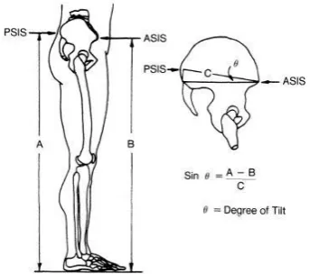
SPT, PPT, and APT angles were calculated from the raw data by using a trigonometric calculator to determine angle theta (θ) using the formula by Sanders and Stavrakas, 1981.Schematic diagram of pelvic-tilt measurement (Fig.3). Side opposite was the height difference between the PSIS and the floor and the ASIS and the floor, and the hypotenuse was the distance between the ASIS and PSIS (Fig. 3). Pelvic-tilt angles with the PSIS above the horizontal were assigned positive degree values and those with the PSIS below the horizontal were assigned negative degree values. The PPT-ROM was calculated as the difference between the PPT angle and the SPT angle, and the APT-ROM was calculated as the difference between the APT angle and the SPT angle. The TPT-ROM was determined as the difference between the PPT and APT angles (Fig.3).
3.
Results
49 candidates, whose age reneges from 20-25, were subjected to the study (n=49). There were () male (n=24) and females (n=25). The male to female ratio was approximately 1:1. The mean age of the subjects was 23.2 years, and their mean weight was 58.1 kg (range, 40-96 kg), and their mean height was 163.6 cm (range, 147-183 cm). Subjects were limited normal muscle strength and normal range of motion the back and lower extremities.A greater part of the sample (55.1%) was elaborated about normal Body Mass Index (BMI) category of the study sample is shown in Table 1. Validity of Tri-goniometric method of Pelvic tilt in neural, anterior and posterior and also Pelvic Range of Motion measurements were correlated with standard digital goniometer method in Right and Left side of the subjects.()Reliability of Tri-goniometric method of Pelvic tilt in neural, anterior and posterior and also Pelvic Range of Motion measurements were correlated in Right and Left side of the samples
4.
Discussion
number of techniques has been reported in the literature to measure pelvic tilt and pelvic range of motion. Mainly the techniques were included using radiography and photography. Although above methods have been used to measure the standing pelvic tilt they are regarded as expensive, potentially harmful and not always available to the subjects Literature also suggested that tri-goniometric method is an inexpensive, safe and simple technique which can use to measure pelvic tilt. Inclinometer, a meter stick with a sliding pointer and bowleg caliper were the necessary equipment to conduct the study. Maintaining the reliability and stability of the measurements is essential during the procedure. Practicing and performing the same movements by all the subjects is needed for this. We could address this problem as we used the subjects who already knew the correct actions. And also it was aided to decrease occurring of repeated movements by the subjects. Before testing, we confirmed that each subject could maintain the relaxed erect posture and perform the anterior, posterior tilt. It was helped to isolate the movements of the pelvis for the subjects during the procedure. Therefore instruction sessions and a special training session before the testing should be needed as anterior and posterior pelvic motions are complex actions. Consistently applying the same action commands ensured that all subjects performed the same movements and reached similar end points of each motion for tests. Few steps were taken for the accuracy of the measurements. Literature suggested that accurate palpation of the bony landmarks is very important when using this method for pelvic tilt measurements. For reliable measurements Anterior Superior Iliac Spine and Posterior Superior Iliac Spine of both sides were palpated and marked with removable, small adhesive star stickers when the end points of iliac spines were reached. And also subjects were asked to maintain the erect posture on the foot tracings to increase the consistency of the subjects’ stance. In some cases it was difficult to palpate the PSIS. Literature also suggests that palpation of the PSIS was more difficult with the subject’s contraction of the gluteaus maximus muscle in performing posterior tilt. Measurements were taken three times to minimize the measurement errors and to get an average measurement. Our study included male and female subjects of a medium to lean built. Variations in subcutaneous connective tissue thickness should be considered because patients may vary in body build from lean to obese. Our results were reliable, measuring this distance on people who are obese or whose body weight changes over time may not be as reliable as reported on the subjects in this study. Therefore at the end of our study we suggest additional studies are needed to examine the effect of subjects’ body type on the reliability of tri-goniometric method for pelvic tilt measurements. In this study the subjects with prolong back dysfunction were excluded. Back dysfunction can be a cause of performing wrong movements by the subjects during the procedure. Then there can be a doubt regarding the reliability and validity of the tri-goniometry measurements. During the data collection each and every subject happened to perform the anterior and posterior pelvic tilt for 12 times to minimize the measurement errors. Although we thought that there may be a possibility to get fatigue, it superficially didn’t affect the majority of the subjects. But a few subjects reported that
they were experiencing fatigue of their paraspinal musculature and gluteus maximus muscle during anterior and posterior pelvic motion respectively. Even with these concerns, strong reliability of the method was documented. The range of standard deviations of the tri-goniometer method validity found in this study is comparable to or smaller than those reported for other clinical goniometric methods of evaluating mobility of the pelvis. The standard deviations using a standard goniometric measurement of platforms to assess for pelvic tilt during stance from 3.2 and 3.4 degrees right and left respectively. Standard deviation of the tri-goniometer method reliability ranged from 2.2 to 5.4 degrees. As might be expected, this range of standard deviations was slightly higher than that reported in other studies for goniometric measurements taken in the extremities. Boone et al reported standard deviations ranging from 1.5 to 4.6 degrees in goniometric measurements.
5.
Clinical Implications
Clinicians may use this method of pelvic-tilt measurement to quantitatively document pelvic tilt. The clinical consequences of the APT and the PPT may represent the standing ROM of lumbar trunk extension and flexion, respectively. The method is easy access to use in any clinical setting non-invasive method and does not require expensive equipment or radiographic examination. The progress of a patient and the effectiveness of treatments used to address dysfunction of the pelvis may be assessed. We offer several suggestions for physical therapists using this method of pelvic-tilt measurement. The initial caliper measurement can be used as a constant for future measurements on the same patient, because the caliper reading is representative of the distance from the ASIS to the PSIS, this measurement should theoretically remain unchanged for a given patient, unless body weight remarkably change, because caliper measurements variability over time if soft tissue changes occurred.
6.
Conclusion
7.
Illustrations
7.1 Figures
Figure 1: Tester measuring distance between ASIS and PSIS with depth caliper
Figure 2: Tester measuring distance between PSIS and floor
Figure 3: Schematic diagram of pelvic-tilt measurement. A-B = side opposite; C = hypotenuse; ASIS = anterior superior iliac spine; PSIS = posterior superior iliac spine
(Sanders and Stavrakas, 1981).
7.2 Tables
Table 1: BMI category of study sample
Table 2: Validity of tri-goniometric method for measuring pelvic tilt and pelvic range of Motion of right and left side
Table 2: Reliability of tri-goniometric method for
measuring pelvic tilt and pelvic range of Motion of right and left side
8.
References
[1] Sanders G and Stavrakas P. A Technique for Measuring Pelvic Tilt. PHYS THER. 1981; 61:49-50.
[2] Gajdosik R, Simpson R, Smith R and Dontigny R. L. Intratester Reliability of Measuring the Standing Position and Range of Motion. PHYS THER.1985; 65:169-174.
[3] Prushansky T, Ezra N, Kurse N, Man L, Schneiderman Y (2008). Reproducibility of sagittal
BMI Category Frequency Percentage
Under weight 13 26.5
Normal 27 55.1
Overweight 09 18.4
Validity of the
tri-goniometric method Mean
Standard
Deviation Correlation
Standing Pelvic Tilt (SPT)
Right Left
11.5 11.3
3.4 3.2
.857 .903
Anterior Pelvic Tilt (APT)
Right Left
20.4 19.2
4.2 4.0
.914 .934
Posterior Pelvic Tilt (PPT)
Right Left
5.2 4.8
2.4 1.9
.859 .896
Total Pelvic Range of Motion (TROM) Right
Left
25.7 23.9
5.6 5.4
.889 .979
Reliability of the tri-goniometric
method
Mean Standard Deviation
Correlation
Standing Pelvic Tilt (SPT)
Right Left
11.1 10.8
3.9 3.4
.732 .822
Anterior Pelvic Tilt (APT)
Right Left
18.8 18.6
4.1 4.1
.744 .683
Posterior Pelvic Tilt (PPT)
Right Left
4.9 5.5
2.2 2.3
.758 .732
Total Pelvic Range of Motion (TROM) Right
Left
23.7 24.1
5.4 5.3
pelvic tilt measurements in normal subjects using digital inclinometry [abstract only] Available at:
http://www.gaitposture.com/article/S0966-6362%2808%2900035-0/fulltext
[4] Alviso D. J, Dong G.T, and Lentell G.L. Intertester Reliability for Measuring Pelvic Tilt in Standing. PHYS THER.1988; 68:1347-1351.
[5] Day J. W, Smidt G. L and Lehmann. Effect of Pelvic Tilt on Standing Posture. PHYS THER. 1984;64:510-516.
[6] Burdett R. G, Brown K. E and Fall M. P . Reliability and Validity of Four Instruments for Measuring Lumbar Spine and Pelvic Positions. PHYS THER.1986;66:677-684.
[7] Preece S, Willan P, Nester J, Smith P, Herrington L, and Bowker P. Variation in Pelvic Morphology May Prevent the Identification of Anterior Pelvic Tilt. J Man Manip Ther. 2008;16(2): 113–117.
[8] Clayson G.F, Newman .IM, Debevec D. Evaluation of mobility of hip and lumbar vertebrae of normal young women. Arch Phys Med Rehabil. 1962; 43(3):1-8.
[9] Troup.JDG,Hood.CA, Chapman.AE. Measurements of the sagittal mobility of the lumbar spine and hips. Annals of Physical Medicine. 1968;9:308-321.
[10]Hart FD, Strickland D, Cliffe P. Measurements of spinal mobility. Ann Rheum Dis. 1974;33(9):136-169.
[11]Israel M. A quantitative method of estimating flexion and extension of the spine A preliminary report. Mil Med. 1989;124(10):181 -186.
[12]Anderson J.A.D, Sweetman B.J. A combined flexi-rule/hydro goniometer for measurement of lumbar spine and its sagittal movement. Rheumatology and Rehabilitation. 1975; 14(11):173-179.
[13]Macrae IF, Wright V. Measurement of back movement. Ann Rheum Dis. 1969;28:584-588.
[14]Moll JMH, Wright V. Normal range of spinal mobility. Ann Rheum Dis. 197;30:381-386.
[15]Moll JMH, Lyanage SP, Wright V. An objective clinical method to measure spinal extension. Rheumatology and Physical Medicine. 1972; 2:293-312.
Author Profile
I graduated B.Sc(Hons) in
Physiotherapy from University of