in an elderly blind patient:
a case report
Henry M Lee,
DC, FCCRS(C)*
Muscle imbalances play an important role in the development of musculoskeletal complaints that are presented in clinical practice. According to Karel Lewit, muscle imbalance usually precedes recurrent joint dysfunction. Chiropractors are uniquely positioned to recognize and treat such imbalances through low tech rehabilitation techniques. A case report is presented of an elderly blind patient and the challenges involved in the rehabilitation of her proximal crossed syndrome.
(JCCA 2000; 44(4):223–229)
K E Y W O R D S: muscle imbalance, rehabilitation, aged, blindness, proximal crossed syndrome, chiropractic, posture.
Les déséquilibres musculaires jouent un rôle important dans les symptômes musculosquelettiques rencontrés en pratique clinique. Selon Karel Lewit, le déséquilibre musculaire précède généralement le dysfonctionnement récurrent des articulations. Les chiropraticiens sont tout désignés pour déceler et traiter ce genre de troubles au moyen de techniques de réadaptation relativement simples. Voici le cas d’une patiente âgrée aveugle et les difficultés rencontrées durant la réadaptation pour correction du syndrome de dysfonctionnement croisé proximal.
(JACC 2000; 44(4):223–229)
M O T S C L É S : déséquilibre musculaire, réadaptation, personne âgée, cécité, syndrome de dysfonctionnement croisé proximal, chiropratique, posture.
* Private practice. 1042 Broadview Avenue, Toronto, Ontario M4K 2S2. Phone (416) 467-1974. Fax (416) 421-3234.
© JCCA 2000.
Introduction
According to Karel Lewit,1 muscle imbalance usually pre-cedes recurrent joint dysfunction. Vladimir Janda2 de-scribed muscle imbalances as the situation in which some muscles become inhibited and weak, while others become tight. Such imbalances lead to tissue (musculotendonous and ligamentous) changes that may result in inappropriate patterns of movement. Such scenarios may eventually lead to symptomatic states of pain and inflammation.
Janda noted predictable patterns that develop largely due to postural positioning in sedentary environments and repetitive work tasks. He named one of the patterns, in-volving the neck, upper thoracic, and shoulder girdle re-gion, the “proximal crossed syndrome.”2 The resultant
posture demonstrates typically elevated, protracted and abducted scapulae with compensatory anterior head car-riage and upper cervical hyperextension. Treatment of this syndrome in a blind patient is more difficult, due to a lack of the optical righting reflex3 that normally aids in the maintenance of a level head and neck posture.
The following case describes the rehabilitation of an elderly blind patient presenting with proximal crossed syn-drome.
Case report
The patient reported that her musculoskeletal condition had existed for at least twenty years. She described her symptoms as being constant.
She related that her condition was aggravated by read-ing or writread-ing braile and by prolonged sittread-ing. She stated that symptoms would improve, but not abate with lying down. Biweekly massage therapy gave her temporary re-lief. She had received chiropractic care consisting of spinal manipulation twenty years previously, but found it brought only temporary relief.
The patient reported that as a result of congenital glau-coma, she has been totally blind for sixty-four years. As well, she had undergone surgical enucleation of her left eye with a prosthetic insert at the age of nine, and her right eye had become totally blinded at the age of thirteen. The
patient has required a cardiac pacemaker for the past twenty years. She currently suffers from periodic angina and transient ischemic attacks. Her medications include Cardizem, Prepulsid, Isosorbide Dinitrate, Aspirin, Losec, Nitro, Senokots and Paxil.
Family history gives one sister who has glaucoma, ma-ternal diabetes and pama-ternal lung cancer.
Previous examinations of this patient were performed by her family physician and by two neurologists. She re-ported that previous nerve conduction studies were unre-markable. Recent plain film radiographs of the cervical spine showed mild to moderate degenerative disc disease and osteoarthritis of the facet joints from C4 to C7. A bone mineral density report revealed moderate risk for femur fracture. She worked most of her career in a secretarial
Table 2
Thoracic Range of Motion
8 WEEKS POPULATION
INITIAL TREATMENT NORM*
FLEXION/EXTENSION 30 deg. 40 deg. 60 deg.
ROTATION – LEFT 25 deg. 30 deg. 45 deg.
ROTATION – RIGHT 25 deg. 30 deg. 45 deg.
* Guides to the Evaluation of Permanent Impairment 4th Ed. (20). Table 1
Cervical Range of Motion
8 WEEKS POPULATION
INITIAL TREATMENT NORM*
FLEXION 30 deg. 40 deg. 50 deg.
EXTENSION 40 deg. 50 deg. 60 deg.
LATERAL FLEXION – LEFT 20 deg. 35 deg. 45 deg.
LATERAL FLEXION – RIGHT 20 deg. 40 deg. 45 deg.
ROTATION – LEFT 60 deg. 70 deg. 80 deg.
ROTATION – RIGHT 50 deg. 70 deg. 80 deg.
capacity but is now retired.
Postural examination revealed marked anterior head carriage with upper cervical hyperextension, elevated and protracted shoulders, a hyperkyphotic thoracic spine and increase of the lumbar lordosis.
Aside from her ocular dysfunction, neurological testing was unremarkable. Her cervical ranges of motion were restricted (see Table 1) with the report of pulling muscular pain elicited at the end ranges. Her thoracic ranges of mo-tion were restricted. (see Table 2).
Orthopaedic testing revealed positive cervical Kemp’s test bilaterally.
Tender myofascial trigger points were detected in the suboccipital, sternocleidomastoid, levator scapulae, and upper trapezii muscle groups bilaterally.
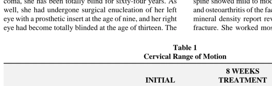
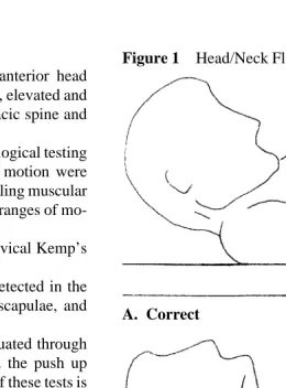
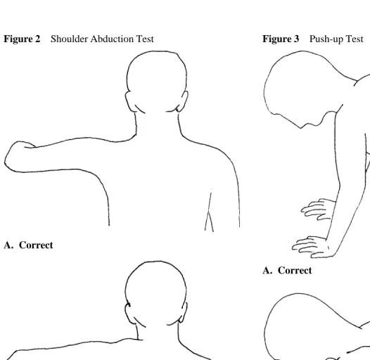
The quality of muscle function was evaluated through head/neck flexion, shoulder abduction and the push up tests as described by Janda.4,5 The purpose of these tests is to detect abnormal movement patterns indicating muscle imbalance. On head/neck flexion a positive test of chin poking indicates tight sternocleidomastoid and suboc-cipitals and inhibited deep neck flexors (see Figure 1). With the shoulder abduction test, shoulder elevation or rotation prior to 60 degrees abduction indicates overactive upper trapezius and/or levator scapulae and inhibited lower scapular stabilizers (see Figure 2). During the push-up test, winging of the scapula indicates inhibited serratus anterior and tight pectoralis muscles (see Figure 3). The patient demonstrated a positive result for all three tests.
On the Neck Disability Index6 she scored 67% disabled. Verbally she rated her pain as 9 out of 10.
Conservative chiropractic rehabilitative management was implemented including spinal mobilizations, myo-fascial trigger point techniques, patient generated muscle stretching and strengthening, proprioceptive neuromuscu-lar facilitation techniques, and postural correction and education.
The most useful spinal mobilization technique for this patient was thoracic extension mobilization performed with the patient seated. The myofascial trigger points were treated by deep direct pressure applied for 10 seconds per point using a “Triggerizer,” a plastic device with a rubber tip designed for this purpose. The intensity of the therapy was kept below the threshold of pain in order not to invoke a defensive reflex contraction.7
The muscles which required stretching were the upper
trapezii, suboccipitals, SCM, levator scapulae, pectoralis major and minor. The muscles which required strengthen-ing were the deep neck flexors, rhomboids, serratus ante-rior, middle and lower trapezii. The deep neck flexors were strengthened by head/neck retractions. Low resist-ance exercise tubing was used to strengthen the lower scapular stabilizers.
It also is important when dealing with the proximal crossed syndrome not to ignore the lumbopelvic region. Lower body imbalances affect the overall posture and if left untreated would contribute to/or sustain an upper body postural disorder. Lewit8 states the most important imbal-ance in the lumbopelvic region is between weak gluteal muscles with hyperactive hip flexors, and hyperactive lumbar erector spinal with weak abdominal muscles. The patient was taught pelvic tilt and pelvic bridge exercises to A. Correct
B. Incorrect
strengthen her abdominal and gluteal muscles which were found to be weak.
In proprioceptive neuromuscular facilitation (PNF), neuromuscular re-education is the goal.2 Some of the PNF techniques used were contract-relax, hold relax and rythmic initiation.9,10 With contract-relax technique, the goal is to increase passive range of motion and facilitate
relaxation. The joint is taken to the end range of passive motion. A concentric contraction of moderate intensity, allowing some motion, is resisted by the clinician for ap-proximately 10 seconds. Following complete relaxation, A. Correct
B. Incorrect
Figure 2 Shoulder Abduction Test
B. Incorrect
Figure 3 Push-up Test
the segment is moved into an increased range. The pattern is repeated usually until no further motion is gained. Hold-relax is similar to contract-Hold-relax except there is no move-ment produced. A light isometric contraction is produced instead. The goals are to reduce pain and to increase pas-sive range of motion. These two techniques were used to increase the range of motion of the cervical spine by stretching the sternocleidomastoid, upper trapezii, levator scapulae and suboccipital muscles. With rythmic initia-tion, the goals are to improve control and co-ordination and to teach a desired direction of motion. The clinician actively moves the target body part. The technique progresses from passive, to active assisted, to active re-sisted motion. The movements taught to the patient by this technique were head/neck retraction, shoulder retraction, chest up position and performing a posterior pelvic tilt. These movements are important in achieving proper postural control and awareness.
Postural correction was achieved through in-office re-petitive repositioning of the patient and prescribed home exercises. Specific postural exercises included:
1 Bruegger’s Position2,11 of external arm rotation, shoulder abduction and retraction of the scapulae (see Figure 4).
This position with simultaneous head and neck re-traction is a strengthening exercise for deep neck flexors and lower scapular stabilizers.
2 Postural Realization Exercise.12
The patient is positioned with their back against a wall. Their shoulder blades and buttocks should be in contact with the wall. They are then instructed to retract their head and neck until the posterior occiput contacts the wall. They are to hold for 10 seconds.
Hourly repetition of these exercises is recommended. The patient was also placed on a daily walking program, stressing proper gait and posture. During walking, the pa-tient was instructed to focus on maintaining the move-ments learned through PNF. These were head retraction, shoulder retraction, chest up and maintaining a posterior pelvic tilt.
The patient was seen 3 times per week for 2 weeks followed by 2 times per weeks for 6 weeks and once per week for 4 weeks.
After eight weeks she reported symptomatic relief and
objective findings of improved posture, increased ranges of motion and improvement of muscle imbalances were noted. She reported good exercise compliance. However, the patient reported that she could not sustain a proper head and neck posture due to her blindness. This was prob-lematic as her head and neck positioning resulted in con-tinued upper cervical hyperextension (see Figure 5 ) and improper head/neck flexion. At this point the patient was instructed to make a fist, placing her fifth finger on top of her manubrium. She was then instructed to lower her chin down to meet the top of her fist and perform an isometric contraction. This approximated a level head posture for her. (see Figure 6). The patient was instructed to perform this manoeuvre at least once per hour, everyday. After four weeks the patient reported, and it was observed, that she was able to maintain a level head position.
At twelve weeks, subjectively she reported a decrease in her pain score from 9 to 4 out of ten. Her rating on the Neck Disability Index dropped from 67% to 30%, indicating improvement in self perceived disability. The range of mo-tion of her cervical (see Table 1) and thoracic (see Table 2) spine increased. The quality of muscle movement im-proved on her head/neck flexion, push up and shoulder abduction tests.
follow up assessment at 1, 2 and 3 months post discharge, with no significant change in symptoms or function. The patient chose to continue further treatment on an as needed basis.
Discussion
Special circumstances taken into consideration for the re-habilitation of this patient were that she was both elderly and blind. Ammendolia13 states that contraindications to exercise are more prevalent in older patients. Proper screening of the older patient is important but so is the ability to modify a treatment program. Ammendolia also states that the key to rehabilitating the older patient is the emphasis on early activation.
Grod14 outlined a few of the goals of chiropractic treat-ment for the elderly as:
•
relief of pain and discomfort•
improvement of quality of life•
prevention of disability.He also states that when spinal manipulation is contra-indicated, treatment may include mobilization, soft tissue treatment, trigger point therapy, stretching and exercise as alternatives.
In the present study, consideration was given regarding the patient’s chronological age and medical history in de-vising her treatment program. Treatment modifications included soft tissue therapy, mobilizations, and doctor and patient generated PNF stretches. Modifications to exercise included a prescribed walking program and use of low resistance exercise tubing instead of more vigor-ous exercise.
Blind patients lack the optical righting reflex that, along with the tonic labyrinthine reflex, helps to maintain the upright and vertical posture of the head. Chusid3 outlines the neuroanatomy of the optic nerve connection to the su-perior colliculi to the pontine nuclei, via the corticopontine tract, for postural reflexes. The presented patient exhibited this inability to maintain postural integrity of her head. Welsh and Blasch15 observed that anterior head carriage and an increased thoracic kyphosis were common postural findings in the blind. These postural findings, observed in the presented patient, are also characteristic of the proxi-mal crossed syndrome.
Baker-Nobles and Bink’s16 literature review provides evidence that tactile and vestibular deficiencies are preva-lent in the blind population. They presented three case studies of blind patients undertaking sensory integrative treatment. Improvements were noted in mobility, activities Figure 5 Anterior head carriage with upper cervical
hyperextension
of daily living, writing and behaviour.
A study by Potter and Muzzin17 at CMCC suggested that blindness, however, does not significantly affect cer-vical ranges of motion.
Van Benschoten18 introduced a sensory integration pro-gram to 18 blind children. The children’s performance over six weeks improved in vestibular integration, lessen-ing of fear of movement, and in some, improved integra-tion of reflexes.
These studies illustrate how increased proprioceptive input as a treatment modality may be useful in increasing that functional abilities of the blind.
With regard to the present case study, PNF was used to improve posture and help to correct the proximal crossed syndrome of a blind 77-year-old patient. One procedure that was found to be most helpful was the proprioceptive input of repetitive placement of the fist under the chin for approximation of a level head posture. A possible hypoth-esis is that this manoeuvre would increase stimulation of the Type I and Type II mechanoreceptors in the cervical spine. The Type I mechanoreceptors are found in the superfical layer of the joint capsule.19 The greatest number of these receptors are located in the cervical spine. The Type II mechanoreceptors are found in the deeper layer of the joint capsule.19 They function to help maintain static posture and modify neurological reflexes involved in ac-tive joint movement. It is possible that repetiac-tive proprio-ceptive stimulation of these receptors increases kinesthetic awareness, compensating for the lack of the optical right-ing reflex in the blind. Further studies are required, how-ever, to determine if such result may be replicated, prior to drawing such as conclusion.
Conclusions
Low tech rehabilitation may be successfully used to treat the challenging patient, in this case, an elderly blind woman with proximal crossed syndrome. Very little is known about posture, postural pain or musculoskeletal habilitation in the blind or the aged. Further study is re-quired on these populations. An observation by Welsh and Blasch15 that anterior head carriage and an increased tho-racic kyphosis were common findings in the blind suggests that postural problems may be more prevalent in this popu-lation. Further study is also needed on the special needs of the blind for rehabilitation of postural problems, given their lack of visual and environmental cues.
References
1 Lewit K. The functional approach. J Orthopedic Medicine 1994; 15:73–74.
2 Liebenson C. Rehabilitation of the Spine: A Practitioner’s Manual. Baltimore: Williams and Wilkins, 1996;
97–112,196.
3 Chusid JG. Correlative Neuroanatomy and Functional Neurology, Nineteenth Edition. California: Lange Medical Publications, 1985; 55,56,113.
4 Janda V. Muscle Function Testing. London: Butterworth, 1983.
5 Chapman-Smith D. The Chiropractic Report. Rehabilitation and Chiropractic Practice, Toronto, July 1996:4.
6 Vernon H, Mior S. The Neck Disability Index: A study of reliability and validity. JMPT 1991; 14(7):409.
7 Schater RC. Chiropractic Management of Sports and Recreational Injuries, Second Edition. Baltimore: Williams and Wilkins, 1986, 235,236.
8 Lewit K. Manipulative Therapy in Rehabilitation of The Locomotor System. London: Butterworth & Co., London, 1985.
9 Adlers S, Beckers D, Buck M. Proprioceptive Neuromuscular Facilitation in Practice; An Illustrated Guide. Springer-Verlag, 1993.
10 Knott M, Voss D. Proprioceptive Neuromuscular Facilitation, Harper and Row, 1968.
11 Skaggs CD. Rehabilitative Management of Orofacial Pain. Chiropractic Rehabilitation Lecture, CMCC, Toronto, 1998.
12 Waerlop IF. Lecture on Whiplash, Headache and Vertigo Diagnosis, Management and Rehabilitation 1997 CMCC, Toronto.
13 Ammendolia C. Rehabilitation of the older patient: a case report. JCCA 1998; 42(1):42–45.
14 Grod JP. Chiropractic and the aging population. Canadian Chiropractor. Toronto: PIC Publishing, February 1997; 23–24.
15 Welsh RL, Blasch BB. Foundations of Orientation and Mobility, New York: American Foundation for the Blind Inc., 1980; 66–68.
16 Baker-Nobles L, Bink MP. Sensory integration in the rehabilitation of blind adults. Am J Occup Ther 1979; 33(9):559–564.
17 Potter BA., Muzzin MA. Cervical Motion Assessment of the Legally Blind: Investigative Project 1988 CMCC, Toronto.
18 Van Benschoten R. A sensory integration program for blind campers. Am J Occup Ther 1975; 29(10):615–617. 19 Wyke B. The neurology of joints. Ann R Coll Surg
England 41, 1967.