RVC OPEN ACCESS REPOSITORY – COPYRIGHT NOTICE
This is the peer-reviewed, manuscript version of the following article:
Mehl, N. S., Khalid, M., Srisuwatanasagul, S., Swangchan-uthai, T. and Sirivaidyapong, S. (2016) 'GnRH-agonist implantation of prepubertal male cats affects their reproductive performance and testicular LH receptor and FSH receptor expression', Theriogenology, 85(5), 841-848.
The final version is available online via
http://dx.doi.org/10.1016/j.theriogenology.2015.10.031.
© 2016. This manuscript version is made available under the CC-BY-NC-ND 4.0 license http://creativecommons.org/licenses/by-nc-nd/4.0/.
The full details of the published version of the article are as follows:
TITLE: Prognostic factors for 1-week survival in dogs diagnosed with meningoencephalitis of unknown aetiology
AUTHORS: N.S. Mehl, M. Khalid, S. Srisuwatanasagul, T. Swangchan-uthai, S. Sirivaidyapong
JOURNAL TITLE: Theriogenology VOLUME/EDITION: 85/5
PUBLISHER: Elsevier
GnRH-agonist implantation of pre-pubertal male cats affects their reproductive 1
performance and testicular LHR and FSHR expression 2
3
4
5
N.S. Mehla, M. Khalidb, S. Srisuwatanasagulc, T. Swangchan-uthaia, S. Sirivaidyaponga* 6
aDepartment of Obstetrics, Gynaecology and Reproduction, Faculty of Veterinary Science, Chulalongkorn 7
University, Bangkok, Thailand. 8
b Department of Production and Population Health, The Royal Veterinary College, Hertfordshire, The United 9
Kingdom. 10
c Department of Anatomy, Faculty of Veterinary Science, Chulalongkorn University, Bangkok, Thailand. 11
12
13
14
*Corresponding author: S.Sirivaidyapong, Tel: +662 2189776, Email: sudson.s@chula.ac.th
15
Abstract 17
This study was conducted to investigate the effect of GnRH-agonist implantation in
18
pre-pubertal tomcats on sexual behavior, reproductive performance and expression of
19
testicular LHR and FSHR, and also to compare the testicular characteristics, LHR and FSHR
20
expression between pre-pubertal and adult tomcats. In Exp1, 3 months-old tomcats
21
(n=6/group) were either treated with or left without 4.7 mg Deslorelin implants. Semen
22
collection and evaluation were performed just before castration at 48 wks after treatment;
23
removed testes were analyzed for mRNA and protein expression of LHR and FSHR. We
24
were able to collect semen from six non-treated cats, whereas in treated cats, semen was
25
uncollectable. The results revealed that sexual behavior was absent in the implanted cats
26
throughout the study period. Testicular volume was decreased found from 30 wks after
27
treatment onwards in the implanted cats compared to the controls (P < 0.05). Semen
28
production was found only in non-implanted cats. Testicular tissue score, seminiferous tubule
29
diameter and LHR protein expression was found lower in the implanted cats (P < 0.05) but no
30
differences were observed in mRNA expression of LHR and protein expression of FSHR
31
between groups. The mRNA expression of FSHR was higher in the implanted (P < 0.05)
32
compared to control cats. In Exp2, testes from pre-pubertal (n=6) and adult (n=6) male cats
33
were collected after castration and analyzed for mRNA and protein expression of LHR and
34
FSHR. No differences were observed in the protein expression of LHR and FSHR between
35
the two groups, while mRNA expression of FSHR was higher in pre-pubertal cats (P < 0.05).
36
Testicular and epididymal weight, diameter of seminiferous tubules and the testicular grade
37
were higher in the adult compared to pre-pubertal cats (P < 0.05). In conclusion, deslorelin
38
implants suppressed protein expression of LHR and enhanced mRNA expression of FSHR
39
along with suppression of reproductive function without any adverse effects for at least 48
40
wks in male cats.
42
Key Words: GnRH-agonist, Pre-pubertal cat, LHR, FSHR, Reproductive behaviour
43
Introduction 44
Overpopulation of cats is a serious global problem and in big towns/cities roaming of
45
tomcats is reported to be out of owners’ control. The result is unwanted pregnancies with
46
undesirable consequences in this species. Free roaming cats without any care are also
47
subjected to higher risks like suffering from diseases (including zoonotic diseases),
48
malnutrition and accidents. The number of cats euthanized in shelters is also on the rise every
49
year mainly due to overpopulation [1] which needs to be controlled in order to address the
50
welfare problems associated with it.
51
Contraception is one of the most successful methods for population control in many
52
animal species. Traditional way of contraception by castration is presently in practice in cats
53
as well. However, castration is an invasive surgical procedure and can only be performed on
54
anesthetized animals, whereas anesthesia poses serious problems in juvenile and senile cats
55
and in cats with health problems. Cats reach puberty by the age of 4 month [2] with a
56
possibility of mating soon after. However, surgical neutering in early age may pose risks like
57
higher sensitivity to many drugs including the anesthetics [3]. Therefore, nonsurgical
58
neutering could be a welfare-friendly and viable alternative to surgical methods of neutering
59
[4].
60
Reproduction in mammals is controlled by the hypothalamic-pituitary-gonadal (HPG)
61
axis and it has been shown that long-term continuous administration of GnRH
62
desensitizes/downregulates the pituitary gland, profoundly suppresses the gonadotrophins
63
release and impairs the reproductive function [5]. Accordingly, a contraceptive method has
64
been developed; it is employed by GnRH-agonist implantation (Suprelorin®; Peptech Animal
65
Health), and has been proven effective in pubertal tomcats [5, 6] and female domestic cats
8]. This method results into long-term reversible contraception without any negative effects
67
to the animals. The contraceptive effects of GnRH-agonist have also been reported in other
68
species such as dogs, wild felids, gilts, flying fox and giraffes [9-15]. Moreover, Trigg, Doyle
69
[16] have reported that when 4 months old female pups were implanted with 9.4 mg of
70
deslorelin, contraceptive effect was prolonged and lasted for at least 36 wks while the
71
contraceptive effect in pubertal dogs was varied from 24 to 48 wks. It is a possibility that this
72
longevity effect might have been achieved by a delay in the age of puberty in these animals.
73
Moreover, there are reports to suggest that early-age neutering could reduce undesirable
74
behaviour of cats especially in adopted cats and could help reduce the unwanted litters in
75
many pet shelters. Although GnRH implantation has been used in cats to suppress the
76
reproductive function but the studies in pre-pubertal cats are rare and with variable results
77
[17, 18].
78
The effects of GnRH implantation on the gonadotrophins’ release along with the
79
suppression of reproductive function are well documented [5, 19]. However, it is not known
80
whether such effects are achieved through an alteration in the gonadal expression of receptors
81
for LH and FSH and/or testosterone production. The present study was, therefore, designed
82
to investigate the effects of long-term GnRH implantation [4.7 mg GnRH-agonist
83
(Deslorelin)] on the reproductive performance, testicular morphology and expression of LHR
84
and FSHR in pre-pubertal male cats. Testicular morphology and expression of LHR and
85
FSHR were also compared between pre-pubertal and adult male cats.
86
87
2. Materials and methods 88
2.1Experiment design and animals 89
Experiment 1: Three months old tomcats that were proven to be clinically healthy and
90
had attended a complete vaccination program were either implanted with 4.7 mg deslorelin
GnRH-agonist (Suprelorin® 4.7mg, Virbac Animal Health, France) in the interscapular area
92
(Deslorelin implanted; n=6) or left without any implantation and served as controls
(Non-93
implanted; n=6). The cats were housed together in an open-air room with natural daylight in
94
the Department of Obstetrics, Gynaecology and Reproduction, Faculty of Veterinary Science,
95
Chulalongkorn University, Thailand. During the study period animals were fed with a
96
commercial diet twice daily with water always available ad libitum. The study had ethical
97
approval and was performed under the license of Chulalongkorn University Laboratory
98
Animal Center number13310056.
99
Implanted animals were monitored for any potential adverse effects like tissue reaction at
100
the implantation site and/or infection, rashes, oedema, erythema of implantation area etc for a
101
period of one week. Body temperature was measured daily for one week after the hormonal
102
implantation to monitor any infection and if found, blood was collected for profile
103
monitoring.
104
Body weight of all the cats in both the groups was recorded fortnightly until the end of
105
the experiment (48 wks) when both the testes were collected after surgical castration.
106
Throughout the experimental period, functional evaluations of the reproductive organs such
107
as penile spines, testicular volume and consistency, and male sexual behavioural
108
characteristics were monitored at 2-weekly intervals in all the cats. Presence of penile spine
109
was taken as a criterion of puberty [5]. Length, width and depth of the scrotum/testis were
110
measured using vernier calipers and testicular volume was calculated with a modified
111
spherical equation; volume (cm3) = 4/3 x π x (1⁄2 length x 1⁄2 width x 1⁄2 depth) [5].
112
Testicular consistency was recorded by palpation by one observer and was noted as soft, firm
113
or hard.Male sexual behaviour such as marking, mounting (with or without intromission),
114
and fighting [20] were observed for at least 30 min at 2-weekly intervals in all the cats.
115
Faeces were collected at 2-weekly intervals to measure testosterone concentrations. An
attempt was made to collect semen from all the cats before surgical castration by using the
117
electro-ejaculator; which was performed 48 wks after implantation. Soon after collection,
118
semen was evaluated for its volume, colour, motility, concentration, viability and sperm
119
morphology. If semen ejaculation could not be accomplished, epididymal sperms were
120
collected immediately after castration and evaluated.
121
Experiment 2: Testes were collected from 4 to 6 months old (pre-pubertal, n = 6) or 1 to
122
3 years old (adult, n=6) normal healthy male cats after surgical castration at the Small Animal
123
Hospital, Faculty of Veterinary Science, Chulalongkorn University, Thailand.
124
In both the experiments, after castration, weight of each testis and epididymis was
125
recorded. Each testis was divided into two parts; one part was fixed in 4% (w/v)
126
paraformaldehyde for 48 to 72 hours and then stored in 70% ethanol until processing for
127
cytology/morphology and immunohistochemistry, whereas the other part was snap frozen in
128
liquid nitrogen and stored in -80 ºC until RNA extraction.
129
2.2Luteinizing hormone receptor (LHR) and Follicle stimulating hormone receptor (FSHR) 130
Expression 131
Fixed testicular tissues were embedded in paraffin wax and cut into 5µm sections by a
132
rotor microtome, applied to gelatin-coated slides and left to dry in an incubator at 37ºC.
133
Sections were deparaffinized with Xylene (J.T. Baker, PA, USA) and rehydrated through
134
ascending concentrations of alcohol (50%, 70%, 90%, 99.7% and 100%). The
135
immunohistochemical staining was performed as described previously by Ponglowhapan et al. 136
[21]. Briefly, the tissue sections/slides were placed in boiling 0.01M sodium citrate solution,
137
then cooled down to room temperature for 35 mins to de-mask epitopes. Slides were then rinsed
138
three times in phosphate buffered saline (PBS). Endogenous peroxidase activity was
139
inactivated by immersing slides in 1% (v/v) hydrogen peroxide in methanol for 10 min, then
140
rinsed again three times in PBS. Sections were subsequently blocked for 60 min in a humidified
chamber using a blocking solution, comprising 1% normal horse serum (Vector Laboratories,
142
CA, USA) diluted in PBS and 20% (v/v) avidin solution (Avidin/Biotin blocking kit; Vector
143
Laboratories, CA, USA). After washing slides three times in PBS, the slides were incubated
144
overnight at 4ºC in a humidified chamber with LHR (H–50) polyclonal antibody (Santa Cruz
145
biotechnology, Inc., USA) at a dilution of 1:50 or with FSHR (N–20) polyclonal antibody
146
(Santa Cruz biotechnology, Inc., USA) at a dilution of 1:50. The primary antibodies were
147
diluted in PBS to which 20% (v/v) biotin solution (Avidin/Biotin blocking kit; Vector
148
Laboratories, CA, USA) was added. The negative control sections were treated in the same
149
manner with PBS and biotin mixture in the absence of primary antibodies. After incubation,
150
sections were washed with PBS three times (3 x 10 minutes). Then, secondary antibody
151
(Biotinylated anti-mouse anti-rabbit IgG, Vector Laboratories, Inc., USA for LHR localization
152
and Biotinylated anti-goat IgG, Vector Laboratories, Inc., USA for FSHR localization) were
153
applied to the sections and incubated for 30 min. Sections were washed again three times in
154
PBS and incubated at room temperature with 20% (v/v) avidin-biotin complex solution
155
(VECTASTAIN® Vector Laboratories, Inc., USA) for 30 min. Tissue sections were then
156
incubated with DAB peroxidase substrate (Vector Laboratories, Inc., USA) until colour
157
development. All slides were counterstained with Mayer’s hematoxylin. Brown staining was
158
observed on tissue sections with positive staining for both LHR and FSHR and no staining was
159
observed for negative controls for either receptor.
160
At least two sections for both positive antibody staining and negative controls were
161
examined from each animal.
162
163
2.3Quantification of immunohistochemical staining 164
The pattern and intensity of protein staining for LHR and FSHR were determined
165
semi-quantitatively using a histochemical score (HSCORE) method. Ten fields per
section of each tissue sample were assessed blind by one assessor using a light
167
microscope at X 200 magnification. The intensity of staining was classified on a scale of
168
1-3, where 1 = weak staining, 2 = moderate staining and 3 = strong staining [21, 22].
169
Histochemical score (H-SCORE) was assessed as percent of each level (weak, moderate
170
or strong) of staining in each tissue area with the Image-pro plus 7.0 program (Media
171
Cybernetics, Inc. MD, USA). An expression index (EI) was calculated for each tissue
172
sample based on the percentage of positively stained cells and the intensity of staining
173
using the following formula:
174
EI = % total stained cells x [(1 x % weak) + (2 x % medium) + (3 x %
175
strong)]/100
176
A mean expression index was calculated to represent the protein expression of LHR
177
or FSHR in each testicular section of every testis from an individual animal [22-24].
178
179
2.4Morphology of Testes 180
Testes collected in both the experiments were fixed, embedded in paraffin wax, cut into
181
5µm sections and stained with hematoxylin and eosin. The stained sections (5 sections/testis)
182
were evaluated for seminiferous tubules; those with normal basement membrane (basement
183
membrane with a continuous line and germ cells well attached) [19] were considered as normal
184
and functional. Diameter of seminiferous tubules in each tissue section was measured using
185
ocular micrometer at X 200 magnification. Using the criteria of Novotny et al. [19] a total of
186
200 seminiferous tubules per section were classified as st0, st1, st2, st3 and st4 if they had only
187
spermatogonia (st0), only spermatogonia and spermatocytes (st1), or with spermatids (st2), or
188
with elongating spermatids (st3) or with elongated spermatids (st4) present in the lumen. Each
189
tissue section was graded into 5 grades (0 to 4), based on the majority of seminiferous tubules
found in the tissue sections; Grade 0: Testicular tissue with the majority of st0 seminiferous
191
tubules, Grade 1: Testicular tissue with the majority of st1 seminiferous tubules, Grade 2:
192
Testicular tissue with the majority of st2 seminiferous tubules, Grade 3: Testicular tissue with
193
the majority of st3 seminiferous tubules, and Grade 4: Testicular tissue with the majority of st4
194
seminiferous tubules. Any pathological changes in the testes, if present, were investigated and
195
recorded.
196
197
2.5Quantitative real-time polymerase chain reaction (qPCR) for the LHR and FSHR mRNA 198
in the testicular tissue 199
2.5.1 Extraction and reverse transcription of mRNA 200
Frozen testicular tissue was ground with a homogenizer at 10,000 to 20,000 RPM for 10
201
to 20s and used to extract the total RNA by the RNeasy mini kit (QIAGEN®, Alameda, CA,
202
USA) following the manufacturer’s instructions. Concentration and purity of the extracted
203
RNA were assessed by spectrophotometer (ND-2000, NanoDrop,Wilmington, DE, USA).
204
The RNA samples were stored at -70ºC before qPCR analysis.
205
2.5.2 Quantitative real-time PCR 206
Conventional PCR was performed and the PCR product was used for the preparation of
207
standards and analyzing the optimal melting and annealing temperature for each gene [LHR,
208
FSHR and GAPDH (reference gene)]. The thermal cycler (G-Storm Thermal Cycler,
209
Somerset, United Kingdom) was set at the conditions of 15 min at 95ºC to activate Taq DNA
210
polymerase, 30 cycles of 30s at 94ºC for denaturing, 90s at 57ºC for annealing, 30s at 72ºC
211
for extension and 10 min at 72º C for the final extension. Previously published sequences of
212
forward and reverse primers for feline LHR and FSHR, and GAPDH were used [25, 26] and
213
are shown in Table 1. Each reaction was contained with Qiagen Multiplex PCR Kit
214
(QIAGEN®, Alameda, CA, USA). Amplified products were run on 1.2% agarose gel
(SIGMA-ALDRICH®, St, Louis, MO, USA) and visualized under UV gel document and
216
analysis (SYNGENE® Cambridge, United Kingdom)to confirm the presence of single
217
products without dimers. Purification of the amplified products was performed with the
218
QIAquik PCR purification kit (QIAGEN®, Alameda, CA, USA). Purified products were
219
quantified by spectrophotometer (ND-2000, NanoDrop,Wilmington, DE, USA) and used to
220
prepare standards for use in qPCR assessment.
221
Real-time qPCR amplification was performed using CFX96 Thermal cycler (Bio-Rad
222
Laboratories, Inc., Hercules, CA, USA) with the Bio-Rad CFX manager 3.1 software (Bio-Rad
223
Laboratories, Inc., Hercules, CA, USA). Each reaction (20µl) was contained with 10µl of 2x
224
qPCR BIO SyGreen Mix Lo-ROX (PCR Biosystems Ltd, London, United Kingdom), 0.8µl of
225
each forward and reverse primer, 5µl of a DNA template (5ng/µl), and the volume made up to
226
20µl with RNase free water. RNase free water was added instead of cDNA template in the
227
Non-template control (NTC). Thermocycler was set for 38 cycles of denaturing at 95ºC for 5s
228
following with the optimum annealing temperature of 61.4ºC, 60ºC and 61.4ºC for 25s and
229
melting temperature of 82ºC, 80ºC and 76ºC for 10s for GAPDH, FSHR and LHR, respectively
230
with a gradient from 50 to 95ºC to investigate the gene expression. Standards of each gene
231
were used as controls to determine the absolute quantity of mRNA (fg/μg of total RNA).
232
2.6Statistical analysis 233
Body weight and testicular volume were compared between the deslorelin implanted and
234
non-implanted (Expt 1) animals using Independent T-test.
235
General linear model (GLM) was performed to compare the protein and mRNA expression
236
of LHR and FSHR and the epididymal weight between the deslorelin implanted and
non-237
implanted (Expt 1) and between the pre-pubertal and adult (Expt 2) animals. Wilcoxon rank
238
sum test was performed to compare the testicular weight, the mean diameter of seminiferous
tubules and the grade of seminiferous tubules between the deslorelin implanted and
non-240
implanted cats (Expt 1) and between the pre-pubertal and adult cats (Expt 2)
241
3. Results 242
No tissue reaction and/or infection were observed after deslorelin implantation.
243
No difference in body weight was recorded between deslorelin implanted and non-implanted
244
cats (Figure 1). The implanted cats had significantly lower (P<0.05) testicular volume from wk
245
30 of study onwards (Figure 2). Male sexual behaviour was absent in implanted cats but was
246
present in non-implanted cats from 28 wks onwards of the study period. However, testicular
247
consistency was soft in both groups and remained soft in deslorelin implanted cats throughout
248
the study while from the 28th wk until the end of study period non-implanted cats had
249
comparatively firmer testicular consistency. Penile spines in non-implanted cats were present
250
from the 28th wk onwards of study period and were absent in implanted cats throughout the
251
study period of 48 wks. Higher faecal testosterone levels (P<0.05) were observed in
non-252
implanted compared to deslorelin implanted cats from 20 wks onwards of study period (Figure
253
3). It was possible to collect semen from all of non-implanted cats., semen volume were 10 –
254
73 μl with 40 – 60% of sperm motility, 49 – 72% of sperm viability and sperm concentration
255
at 0.15 x 10⁶ –18 x 106 sperms/ml. Not only implanted cats failed to ejaculate but also we were
256
not able to collect epididymal sperm from them.
257
Testicular and epididymal weight, testicular grade and mean seminiferous tubule diameter were
258
higher in the adult as well as non-implanted cats compared to pre-pubertal or implanted cats (P
259
< 0.05) (Table 2).
260
LHR and FSHR were localized in the cytoplasm of Leydig and germ cells, respectively. LHR
261
expression was significantly higher (p<0.05) in the non-implanted compared to deslorelin
262
implanted tomcats whereas no difference was found between adult and pre-pubertal animals.
FSHR expression was not different between adult and pre-pubertal cats or between implanted
264
and non-implanted cats (Figures 4, 5).
265
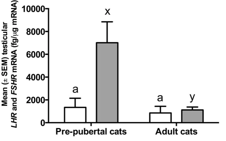
LHR and FSHR mRNA were expressed in all the testicular samples collected in both the
266
experiments. No differences were observed in the expression of LHR mRNA expression
267
between the groups in either experiment. The expression of FSHR mRNA, however, was
268
significantly higher (P < 0.05) in the pre-pubertal and deslorelin implanted cats compared to
269
adult and non-implanted cats, respectively (Figures 6 and 7).
270
271
4. Discussion 272
The objectives of this study were to compare 1) the testicular charateristics, and both
273
protein and mRNA expression of LHR and FSHR between pre-pubertal and adult tomcats and
274
2) to investigate the effect of GnRH-agonist implantation on the sexual behavior, reproductive
275
performance and the testicular LHR and FSHR expression in pre-pubertal tomcats.
276
Deslorelin implantation which was done without any anesthesia, local or general, was
277
very well tolerated by male pre-pubertal cats as has been reported in previous studies [5, 17].
278
The sexual behaviour of implanted cats was suppressed and many unwanted behaviours such
279
as spraying, fighting and roaming were totally absent in these cats. This suppression of
280
behaviors resulting from Deslorelin implantation was comparable with behaviours eliminated
281
by surgical castration [27]. Moreover, suppression of physiology of the reproductive organs
282
such as, the grade of testicular tissue, the seminiferous tubules diameter, and the weight of
283
testes and epididymides of implanted cats, which were significantly lower compared to
non-284
implanted cats, confirms the action of Deslorelin implantation on suppressing the function of
285
male reproductive tract.
286
The physiology and sexual behavior of male mammals is mainly controlled by
287
testosterone which is produced by the activation of Leydig cells by the LH released from the
pituitary gland [28, 29]. Aromatase and 5-α reductase transform testosterone into estrogen and
289
dihydrotestosterone, respectively and these two hormones are considered to be responsible for
290
the change in the male behaviour. However, testosterone itself acts on the Sertoli cells of the
291
testis to support spermatogenesis. FSH is believed to have an important role in the first wave
292
of spermatogenesis in pre-pubertal mammals but its role in spermatogenesis in adults remains
293
to be confirmed. However, FSH is considered to induce meiosis during spermatogenesis
294
process and therefore, is responsible for increasing the number of spermatogonia in the
295
seminiferous tubules [30].
296
The HPG axis is activated by the release of GnRH from the hypothalamus which
297
stimulates the release of gonadotropins from the pituitary gland to regulate the reproductive
298
function. The chronic administration of GnRH down regulates the pituitary GnRH receptors
299
and suppresses the release of gonadotropins and the reproductive function [31]. It is for this
300
reason that GnRH-agonist implantation is used as an alternative to surgical castration in a
301
number of species including felines. In the present study, we have tried to confirm this in
pre-302
pubertal male cats and to explore whether such a GnRH therapy suppresses the reproductive
303
function via an involvement of testicular expression of LHR and FSHR. FSHR is expressed in
304
the Sertoli cells of the testes and is responsible to control spermatogenesis after activation by
305
the FSH [32], whereas LHR being expressed in the Leydig cells, is responsible to stimulate
306
androgen secretion [33] by activating the biosynthetic pathway that changes cholesterol into
307
testosterone [34].
308
In the present study, the significantly higher expression of FSHR mRNA in the testicular
309
tissue of deslorelin implanted male cats compared to untreated controls could be a result of the
310
compensatory mechanism resulting from the suppression of the endogenous release of GnRH
311
(and/or FSH) due to implantation of deslorelin (GnRH-agonist). It seems that translational
312
pathway has also been affected by deslorelin implantation as no difference was observed in the
protein expression of FSHR, even though the mRNA expression of the FSHR was significantly
314
higher in the implanted group. As in pre-pubertal mammals FSH is known to plays a major
315
role in the first wave of spermatogenesis [30, 35] but in pubertal mammals, spermatogenesis is
316
mainly androgen-dependent and the effect of FSH is limited mainly to support the production
317
of spermatogonia [30], the absence of sperm production observed in deslorelin-implanted cats
318
therefore seems to result from the suppression of testosterone production due to the
319
downregulation of LHR in the Leydig cells of deslorelin-treated cats.
320
In this study, we observed that faecal testosterone concentrations and
testosterone-321
dependent sexual behaviour were both suppressed in deslorelin implanted compared with the
322
non-implanted cats. We also found that the LHR protein expression in deslorelin implanted
323
cats was suppressed compared with the non-implanted cats. As testosterone production
324
depends on the activation of LHR in the Leydig cells, it therefore, seems highly likely that the
325
observed suppression of faecal testosterone and the testosterone-dependent behaviours may be
326
the result of observed testicular suppression of LHR protein in the implanted cats.
327
Both mRNA and protein expression of the LHR were studied in deslorelin-implanted and
328
control cats. Deslorelin suppressed the LHR protein expression but was without any effect on
329
the LHR mRNA. These results suggest that deslorelin downregulates the LHR by interfering
330
at the translational level but do not interfere transcription of the gene. Surprisingly, no
331
difference was observed in the protein or mRNA expression of LHR between the adult and
pre-332
pubertal cats. This may indicate that at the age of 3 months cats already had active Leydig cells
333
that are capable to produce testosterone. It is difficult to estimate the exact time period after
334
which deslorelin might have been effective to suppress the LHR in the implanted cats because
335
the testes were collected only 48 wk after the implantation. However, there was no difference
336
in the faecal testosterone concentrations between the two groups at the time of implantation
337
but from wk20 onwards testosterone concentrations in the implanted group started to be
significantly lower than the controls. This may suggest that the LHR suppression might have
339
resulted sometimes within the 20 wks period of deslorelin implantation. However, it remains
340
to be determined whether testosterone suppression in deslorelin-implanted cats was the result
341
of LHR suppression only or was a result of the combined effect of suppression in the LH
342
concentrations and LHR. Moreover, this study was not designed to investigate how the
343
deslorelin implantation directly or indirectly might have affected the biosynthetic pathway of
344
testosterone production that may involve changes in the cAMP to stimulate the transport of
345
cholesterol into the mitochondria and changes in the activities of different enzymes responsible
346
for the pregnenolone, progesterone, androstenone and finally testosterone production [29]. We
347
speculate that the effect of deslorelin may not be only at the level of LH production and/or
348
testicular LHR expression but may also be at other sites in the biosynthetic pathway of
349
testosterone production. A suppression of pulsatile LH secretion from long-term GnRH-agonist
350
(Goserelin) treatment has already been reported in gilts [15] and several male characteristics
351
such as the presence of penile spines and male behaviours such as roaming, fighting and
352
spraying are gonadal steroid hormones (especially testosterone) dependent and could be
353
eliminated via the suppression of testosterone [27].
354
Normally GnRH-agonist implantation presents an upregulation effect in the first period
355
after hormonal implantation followed by a downregulation effect after long-term
356
administration in pubertal tomcats [5]. However, in this study we did not observe any
357
upregulation effect when male cats were implanted with GnRH-agonist at the age of 3 months
358
possibly because of an immature HPG axis at this age [17].
359
This study was not designed to investigate whether the observed suppression of the
360
reproductive function could be reversed or not. However, non-reversibility of reproductive
361
function has been reported in dogs that were implanted with GnRH agonist before the age of 4
362
months [16, 36]. Reversibility of reproductive function has been reported in studies using 1.6
mg deslorelin in post-natal cats and 4.7 mg deslorelin in 114 days old female cats, puberty was
364
postponed until the age of 16 months and 134 to 286 days, respectively [17, 18] . However, in
365
this study it remains to be seen whether reproductive function could be reinstated or not,
366
nevertheless, the results obtained show that reproductive function remained suppressed for a
367
period of at least 48 wks after 4.7 mg deslorelin administration at the age of 3 months.
368
In conclusion, the results of the present study have shown that implantation of 4.7 mg
369
GnRH-agonist (Deslorelin®) in male cats at the age of 12 wks suppresses the reproductive
370
function for at least for 48 wks without any adverse effects on the general health. Moreover,
371
this suppression of reproductive function may be achieved partly by down-regulation of LHR
372
in the Leydig cells while maintaining the FSHR expression at the pre-pubertal levels in the
373
Sertoli cells of the testis.
374
375
5. Acknowledgement 376
This study was supported by the Royal Golden Jubilee Ph.D. programme, the 90th
377
Aniversary of Chulalongkorn University (Ratchadaphiseksomphot Endowment Fund) and the
378
Research Unit of Obstetric and Reproduction in animals of Chulalongkorn University,
379
Bangkok, Thailand. We are thankful to Dr Zhangrui Cheng for laboratory assistance and Dr
380
Em-on Olanratmanee for help in statistical analysis of the data.
381
382
References 383
[1] Joshua MF, Pamela LC. Analysis of programs to reduce overpopulation of companion
384
animals: Do adoption and low-cost spay/neuter programs merely cause substitution of
385
sources? Ecol Econ. 2007;62:740 - 6.
386
[2] Johnston SD, Root Kustritz MV, Olson PNS. Sexual differentiation and Normal Anatomy
387
of the Tom Cat. Canine and Feline Theriogenology. 1 ed. Philadelphia, PA 19106:
388
Saunders Elsevier; 2001. p. 497-507.
389
[3] Joyce A, Yates D. Help stop teenage pregnancy! Early-age neutering in cats. J Feline Med
390
Surg. 2011;13:3-10.
391
[4] Spain CV, Scarlett JM, Houpt KA. Long-term risks and benefits of early-age
392
gonadectomy in cats. J Am Vet Med Assoc. 2004;224:372-9.
[5] Goericke-Pesch S, Georgiev P, Antonov A, Albouy M, Wehrend A. Clinical efficacy of a
394
GnRH-agonist implant containing 4.7 mg deslorelin, Suprelorin, regarding
395
suppression of reproductive function in tomcats.
Sandra.Pesch@vetmed.uni-396
giessen.de. Theriogenology. 2011;75:803-10.
397
[6] Munson L. Contraception in felids. Theriogenology. 2006;66:126-34.
398
[7] Munson L, Bauman JE, Asa CS, Jochle W, Trigg TE. Efficacy of the GnRH analogue
399
deslorelin for suppression of oestrous cycles in cats. J Reprod Fertil Suppl.
400
2001;57:269-73.
401
[8] Toydemir TS, Kilicarslan MR, Olgac V. Effects of the GnRH analogue deslorelin
402
implants on reproduction in female domestic cats. Theriogenology. 2012;77:662-74.
403
[9] Wright PJ, Verstegen JP, Onclin K, Jochle W, Armour AF, Martin GB, et al. Suppression
404
of the oestrous responses of bitches to the GnRH analogue deslorelin by progestin. J
405
Reprod Fertil Suppl. 2001;57:263-8.
406
[10] Trigg TE, Wright PJ, Armour AF, Williamson PE, Junaidi A, Martin GB, et al. Use of a
407
GnRH analogue implant to produce reversible long-term suppression of reproductive
408
function in male and female domestic dogs. J Reprod Fertil Suppl. 2001;57:255-61.
409
[11] Herbert CA, Trigg TE. Applications of GnRH in the control and management of fertility
410
in female animals. Anim Reprod Sci. 2005;88:141-53.
411
[12] Wiebe VJ, Howard JP. Pharmacologic advances in canine and feline reproduction.
412
Topics in companion animal medicine. 2009;24:71-99.
413
[13] Patton ML, Bashaw MJ, del Castillo SM, Jochle W, Lamberski N, Rieches R, et al.
414
Long-term suppression of fertility in female giraffe using the GnRH agonist deslorelin
415
as a long-acting implant. Theriogenology. 2006;66:431-8.
416
[14] Melville DF, O'Brien GM, Crichton EG, Theilemann P, McKinnon A, Johnston SD.
417
Reproductive seasonality and the effect of the GnRH agonist deslorelin as a
418
contraceptive in captive male Black Flying-foxes (Pteropus alecto). Theriogenology.
419
2012;77:652-61.
420
[15] Peltoniemi OAT, Easton BG, Love RJ, Klupiec C, Evans G. Effect of chronic treatment
421
with a GnRH agonist (Goserelin) on LH secretion and early pregnancy in gilts. Anim
422
Reprod Sci. 1995;40:121 - 33.
423
[16] Trigg TE, Doyle AG, Walsh JD, Swangchan-uthai T. A review of advances in the use of
424
the GnRH agonist deslorelin in control of reproduction. Theriogenology.
425
2006;66:1507-12.
426
[17] Carranza A, Faya M, Merlo ML, Batista P, Gobello C. Effect of GnRH analogs in
427
postnatal domestic cats. Theriogenology. 2014;82:138-43.
428
[18] Risso A, Corrada Y, Barbeito C, Diaz JD, Gobello C. Long-term-release GnRH agonists
429
postpone puberty in domestic cats. Reproduction in Domestic Animals.
2012;47:936-430
8.
431
[19] Novotny R, Cizek P, Vitasek R, Bartoskova A, Prinosilova P, Janosovska M. Reversible
432
suppression of sexual activity in tomcats with deslorelin implant. Theriogenology.
433
2012;78:848-57.
434
[20] Rosenblatt JS, Aronson LR. The decline of sexual behavior in male cats after castration
435
with special reference to the role of prior sexual experience. Journal of Behviour.
436
1958;12:285 - 338.
437
[21] Ponglowhapan S, Church DB, Scaramuzzi RJ, Khalid M. Luteinizing hormone and
438
follicle-stimulating hormone receptors and their transcribed genes (mRNA) are
439
present in the lower urinary tract of intact male and female dogs. Theriogenology.
440
2007;67:353-66.
[22] Chowdhury MWH, Scaramuzzi RJ, Wheeler-Jones CPD, Khalid M. The expression of
442
angiogenic groeth factors and their receptors in ovarian follicle throughout the estrous
443
cycle in the ewe. Theriogenology. 2010;73:856 - 72.
444
[23] Ishibashi H, Suzuki T, Suzuki S, Moriya T, Kaneko C, Takizawa T, et al. Sex steroid
445
hormone receptors in human thymoma. J Clin Endocrinol Metab. 2003;88:2309-17.
446
[24] Ponglowhapan S, Church DB, Khalid M. Differences in the expression of luteinizing
447
hormone and follicle-stimulating hormone receptors in the lower urinary tract
448
between intact and gonadectomised male and female dogs. Domest Anim Endocrinol.
449
2008;34:339-51.
450
[25] Tharasanit T, Thongkittidilok C, Sananmuang T, Techakumphu M. Recombinant Human
451
Follicle Stimulating Hormone and Growth Factors Improve the Meiotic and
452
Developmental Competence of Cat Oocyte. Thai J V M. 2014;44:107 - 15.
453
[26] Sano J, Nagafuchi S, Yamazaki J, Oguma K, Kano R, Hasegawa A. Effect of
454
antineoplastic drugs on the expression of Bcl-2 and Bcl-xL genes in the feline T-cell
455
leukemia cell line. Res Vet Sci. 2005;79:197-201.
456
[27] Hart BL, Eckstein R. The role of gonadal hormones in the occurrence of objectionable
457
behaviours in dogs and cats. . Appl Anim Behav Sci. 1997;52:331 - 41.
458
[28] Smith LB, walker WH. The regulation of spermatogenesis by androgens. Semin Cell
459
Dev Biol. 2014;30:2 - 13.
460
[29] Midzak AS, Chen H, Papadopoulos V, Zirkin BR. Leydig cell aging and the mechanisms
461
of reduced testosterone synthesis. Mol Cell Endocrinol. 2009;299:23-31.
462
[30] O'Shaughnessy PJ. Hormonal control of germ cell development and spermato genesis.
463
Semin Cell Dev Biol. 2014;29:55 - 65.
464
[31] Goericke-Pesch S, Georgiev P, Fasulkov I, Vodenicharov A, Wehrend A. Basal
465
testosterone concentrations after the application of a slow-release GnRH agonist
466
implant are associated with a loss of response to buserelin, a short-term GnRH
467
agonist, in the tom cat. Theriogenology. 2013;80:65-9.
468
[32] Nieschlag E, Simoni M, Gromoll J, Weinbauer GF. Role of FSH in the regulation of
469
spermatogenesis: clinical aspects. Clin Endocrinol (Oxf). 1999;51:139-46.
470
[33] Chauvigne F, Zapater C, Gasol JM, Cerda J. Germ-line activation of the luteinizing
471
hormone receptor directly drives spermiogenesis in a nonmammalian vertebrate. Proc
472
Natl Acad Sci U S A. 2014;111:1427-32.
473
[34] Bjelic MM, Stojkov NJ, Baburski AZ, Sokanovic SJ, Mihajlovic AI, Janjic MM, et al.
474
Molecular adaptations of testosterone-producing Leydig cells during systemic in vivo
475
blockade of the androgen receptor. Mol Cell Endocrinol. 2014;396:10-25.
476
[35] Meehan T, Schlatt S, O'Bryan MK, de Kretser DM, Loveland KL. Regulation of germ
477
cell and Sertoli cell development by activin, follistatin, and FSH. Dev Biol.
478
2000;220:225-37.
479
[36] Sirivaidyapong S, Mehl NS, Trigg TE. Delay of puberty and reproductive performance
480
in male dogs following the implantation of 4.7 and 9.4 mg GnRH-agonist deslorelin at
481
an early pre-pubertal age. Reproduction in Domestic Animals. 2012;47 Suppl
6:400-482
2.
483 484
Table 1: Description of forward and reverse primers for GAPDH as housekeeping gene and
486
feline LH receptor (LHR) and FSH receptor (FSHR) as target genes.
487
Gene Primer sequence (5′–3′)
Length
(bp) Reference
GAPDH
F: GGAGAAAGCTGCCAAATATG 20
[25] and [26]
R: AGGAAATGAGCTTGACAAAGTGG 23
LHR
F: CTAATGCCTTTGACAACCTAATA 23
[25]
R: CCCATTGAATGCATGACTTTGTA 23
FSHR
F: CATGCTGCTAGGCTGGATCTT 21
[25]
R: CTTGGCGATCTTGGTGTCACT 21
488
489 490
Table 2: Mean ± SEM values for weight (g) of the testes and epididymides, seminiferous
491
tubule diameter (μm), and grade of testicular tissues obtained from tomcats in both the
492
experiments.
493
Expt Groups
Weight (g)
Seminiferous tubule diameter (μm)
Testicular tissue grade Testes Epididymides
1
Implanted
0.09 ±
0.02a 0.03 ± 0.01a 62.02 ± 2.88a 0.00 ± 0.00a
Non-implanted
1.54 ±
0.20b 0.26 ± 0.03b 98.48 ± 3.59b 3.86 ± 0.17b
2
Adult
1.24 ±
0.20x 0.24 ± 0.03x 95.31 ± 3.94x 3.83 ± 0.41x
Prepubertal
0.19 ±
0.04y 0.08 ± 0.02y 57.85 ± 8.50y 1.33 ± 0.21y 494
Values within a column with different superscript letters (a, b and x, y) in an experiment differ
495
significantly (P < 0.05).
Figure legends
497498
Figure 1: The mean (± SEM) body weight (kg) in cats (n=6/group) with or without deslorelin
499
implantation. Cats were implanted with deslorelin at the age of 3 months for a period of 48
500
weeks.
501
503
Figure 2: The mean (± SEM) testicular volume (cm3) in cats (n=6/group) with or without
504
deslorelin implantation. Cats were implanted with deslorelin at the age of 3 months for a
505
period of 48 weeks. Black arrow indicates the week that the control group has significantly
506
higher (p < 0.05) testicular volume than the implanted group.
507
509
Figure 3. The mean (± SEM) faecal testosterone levels (ng/g of faeces) in cats (n=6/group)
510
with or without deslorelin implantation. Cats were implanted with deslorelin at the age of 3
511
months for a period of 48 weeks. Black arrow indicates the week when the control group
512
started to have significantly higher faecal testosterone levels than the implanted group (P <
513
0.05).
514
515
517
Figure 4. The mean expression index (± SEM) for LHR and FSHR in testicular tissue of
518
pubertal and pre-pubertal cats (n=6/group). Different letters on bars for a certain receptor
519
indicate significant differences (P ≤ 0.05).
520
522
523
524
525
Figure 5. The mean expression index (± SEM) for LHR and FSHR in testicular tissue of cats
526
(n=6/group) with or without deslorelin implantation. Cats were implanted with deslorelin at
527
the age of 3 months for 48 weeks. Different letters on bars for a certain receptor indicate
528
significant differences (P ≤ 0.05).
529
531
Figure 6. The mean mRNA concentration (fg/μg mRNA) (± SEM) for LHR and FSHR in
532
testicular tissue of pubertal and pre-pubertal cats (n=6/group). Different letters on bars for a
533
certain receptor indicate significant differences (P ≤ 0.05).
534
536
Figure 7. The mean mRNA concentration (fg/μg mRNA) (± SEM) for LHR and FSHR in
537
testicular tissue of cats (n=6/group) with or without deslorelin implantation. Cats were
538
implanted with deslorelin at the age of 3 months for 48 weeks. Different letters on bars for a
539
certain receptor indicate significant differences (P ≤ 0.05).
540