1
Dynamics of Bcl-2Protein Phosphorylation:
A
Mini Review
Abdul Manan1*, Sidra Ilyas2
1- Institute of Molecular Biology and Biotechnology (IMBB), The University of Lahore, Lahore, Pakistan
2- University of the Punjab, Dept. of Microbiology and Molecular Genetics (MMG), Lahore, Pakistan.
*Corresponding Author: [email protected]
2 Abstract
The regulation of apoptosis depends upon the Bcl-2 protein family. The process of cell death and
survival is highly complicated and regulated by various types of extrinsic as well as intrinsic
network of biological system. Several enzymes and regulators play crucial role in cell death and
survival cycle not only in healthy but also in pathological state particularly in cancer. In
cancerous cells, various proto-oncogenes and anti-apoptotic proteins are activated and
responsible for the cell survival and longevity. The mechanism of activation and inactivation of
various proteins in cell survival is regulated by the process of phosphorylation (kinases) and
dephosphorylation (phosphatases). The current review will summarize the dynamics of Bcl-2
phosphorylation and its role in apoptosis and cell survival.
______________________________________________________________________________
Keywords: apoptosis, cancer, phosphorylation, kinases, Bcl-2
Introduction
Apoptosis or cellular suicide is a genetically programmed process that is important for
recycling of damaged cells, tissue homeostasis and embryonic development. The activation
mechanism of apoptosis requires extrinsic and intrinsic pathways where various enzymes
including cysteine proteases called as “caspases” play a crucial role (Poreba et al., 2019). The
activation of caspases at molecular level depends upon the phosphorylation (Parrish et al., 2013).
In extrinsic pathway, activated death receptors like tumor necrosis factor receptor (TNFR),
TNF-related apoptosis-induced ligand (TRAIL) and first apoptosis signal (Fas) recruits the initiator
caspases (caspase8 and caspase10) (Wang and El-Deiry, 2003; Tummers and Green, 2017).
Contrarily, in intrinsic pathway, stress releases cytochrome c from mitochondria leading to the
formation of a complex called “apoptosome” that in turn activate caspase 9 (initiator
caspase)which recruit caspase 3 (executioner caspase) in the same way asin extrinsic pathway
(Parrish et al., 2013).
Apoptosis is regulated positively by pro-apoptotic protein Bax and negatively by
anti-apoptotic Bcl-2 family proteins both containing Bcl-2homology (BH) domain (Chipuk and
Green, 2008; Brunelle and Letai, 2009). Bcl-2 not only reverses the effect of Bax but also halts
the release of cytochrome c from mitochondria (Greenhalf et al., 1996; Gross et al., 2000).
3
phosphorylation in which phosphate group from ATP is transferred to one of the amino acid
residues of serine, tyrosine or threonine residues by protein kinases (PKs). Imbalance between
anti- and pro-apoptotic proteins may lead to inability to respond appropriately to apoptotic
stimuli and growth of malignant cells and ultimately cancer that is why cancerous cells do not
respond well to treatment (Petros et al., 2004).
In biological system, the regulation of various cellular processes such as signal
transduction pathways, apoptosis, cell cycle and growth depend upon proteins. After translation,
protein undergoes various changes collectively called post-translational modifications (PTMs)
(Hill et al., 2019). These modifications include glycosylation, nitrosylation, methylation,
ubiquitination, acetylation, proteolysis, lipidation as well as most important phosphorylation
(main topic of the current review). These modifications are not only associated with normal
cell’s physiology but also the pathological state. Understanding the role of PTMs is very critical
for the investigation of disease prevention and treatment (Wang et al., 2014; Zhao and Jensen,
2009).
Phosphorylation
Phosphorylation and dephosphorylation are two important biological processes catalyzed
by protein kinases (PKs) and phosphatases that add or remove a phosphate group of proteins
respectively. Study of these momentous processes helps in understanding of not only human
health but also the pathological aspects of various disorders including cancer (Seternes et al.,
2019). Unique and complete balancing of phosphorylated proteins within a biological system
defines its phosphoproteome which is more dynamic than its proteome (Franck et al., 2015).
Phosphorylation acts as a molecular switch to turn the protein in on/off state (Ardito,
Fatima, et al. 2017; Colin et al., 2008). Under the influence of phosphorylation three crucial
aspects such as interactions, degradation and location control of proteins have been investigated.
About one third of proteins undergo phosphorylation and majority is phosphorylated at multiple
sites in a single protein. Most common residues are serine and threonine amino acids whereas
other amino acids such as tyrosine, glutamic acid, aspartic acid and histidine are also subjected to
phosphorylation (Puttick et al., 2008; Hunter, 2009). Higher magnitude of tyrosine
phosphorylation has been observed in activated platelets (Ferrell and Martin, 1988). However,
most of the cells exhibit highest level of phosphorylation on serine residue and threonine amino
4 Bcl-2 family proteins
The Bcl-2 family protein members and contain one to four functional homology domains
(BH1 to BH4) (Petros et al., 2004). Interestingly, Mcl-1 and Bcl-xL show anti-apoptotic role
similar to Bcl-2 protein, whereas Bad, Bid and Bax show pro-apoptotic activity. Several
members of anti-apoptotic proteins contain all four BH regions including Ced-9, Bcl-xL, Bcl-w
and Bcl-2. Moreover, BHRF1 protein, KSHV-Bcl-2 and Mcl-1 exhibit strong homology
sequence in three BH regions including BH1, BH2 and BH3 domains. BH3 region of
pro-apoptotic proteins exhibit interaction with anti-pro-apoptotic members and are responsible for
promoting programmed cell death (Letaiet al., 2002; Kvansakul and Hinds, 2013).
The 3D structure of Bcl-2 family members exhibit two central α-helices (hydrophobic in
nature) that are surrounded by six or seven amphipathic α-helices having varying lengths.
Interestingly, a prominent hydrophobic groove can be observed on the surface of anti-apoptotic
proteins and the residues of this groove are the binding sites for various pro-apoptotic proteins
like Bad and Bax containing peptides that mimic BH3 region. Several Bcl-2 family proteins
contain a carboxy-terminal hydrophobic region that is accepted for mitochondrial membrane
localization. Moreover, membrane localization behavior of anti- and pro-apoptotic proteins is not
similar. Bcl-2 and Bcl-xL are localized at cytoplasmic surface of mitochondrial outer membrane;
anchoring of these proteins may play a critical role associated with the integrity of mitochondria.
Pro-apoptotic members that are present in cytoplasm only localize to outer membrane of
mitochondria when they get activated (Petros et al., 2004).
Bcl-2 protein undergoes several types of phosphorylation. One important type occurs
during mitosis in un-stressed cells, involving a small proportion of Bcl-2 (Choi and Zhu, 2019).
Moreover, investigations by researchers revealed that Thr56, CDK1/cyclin B and JNK were
found to be associated with phosphorylation of Bcl-2 protein in mitosis (Yamamoto et al., 1999;
Furukawa et al., 2000; Du et al., 2004). Cells treated with anti-mitotic drugs undergo stress
which lead to high levels of phosphorylation as observed in all Bcl-2 members and several
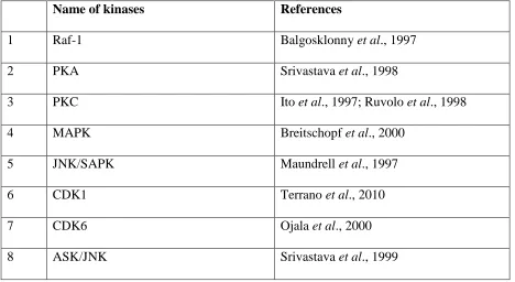
5 Table 1: Various kinases involve in Bcl-2 phosphorylation
Name of kinases References
1 Raf-1 Balgosklonny et al., 1997
2 PKA Srivastava et al., 1998
3 PKC Ito et al., 1997; Ruvolo et al., 1998
4 MAPK Breitschopf et al., 2000
5 JNK/SAPK Maundrell et al., 1997
6 CDK1 Terrano et al., 2010
7 CDK6 Ojala et al., 2000
8 ASK/JNK Srivastava et al., 1999
Phosphorylation of Bcl-2 protein
Under the influence of apoptotic stimuli, Bcl-2 protein performs survival function
through inhibition of cytochrome c release from mitochondria. Multiple sites within the Bcl-2
protein have been phosphorylated including Serine at position (70 and 87) and Threonine (56 and
75). Phosphorylation of these sites may be a marker for mitotic cell division as well as targets for
ASK/MKK7/JNK1 pathway in anti-cancer therapies (Park et al., 2019).
Bcl-2 phosphorylation in response to a range of treatments is specific to stimuli as well as
cell type which lead to the activation or inactivation of anti-apoptotic behavior (Table 2; Figure
1). Role of Bcl-2also depends upon mitochondria, movement of ions and small molecules
necessary for induction of cell death. Bcl-2 is present in mitochondria, endoplasmic reticulum
and nuclear envelope (Krajewski et al., 1993; Popgeorgiev et al., 2018). Association of Bcl-2
with nuclear matrix is reflecting its important feature in genomic organization (Wang et al.,
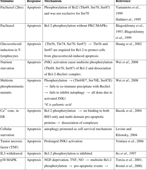
6 Table 2: Bcl-2 phosphorylation, apoptosis and cell survival
Stimulus Response Mechanism Reference
Paclitaxel (2hrs) Apoptosis Phosphorylation of Bcl2 (Thr69, Ser70, Ser87)
and was not exclusive for Ser70
Yamamoto et al.,
1999
Haldaret al., 1995
Paclitaxel Apoptosis Bcl-2 phosphorylation without PKC/MAPKs Blagosklonny et al.,
1997; Blagosklonny
et al., 1999
Glucocorticoid
induction in
T-lymphocytes
Apoptosis (Thr56, Thr74, Ser70, Ser87) → Thr56 and
Ser87 are required for Bcl-2 to protect cells
from glucocorticoid-induced apoptosis
Huang et al., 2002
Nutrient
starvation
Apoptosis JNK1 activation cause multisite phosphorylation
(Thr69, Ser70, Ser87) of Bcl-2 and dissociation
of Bcl-2-Beclin1 complex
Wei et al., 2008
Multisite
phosphomimetic
mutants
Apoptosis Phosphorylation → (Thr69E*, Ser70E, Ser87E)
→ fails to co-immuno precipitate with Beclin1 → fails to inhibit autophagy → all done due to
activated JNK1
*E is gultamic acid
Wei et al., 2008
Ca2+ conc. in
ER
Apoptosis Bcl-2 phosphorylation → no binding to both
BH3-only and multi-domain pro-apoptotic
proteins → dissociation of complexes
Bassik et al., 2004
Cellular
starvation
Apoptosis autophagy promoted as cell survival mechanism Levine and
Klionsky, 2004
Tumor necrosis
factor (TNF)
Apoptosis Prolonged JNK1 activation Ventura et al., 2006
IL3-withdrawal Apoptosis Bcl-2 phosphorylation is inhibited Ito et al., 1997
p38 MAPK Apoptosis NGF-deprivation, TNF, NO → multisite Bcl-2
phosphorylation → pro-apoptotic events →
Torcia et al., 2001;
7
release of cytochrome c → in both cellular
system and cell free experiments
Rosini et al., 2004;
Ishikawa et al.,
2003; Tamura et al.,
2000
p38 MAPK Cell
survival
Bcl-2 phosphorylation → specific targets
(Thr56 and Ser87) → alter anti-apoptotic
potential of Bcl-2
De Chiara et al.,
2006
Specific kinases Cell
survival
Bcl-2 phosphorylation decreased anti-apoptotic
function
Srivastava et al.,
1999; Fan et al.,
2000; Torcia et al.,
2001; De Chiara et
al., 2006
Nutrient
starvation
Cell
survival
Alanine mutants of Bcl2 (Thr69Ala*, Ser70Ala,
Ser87Ala) lead to binding of Bcl-2-Beclin1
complex
Wei et al., 2008
IL3-induced
model
Cell
survival
Bcl-2 phosphorylation lead to increased
anti-apoptotic potential
Ito et al., 1997
Ginsenoside-Rh2 induction
Cell
survival
Transient JNK1 activation Ham et al., 2003
Taxol-induced
model
Cell
survival
hyperphosphorylation of various kinases →
including Raf-1 kinase → Bcl-2
phosphorylation → strong inhibition of
anti-apoptotic potential
Blagosklonnyet al.,
8 Figure 1 Overview of Phosphorylation, apoptosis and cell survival
Glucocorticoid-induced apoptosis and Bcl-2
Mitochondria are involved in steroid-induced apoptosis which depends largely upon the
phosphorylation of Bcl-2 protein. Cell shrinkage appears to be important feature of apoptosis
(Huang and Cidlowski, 2002). It has been observed that Ser70 mutation did not abolish
protective effect of Bcl-2 against dexamethasone-induced shrinkage, whereas mutation at Thr56,
and 74 and Ser87 abolished the potential of Bcl-2 to inhibit steroid induced cell shrinkage.
Anti-apoptotic behavior of Bcl-2 is inhibited during glucocorticoid-induced apoptosis of
T-lymphocytes under the influence of mutation at Thr56 or Ser87 (Huang and Cidlowski, 2002).
In lymphoid cells, apoptosis is induced by glucocorticoids whereas, over-expression of Bcl-2
block apoptosis induced by glucocorticoids. Huang and Cidlowski (2002) prepared five
transfected Bcl-2 cell lines of WEHI 7.1 cells expressing either alanine mutants of Thr56, Thr74,
Ser70 or Ser87. Mutation at Thr56 and Ser87 completely abolished the anti-apoptotic property of
Bcl-2 whereas mutation at Ser70 did not alter its ability to block glucocorticoid-induced
apoptosis. Interestingly, Thr74 mutation partially disturbed the role of Bcl-2 (Huang and
9 JNK and IL-3 induced phosphorylation of Bcl-2
Anti-apoptotic role of Bcl-2 may be enhanced by JNK and/or IL-3 induced
phosphorylation at Ser70in murine myeloid cells lacking growth factor (Deng et al., 2001;May et
al., 1994).Phosphorylation of Bcl-2 was associated with increased cell survival upon the addition
of IL-3 in chemotherapeutic-induced (drug etoposide) apoptosis (Ruvolo et al., 2001).
Bryostatin-1 induced vigorous Bcl-2 phosphorylation and promotes cell survival by acting as a
strong protein kinase C (PKC) agonist and found alternate for IL-3 in cell survival. PKC was
observed to be specific Bcl-2 kinase responsible for its phosphorylation in the same serine
residues both in vivo and in vitro experiments. Seven common phosphorylation sites were found
in human and murine Bcl-2 at serine 24, 70, 102, 158, 164, 202 and 213. Moreover, it was
investigated that only Ser70 mutation makes the Bcl-2 negative anti-apoptotic in its function
reflecting physiological relevance of Bcl-2 phosphorylation at Ser70 (Kennelly and Krebs, 1991;
May et al., 1994; Ito et al., 1997).
JNK mediated phosphorylation of Bcl-2
JNK mediated phosphorylation of Bcl-2 protein is associated not only with programmed
cell death but also with autophagy. JNK mediated phosphorylation has been observed to interfere
with binding of Bcl-2 to apoptotic BH3 domain-containing proteins such as Bax and
pro-autophagy BH3 domain-containing proteins such as Beclin-1 (Wei et al., 2008). Kinetic
relationship and correlation of 2 with Bax and Beclin-1, caspase-3 activation as well as
Bcl-2 phosphorylation was investigated during nutrient starvation (Wei et al., 2008). The nutrient
conditions are responsible to regulate the binding of Bcl-2 and Beclin-1 proteins (Pattingre et al.,
2005). This binding was minimum during nutrient stress and maximum when the nutrients were
in excess. Dissociation of Beclin-1 from Bcl-2 during starvation implicates phosphorylation of
Bcl-2 by stress-activated signaling molecule, JNK1 (Wei et al., 2008). During nutrient
deprivation, activated JNK induces phosphorylation at multiple sites of Thr69, Ser70 and Ser87
in the non-structured loop of Bcl-2 (located between BH3 and BH4 domains).
Alanine (Thr69Ala, Ser70Ala and Ser87Ala) and non-phosphorylatable Bcl2 mutants,
eliminates the starvation-induced separation of Bcl-2 and Beclin-1 and halts autophagy. While,
Bcl-2 phosphomimetic mutant (Thr69Glu, Ser70Glu and Ser87Glu) failed to
co-immuno-precipitate with Beclin-1 and no autophagy inhibitory activity was observed indicate that JNK
10
autophagy (Wei et al., 2008). Moreover, Bcl-2 phosphorylation suppresses its binding to both
BH3-only and multi-domain pro-apoptotic proteins and encourages apoptosis (Bassik et al.,
2004). It has been reported that autophagy is activated as a cell survival mechanism during
nutrient deprivation (Levine and Klionsky, 2004), while delayed cellular nutrient withdrawal
finally leads to apoptosis. In association with the mechanism of autophagy, temporary JNK
activation encourages cell survival in contrast to prolonged JNK activation mediates cell death
(Ventura et al., 2006; Ham et al., 2003).
Sphingolipid-ceramide and phosphorylation of Bcl-2
Sphingolipid-ceramide is well known for its apoptogenic behavior by playing crucial role
in signal transduction pathways and in cellular stress (Jarvis et al., 1996). Ceramide can activate
mitochondrial protein phosphatase (PP2A) which is responsible for apoptosis via
dephosphorylation of Bcl-2, phosphorylated Bcl-2 is responsible for cell survival. Ceramide is
also responsible for activation of ceramide-activated protein phosphatase (CAPP). However,
dynamic phosphorylation/dephosphorylation of Bcl-2 may act as survival sensor during stress
stimuli (Dobrowsky et al., 1993; Westwick et al., 1995). Interestingly, Bcl-2 protein has been
observed to undergo phosphorylation at various amino acid residues (multiple site
phosphorylation) reflecting both apoptotic and anti-apoptotic role (Ruvolo et al., 2001).
Conclusion
Bcl-2 phosphorylation in response to a range of treatments is specific to stimuli as well as cell
type. Phosphorylation sites for Bcl-2 protein identified by investigators are most commonly
observed at serine (70 and 87), and threonine (56, 69, 74 and 75). Moreover, transient activation
of JNK1 leads to the cell survival while prolonged JNK1 activation leads to cell death reflecting
dual role of Bcl-2 protein due to phosphorylation.
Conflict of interest
The authors declare no conflict of interest.
References
Ardito F, Giuliani M, Perrone D, Troiano G, Lo Muzio L. The crucial role of protein phosphorylation
in cell signaling and its use as targeted therapy. International journal of molecular medicine.
2017; 40(2):271-80.
Bassik MC, Scorrano L, Oakes SA, Pozzan T and Korsmeyer SJ. Phosphorylation of BCL-2
11 Berg JM, Tymoczko JL andStryer L. Covalent Modification Is a Means of Regulating Enzyme
Activity.InBiochemistry. 5th edn., New York: WH Freeman; 2002.
Blagosklonny MV, Chuman Y, Bergan RC, Fojo T. Mitogen-activated protein kinase pathway is
dispensable for microtubule-active drug-induced Raf-1/Bcl-2 phosphorylation and apoptosis
in leukemia cells. Leukemia. 1999 Jul;13(7):1028-36.
Blagosklonny MV, Giannakakou P, el-Deiry WS, Kingston DG, Higgs PI, Neckers L and Fojo T.
Raf-1/bcl-2 phosphorylation: a step from microtubule damage to cell death. Cancer Res.,
1997; 57: 130-135.
Blagosklonny MV, Schulte T, Nguyen P, Trepel J, Neckers LM. Taxol-induced apoptosis and
phosphorylation of Bcl-2 protein involves c-Raf-1 and represents a novel c-Raf-1 signal
transduction pathway. Cancer Research. 1996; 56(8):1851-4.
Breitschopf K, Haendeler J, MalchowP, Zeiher AM and Dimmeler S. Post-translational modification
of bcl-2 facilitates its proteasome-dependent degradation: molecular characterization of the
involved signaling pathway. Mol Cell Biol., 2000; 20: 1886-1896.
Brunelle JK andLetai A. Control of mitochondrial apoptosis by the Bcl-2 family. J Cell Sci., 2009;
122: 437-441.
Chipuk JE and Green DR. How do BCL-2 proteins induce mitochondrial outer membrane
permeabilization? Trends Cell Biol., 2008; 18: 157-164.
Choi HJ, Zhu BT. Upregulated cyclin B1/CDK1 mediates apoptosis following
2-methoxyestradiol-induced mitotic catastrophe: Role of Bcl-XL phosphorylation. Steroids. 2019 Oct
1;150:108381.
Colin E, Zala D, Liot G, Rangone H, Borrell-Pagès M, Li XJ, Saudou F and Humbert S. Huntingtin
phosphorylation acts as a molecular switch for anterograde/retrograde transport in
neurons.EMBO J., 2008; 27(15):2124-34.
De Chiara G, Marcocci ME, Torcia M, Lucibello M, Rosini P, Bonini P, Higashimoto Y, Damonte
G, Armirotti A, Amodei S, Palamara AT. Bcl-2 phosphorylation by p38 MAPK Identification
of target sites and biologic consequences. Journal of Biological Chemistry. 2006 Jul
28;281(30):21353-61.
Deng X, Xiao L, Lang W, Gao F, Ruvolo P and May WS Jr. Novel role for JNK as a stress-activated
Bcl-2 kinase. J Biol Chem., 2001; 276(26):23681-8.
Ding Q, He X, Hsu JM, Xia W, Chen CT, Li LY, Lee DF, Liu JC, Zhong Q and Wang X.
Degradation of Mcl-1 by beta-TrCP mediates glycogen synthase kinase 3-induced tumor
12 Dobrowsky RT, Kamibayasha C, Mumby MC andHannun YA. Ceramide activates a heterotrimeric
protein phosphatase 2A. J Biol Chem., 1993; 268: 15523-15530.
Du L, Lyle CS, Obey TB,Gaarde WA, Muir JA, Bennett BL and Chambers TC. Inhibition of cell
proliferation and cell cycle progression by specific inhibition of basal JNK activity evidence
that mitotic Bcl-2 phosphorylation is JNK-independent.J Biol Chem., 2004;
279(12):11957-66.
Ferrell JE Jr and Martin GS. Platelet tyrosine-specific protein phosphorylation is regulated by
thrombin.Mol Cell Biol., 1988; 8(9):3603-10.
Franck WL, Gokce E, Randall SM, Oh Y, Eyre A, Muddiman DC and Dean RA. Phosphoproteome
Analysis Links Protein Phosphorylation to Cellular Remodeling and Metabolic Adaptation
during Magnaportheoryzae Appressorium Development.J Proteome Res., 2015;
14(6):2408-24.
Fulda S, Gorman AM, Hori O, Samali A. Cellular stress responses: cell survival and cell death.Int J
Cell Biol., 2010; 2010: 214074.
Furukawa Y, Iwase S, Kikuchi J, Terui Y, Nakamura M, Yamada H, Kano Y and Matsuda M.
Phosphorylation of Bcl-2 protein by CDC2 kinase during G2/M phases and its role in cell
cycle regulation.J Biol Chem., 2000;275(28):21661-7.
Greenhalf W, Stephan C and Chaudhuri B. Role of mitochondria and C-terminal membrane anchor
of Bcl-2 in Bax induced growth arrest and mortality in Saccharomyces cerevisiae.FEBS Lett.,
1996; 380: 169-175.
Gross A, Pilcher K, Blachly-Dyson E, Basso E, Jockel J, Bassik MC, Korsmeyer SJ and Forte M.
Biochemical and genetic analysis of the mitochondrial response of yeast to BAX and
BCL-X(L), Mol Cell Biol.,2000; 20: 3125-3136.
Ham YM, Chun KH, Choi JS, Kim DH and Lee SK. SEK1-dependent JNK1 activation prolongs cell
survival during G-Rh2-induced apoptosis. BiochemBiophys Res Commun.,
2003;304:358-64.
Hill SM, Wrobel L, Rubinsztein DC. Post-translational modifications of Beclin 1 provide multiple
strategies for autophagy regulation. Cell Death & Differentiation. 2019 Apr;26(4):617-29.
Hotchkiss RS, Strasser A, McDunn JE and Swanson PE. Cell death. N Engl J Med., 2009; 361:
1570-1583.
Huang ST and CidlowskiJA.Phosphorylation status modulates Bcl-2 function during
13 Hunter T. Tyrosine phosphorylation: thirty years and counting. CurrOpin Cell Biol.,
2009;21(2):140-146.
Ito T, Deng X, Carr BK and May WS. Bcl-2 phosphorylation required for anti-apoptosis function. J
Biol Chem., 1997; 272: 11671-11673.
Jarvis WD, Grant S andKolesnick RN. Ceramide and the induction of apoptosis. Clin Cancer Res.,
1996; 2: 1-6.
Kennelly PJ and Krebs EG. Consensus sequences as substrate specificity determinants for protein
kinases and protein phosphatases. J Biol Chem., 1991; 266: 15555-15558.
Kozopas KM, Yang T, Buchan HL, Zhou P and Craig RW. MCL1, a gene expressed in programed
myeloid cell differentiation, has sequence similarity to BCL-2. Proc Natl Acad Sci., 1993;
90: 3516-3520.
Krajewski S, Tanaka S, Takayama S, SchiblerMJ, Fenton W and Reed JC. Investigation of the
subcellular distribution of the bcl-2 oncoprotein: residence in the nuclear envelope,
endoplasmic reticulum, and outer mitochondrial membranes. Cancer Res., 1993; 53:
4701-4714.
Kvansakul M and Hinds MG. Structural biology of the Bcl-2 family and its mimicry by viral
proteins. Cell DeathDiseas., 2013;4: 909.
Letai A, Bassik MC, Walensky LD, Sorcinelli MD, Weiler S andKorsmeyer SJ. Distinct BH3
domains either sensitize or activate mitochondrial apoptosis, serving as prototype cancer
therapeutics. Cancer Cell, 2002; 2:183-192.
Levine B and Klionsky DJ. Development by self-digestion: molecular mechanisms and biological
functions of autophagy. Dev Cell, 2004;6:463-77.
Maundrell K, Antonsson B, Magnenat E, Camps M, Muda M, Chabert C, Gillieron C, Boschert U,
Vial-Knecht E, Martinou JC and Arkinstall S. Bcl-2 undergoes phosphorylation by c-Jun
N-terminal kinase/stress-activated protein kinases in the presence of the constitutively active
GTP-binding protein Rac1. J Biol Chem., 1997; 272: 25238-25242.
Maurer U, Charvet C, Wagman AS, Dejardin E and Green DR. Glycogen synthase kinase-3 regulates
mitochondrial outer membrane permeabilization and apoptosis by destabilization of MCL-1.
Mol Cell, 2006; 21: 749-760.
May WS, Tyler PG, Ito T, Armstrong DK, Qatsha KA and Davidson NE. Interleukin-3 and
bryostatin-1 mediate hyperphosphorylation of Bcl-2a in association with suppression of
14 Morel C, Carlson SM, White FM and Davis RJ. Mcl-1 integrates the opposing actions of signaling
pathways that mediate survival and apoptosis. Mol Cell Biol., 2009; 29: 3845-3852.
Ojala PM, Yamamoto K, Castanos-Velez E, Biberfeld P, Korsmeyer SJ and Makela TP. The
apoptotic v-cyclin-CDK6 complex phosphorylates and inactivates Bcl-2. Nat Cell Biol.,
2000; 2: 819-825.
Park JG, Aziz N, Cho JY. MKK7, the essential regulator of JNK signaling involved in cancer cell
survival: a newly emerging anticancer therapeutic target. Ther Adv Med Oncol.
2019;11:1758835919875574.
Parrish AB, Freel CD and Kornbluth S. Cellular mechanisms controlling caspase activation and
function. Cold Spring HarbPerspect Biol., 2013; 5(6): a008672.
Pattingre S, Tassa A, Qu X, Garuti R, Liang XH, Mizushima N, Packer M, Schneider MD and
Levine B. Bcl-2 antiapoptotic proteins inhibit Beclin 1-dependent autophagy. Cell,
2005;122:927-39.
Petros AM, Olejniczak ET and Fesik SW. Structural biology of the Bcl-2 family of proteins.
Biochimica et Biophysica Acta, 2004; 1644: 83-94.
Popgeorgiev N, Jabbour L, Gillet G. Subcellular localization and dynamics of the Bcl-2 family of
proteins. Frontiers in cell and developmental biology. 2018; 6:13.
Poreba M, Groborz K, Navarro M, Snipas SJ, Drag M, Salvesen GS. Caspase selective reagents for
diagnosing apoptotic mechanisms. Cell Death & Differentiation. 2019 Feb;26(2):229-44.
Puttick J, Baker EN andDelbaere LT. Histidine phosphorylation in biological systems.
BiochimBiophys Acta. 2008; 1784(1):100-5.
Rechsteiner M and Rogers SW. PEST sequences and regulation by proteolysis. Trends Biochem Sci.,
1996; 21: 267-271.
Ruvolo PP, Deng X and May WS. Phosphorylation of Bcl-2 and regulation of apoptosis. Leukemia,
2001; 15: 515-522.
Ruvolo PP, Deng X, Carr BK and May WS. A functional role for mitochondrial protein kinase C
alpha in Bcl-2 phosphorylation and suppression of apoptosis. J Biol Chem., 1998; 273:
25436-25442.
Seternes OM, Kidger AM, Keyse SM. Dual-specificity MAP kinase phosphatases in health and
disease. Biochimica et Biophysica Acta (BBA)-Molecular Cell Research. 2019 Jan
1;1866(1):124-43.
Srivastava RK, Mi QS, Hardwick JM and Longo DL. Deletion of the loop region of Bcl-2 completely
15 Srivastava RK, Srivastava AR,Korsmeyer SJ, Nesterova M, Cho-Chung YS and Longo DL.
Involvement of microtubules in the regulation of Bcl-2 phosphorylation and apoptosis
through cyclic AMP-dependent protein kinase. Mol Cell Biol., 1998; 18: 3509-3517.
Tamura Y, Simizu S andOsada H. The phosphorylation status and anti-apoptotic activity of Bcl-2 are
regulated by ERK and protein phosphatase 2A on the mitochondria.FEBS Lett., 2004;
569(1-3):249-55.
Terrano DT, Upreti Mand Chambers TC. Cyclin-dependent kinase 1-mediated Bcl-xL/Bcl-2
phosphorylation acts as a functional link coupling mitotic arrest and apoptosis. Mol Cell
Biol., 2010;30(3):640-56.
Tummers B, Green DR. Caspase‐8: regulating life and death. Immunological reviews. 2017
May;277(1):76-89.
Ventura JJ, Hubner A, Zhang C, Flavell RA, Shokat KM and Davis RJ. Chemical genetic analysis of
the time course of signal transduction by JNK. Mol Cell., 2006;21:701-10.
Wang S and El-Deiry WS. TRAIL and apoptosis induction by TNF-family death
receptors.Oncogene, 2003; 22(53):8628-33.
Wang YC, Peterson SE and Loring JF. Protein post-translational modifications and regulation of
pluripotency in human stem cells. Cell Res., 2014; 24:143-160.
Wang ZH, Ding MX, Chew-ChengSB, Yun JP and Chew EC. Bcl-2 and Bax proteins are nuclear
matrix associated proteins. Anticancer Res., 1999; 19: 5445-5449.
Wei Y,Pattingre S, Sinha S, Bassik M and Levine B. JNK1-mediated phosphorylation of Bcl-2
regulates starvation-induced autophagy. Mol Cell., 2008;30:678-88.
Westwick JK, Bielawaska AE, Dbaibo G, Hannun YA and Brenner DA. Ceramide activates the
stress-activated protein kinases. J Biol Chem., 1995; 270: 22689-22692.
Yamamoto K, Ichijo H andKorsmeyer SJ. BCL-2 is phosphorylated and inactivated by an ASK1/Jun
N-terminal protein kinase pathway normally activated at G2/M. Mol Cell Biol.,
1999;19(12):8469-78.
Zhao Y and Jensen ON. Modification-specific proteomics: strategies for characterization of