M i t r a l A n n u l a r P l a n e S y s t o l i c E x c u r s i o n a n d T r i c u s p i d An n u l a r
P l a n e S y s t o l i c E x c u r s i o n i n C a t s w i t h H y p e r t r o p h i c C a r d i o m y o p a t h y
I. Spalla, J.R. Payne, K. Borgeat, A. Pope, V. Luis Fuentes, and D.J. Connolly
Background: Left ventricular (LV) systolic dysfunction is associated with increased risk of death in cats with hypertrophic cardiomyopathy (HCM). Mitral and tricuspid annular plane systolic excursion (MAPSE and TAPSE, respectively) are mea-sures of longitudinal systolic function and are reduced in human patients with HCM.
Hypotheses: Cats with HCM have lower MAPSE and TAPSE compared to control cats; lower MAPSE and TAPSE are associated with the presence of congestive heart failure (CHF) and reduced survival time.
Animals: 64 cats with HCM and 27 healthy cats. Forty-five cats with HCM were not showing clinical signs, and 19 had CHF.
Methods: Retrospective study. Anatomic M-mode from the left apical 4-chamber view was used to record MAPSE from the free wall (MAPSE FW) and septum (MAPSE IVS) and TAPSE.
Results: Compared to controls, cats with HCM had lower MAPSE IVS (controls 5.2 [4.6–5.6] mm, asymptomatic HCM
4.7 [4.1–5.2] mm, HCM with CHF 2.6 [2.5–3.2] mm,P<.001), MAPSE FW (controls 5.9 [5.3–6.2] mm, asymptomatic HCM
4.7 [4.1–5.1] mm, HCM with CHF 2.8 [2.4–3.2] mm) and TAPSE (controls 8.6 [7.4–10.2] mm, asymptomatic HCM 7.2 [6.3–
8.2] mm, HCM with CHF 4.6 [4.1–5.4] mm), with the lowest in the CHF group. Univariate survival analysis showed a
shorter survival in cats displaying lower MAPSE IVS, MAPSE FW, and TAPSE.
Conclusions and Clinical Importance:MAPSE and TAPSE were lower in cats with HCM than in control cats and were lowest in CHF, suggesting that systolic longitudinal dysfunction is present in cats with HCM. MAPSE and TAPSE have potential prognostic significance.
Key words: Echocardiography; Feline; Hypertrophic cardiomyopathy.
H
ypertrophic cardiomyopathy (HCM) is the most
common heart disease in cats.
1,2It is characterized
by a hypertrophied left ventricle in the absence of other
cardiovascular or systemic causes.
3Recently, it has been
reported that increased right ventricular wall thickness
is common in cats with HCM and related to the
sever-ity of left ventricular hypertrophy.
4Cats with HCM can
have variable life expectancy; several clinical and
echocardiographic
prognostic
factors
including
left
atrial (LA) size and function, extreme left ventricular
hypertrophy, measures of left ventricular systolic and
diastolic function, presence of congestive heart failure,
and aortic thromboembolism have been associated with
decreased survival in cats with HCM.
5–9Mitral annular plane systolic excursion (MAPSE) and
the right-sided counterpart tricuspid annular plane
sys-tolic excursion (TAPSE) are M-mode-derived indices of
systolic longitudinal displacement of the atrioventricular
annular plane.
10,11MAPSE and TAPSE can therefore
be considered as markers of left ventricular (LV)
10,12and right
ventricular (RV)
11,13long-axis
function,
respectively. It is recognized, however, that left-sided
heart disease can also influence TAPSE.
14,15Currently, there are no published reference values for
MAPSE and TAPSE in cats and no information on
their potential diagnostic and prognostic utility in
HCM.
Study Aims
1
To
provide
preliminary
reference
intervals
for
MAPSE and TAPSE in healthy cats.
2
To determine whether cats with HCM have lower
values of MAPSE and TAPSE compared to control
cats and whether lower MAPSE and TAPSE are
associated with the presence of CHF.
From the Clinical Science and Services, Royal Veterinary College, Hawkshead Lane, North Mymms, Hatfield, Hertfordshire, UK (Spalla, Payne, Borgeat, Pope, Luis Fuentes, Connolly); Highcroft Veterinary Referrals, Whitchurch, Bristol, UK (Payne, Borgeat).
Where the work was performed: Queen Mother Hospital for Ani-mals, The Royal Veterinary College London (UK)
Is the study supported by a grant? No
Presented as an oral communication at ECVIM congress held in Gothenburg, September 2016.
Corresponding author: I. Spalla, Clinical Science and Services, Royal Veterinary College, Hawkshead Lane, North Mymms, Hatfield, Hertfordshire, UK; email: ispalla@rvc.ac.uk or illispa@ hotmail.com
Submitted November 5, 2016; Revised January 3, 2017; Accepted February 23, 2017.
Copyright © 2017 The Authors. Journal of Veterinary Internal Medicine published by Wiley Periodicals, Inc. on behalf of the Ameri-can College of Veterinary Internal Medicine.
This is an open access article under the terms of the Creative Commons Attribution-NonCommercial License, which permits use, distribution and reproduction in any medium, provided the original work is properly cited and is not used for commercial purposes.
DOI: 10.1111/jvim.14697
Abbreviations:
LA/Ao left atrial-to-aortic root ratio
LAD left atrial diameter in long axis
LAFS left atrial fractional shortening
LVFS left ventricular fractional shortening
MAPSE mitral annular plane systolic excursion
S0FW tissue Doppler-derived S0 wave at the left ventricular
free wall
S0IVS tissue Doppler-derived S0 wave at the interventricular
septum
TAPSE tricuspid annular plane systolic excursion
3
To investigate whether lower values of MAPSE or
TAPSE have prognostic value in cats with HCM.
Materials and Methods
Retrospective study. The electronic patient record of the Queen Mother Hospital for Animals (QMHA) was reviewed for cats diagnosed with HCM between April 2013 and September 2015. The control group comprised healthy cats undergoing cardiac assessment as part of blood donor program.
To be included in the study, a complete case record (owner data, cat/donor signalment and history, complete physical exami-nation, and current medications) and a complete echocardio-graphic examination were required. An additional inclusion criterion was a left apical 4-chamber cineloop of adequate quality to measure MAPSE and TAPSE.
Exclusion criteria included cats diagnosed with other conditions that could affect LV wall thickness such as hyperthyroidism, sys-temic hypertension, acromegaly, cardiomyopathies other than HCM, congenital heart disease, or neoplastic disease, or those with incomplete case record or echocardiographic examination.
A complete standard echocardiographic examination including M-mode, B-mode, and Doppler echocardiography was performed in all cats in accordance with published human and veterinary
guidelines for human and veterinary medicine.11,16
Cats were diagnosed with HCM when end-diastolic left ventric-ular wall thickness measured in B-mode was equal to or greater than 6 mm. In addition, LA size and function were assessed by measuring left atrium-to-aorta ratio (LA/Ao) at the onset of
QRS,8,17 left atrial diameter in long axis (LAD)8,17 measured at
end systole and left atrial fractional shortening (LAFS).8Left
ven-tricular systolic function was assessed by left venven-tricular fractional
shortening (LVFS) on M-mode.8Where available, the S0 wave of
the interventricular septum (S0 IVS) and left ventricular free wall
(S0 FW) were measured from mitral annular septal and free wall
tissue Doppler imaging (TDI), respectively.11,12,18
Cats with HCM were classified as asymptomatic if they were not receiving any cardiac medication and had no signs or history of increased respiratory rate, dyspnea, syncope, or systemic throm-boembolism.
To be included in the CHF group, cats had to have increased respiratory rate, abnormal thoracic auscultation, and evidence of pulmonary edema or pleural effusion by imaging (either thoracic ultrasound or thoracic radiography) at the time of presentation at QMHA. The full echocardiographic examination occurred no later than 24 hours after admission following stabilization where neces-sary. All echocardiographic examinations were performed by a board-certified veterinary cardiologist or a resident under the supervision of a specialist cardiologist.
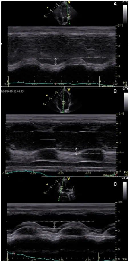
Off-line measurements of MAPSE and TAPSE were performed
by anatomic M-modea from the left apical 4-chamber view as
described in human guidelines and dogs.10–12,19,20 Briefly, the
anatomic M-mode cursor was aligned parallel to the interventric-ular septum (for MAPSE IVS), free wall (for MAPSE FW), or the right ventricular free wall (for TAPSE), and an M-mode trac-ing was obtained. Both MAPSE and TAPSE were measured in mm with electronic calipers between the most basilar position of the tricuspid annulus in end diastole and its most apical displace-ment at end systole by the leading edge method (Fig 1). All TAPSE and MAPSE measurements were performed by a single observer (IS). Five consecutive measurements were performed and the results were averaged. Intra- and interobserver variability for the acquisition of the anatomic M-mode image and subse-quent measurement was assessed in a randomly selected 10% of the population. Measurements for intraobserver repeatability were performed by a single observer (IS) on 2 occasions 1 week
apart from each other. Measurements for interobserver
repeatability were performed independently by 2 observers (IS and JRP).
Survival information for the cats diagnosed with HCM was obtained by reviewing the electronic clinical archive and by contact-ing referrcontact-ing veterinarians. All-cause mortality was the end-point in the survival analysis. Cats still alive were censored in the statistical analysis; subjects lost to follow up were included in the survival analysis up until the last time point at which they were known to be alive and then were thereafter censored in the analysis.
Statistical Analysis
Statistical analysis was performed by a commercially available
statistical softwareb , and in all cases, statistical significance was
set at P<.05. The Shapiro-Wilk test was used to verify normal
distribution of variables. Non-normally distributed data are reported as median (interquartiles range, IQR1-3, 25th percentile to 75th percentile).
Mann-Whitney Uor Kruskal-Wallis test was used to compare
ordinal and continuous, non-normally distributed data as appro-priate. Posthoc comparisons were performed by Dunn’s method.
For the repeatability study, intra- and interobserver coefficients of variation (CV) were calculated and Bland-Altman plots were obtained.
Reference intervals were calculated based on 90% confidence intervals, as recommended for normally distributed populations
with 20–40 individuals.21
Survival was calculated as the days between diagnosis and death or last visit. The Kaplan-Meier method was used to estimate sur-vival function and plot time to event curves in the different groups. Continuous variables were explored by division into groups based on tertiles. A log-rank test with right censoring was used to determine whether a significant difference existed among groups.
Results
From April 2013 to September 2015, 100 cats were
diagnosed with left ventricular hypertrophy. After
exclusion for concurrent systemic disease (n
=
22), lack
of a suitable 4-chamber apical cineloop (n
=
8) or both
(n
=
6), 64 cats with HCM were included in the study.
Of these 64 cats, 45 were asymptomatic and 19 had
CHF. The control group comprised 27 healthy blood
donor cats.
The median age of the population was 5.6 (3.8
–
7.0)
years, and the median body weight was 4.6 (4.2
–
5.0) kg.
There were 56 males and 35 females. The majority of
cats were domestic shorthairs (n
=
61), followed by
Bengals (n
=
6), Persians (n
=
6), and Domestic Long
Hairs (n
=
5). Other breeds included were British Short
Hairs and Ragdolls (n
=
3), Maine Coons, Scottish
Fold and Birman (n
=
2), Selkirk Rex (n
=
1).
Reference intervals were generated for healthy control
cats (Table 1).
There was no significant difference between controls
and cats with HCM with regard to age (P
=
.15), sex
(P
=
.45), body weight (P
=
.46), or LVFS (P
=
.45).
Cats with HCM had larger LAD (P
<
.001) and LA/
Ao (P
<
.001) than control cats. Cats with HCM had
lower LAFS (P
<
.001), TAPSE (P
<
.001), MAPSE
IVS
(P
<
.001),
MAPSE
FW
(P
<
.001),
S
0IVS
(P
=
.037),
and
S
0FW
(P
<
.001)
than
controls
(Table 2).
When comparing control cats, asymptomatic cats
with HCM, and cats with HCM and CHF, cats with
HCM and CHF had significantly lower MAPSE IVS,
MAPSE FW, and TAPSE than asymptomatic cats with
HCM (P
<
.001,
P
<
.001, and
P
<
.001, respectively)
and significantly lower MAPSE IVS, MAPSE FW, and
TAPSE (P
<
.001,
P
<
.001, and
P
<
.001, respectively)
than healthy control cats. Asymptomatic cats with
HCM had a significantly lower MAPSE IVS (P
<
.001),
MAPSE FW (P
<
.001), and TAPSE (P
<
.001)
com-pared to healthy control cats (Figs 2-4, Table 2).
At the end of the study period, 38 of the 64 cats with
HCM were still alive, 6 were lost to follow up, and 20
had died. At the univariable level, increased LAD and
LA/Ao and decreased LAFS, MAPSE IVS, MAPSE
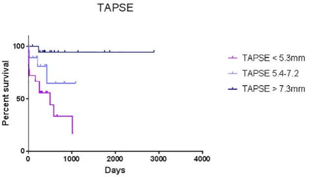
FW, and TAPSE (Table 3, Figs 4-7) were all associated
with reduced survival times.
Intra-
and
interobserver
coefficient
of
variation
showed good repeatability for acquisition and
measure-ment of MAPSE and TAPSE (Table 4) with minimal
bias (Figs 8-10).
Discussion
The results of our study indicated that cats with
HCM have lower MAPSE and TAPSE values as
com-pared to healthy control cats. Furthermore, cats with
CHF showed the lowest values of MAPSE and TAPSE.
Although the number of cats in the analysis was small,
lower MAPSE and TAPSE were also associated with a
decreased survival time for all-cause mortality,
suggest-ing potential prognostic value. Further studies with a
greater number of cats should enable a greater
appreci-ation of the prognostic utility of MAPSE and TAPSE
for both cardiac and all-cause mortality. The intra- and
interobserver CV as well as the Bland-Altman limits of
agreement indicated that the technique is easily
achiev-able and repeatachiev-able among different observers.
Shortening of the left ventricle in the longitudinal
axis is one of the major components of cardiac
contrac-tion as the heart base displaces toward the apex.
MAPSE and TAPSE measure the longitudinal
displace-ment of the annular plane during the cardiac cycle
and can therefore be considered as markers of systolic
long-axis function. It has been estimated that the
contri-bution of longitudinal contraction as assessed by
atri-oventricular plane displacement is responsible for up to
60% of the total cardiac stroke volume in healthy
human adults.
22A decrease in longitudinal function has
been identified by speckle-tracking echocardiography in
the early stages of HCM in people.
23,24Furthermore,
MAPSE measured by MRI is decreased in people with
obstructive and nonobstructive HCM and correlates
with the presence of fibrosis as assessed by late
gadolin-ium enhancement.
25Similarly, the findings of our study indicate that cats
with HCM have lower MAPSE compared to normal
cats, confirming reduced systolic longitudinal function
even in those cats not showing clinical signs. This
paral-lels the findings in human medicine.
23,24Left ventricular functional abnormalities might be
expected in cats with HCM, but our study showed that
RV longitudinal displacement is also reduced in cats
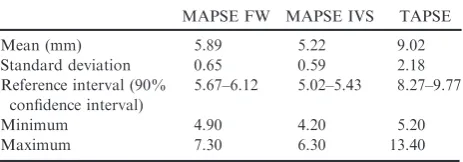
Table 1.
Distribution of data for MAPSE and TAPSE
in 27 healthy cats.
MAPSE FW MAPSE IVS TAPSE
Mean (mm) 5.89 5.22 9.02
Standard deviation 0.65 0.59 2.18
Reference interval (90% confidence interval)
5.67–6.12 5.02–5.43 8.27–9.77
Minimum 4.90 4.20 5.20
with HCM. This parallels findings in people with
HCM, where a decrease in TAPSE has been
docu-mented.
15It remains unclear whether the decrease in
TAPSE can be purely attributed to concomitant right
ventricular cardiomyopathy, which has been identified
in one-third of human patients and about half of cats
with HCM.
4,26Alternatively left-sided heart disease
may provoke right ventricular dysfunction through a
number of pathophysiological mechanisms including the
development of pulmonary hypertension,
27,28reduction
in right ventricular compliance due to ventricular
inter-dependence,
15,28or cause detrimental alteration in right
ventricular coronary perfusion pressure. One or more of
these mechanisms may explain the reduction in TAPSE
identified in this study.
12,15,27,28In this report, cats with CHF had the lowest values
for MAPSE and TAPSE, which probably reflects
dis-ease progression and worsening systolic dysfunction. A
similar finding was reported for LVFS in cats with
more advanced disease.
8,9Furthermore, cats in the lowest tertile for each of
MAPSE IVS, MAPSE FW, and TAPSE were more
likely to reach the final end-point of all-cause mortality,
suggesting that these echocardiographic parameters
have potential prognostic value. Left atrial size and
function was also shown to be of prognostic importance
in our population, as identified in previous studies.
6–9Because of the low number of events, it was not
pos-sible to assess the prognostic utility of these variables
for cardiac mortality and there were too few events to
Fig 2. Box and whiskers plot for MAPSE FW in control cats,asymptomatic cats with HCM, and cats with HCM and CHF.
Fig 3. Box and whiskers plot for MAPSE IVS in control cats, asymptomatic cats with HCM, and cats with HCM and CHF.
Fig 4. Box and whiskers plot for TAPSE in control cats, asymp-tomatic cats with HCM, and cats with HCM and CHF.
Table 3.
Univariable
survival
analysis.
Median
survival time (MST) and ranges are displayed. Bold
values indicate statistically significant results.
Variables Tertiles Median survival time Pvalue
LAD <15.1 mm >2884 day (0–2884 day) <.001
15.1–18.8 mm >1748 day (0–1748 day)
>18.8 mm 257 day (0–1865 day)
LA/Ao <1.4 mm >1748 day (0–1748 day) <.001
1.4–1.9 mm >2884 day (0–2884 day)
>1.9 mm 500 day (0–1865 day)
LAFS <20.2% 160 day (0–500 day) <.001
20.2–27.3% 208 day (12–1014 day)
>27.3% >2884 day (0–2884 day)
LVFS <44.0% 500 day (0–1086 day) .093
44–58.4% >2884 day (0–2884 day)
>58.4% 1014 day (0–1139 day)
MAPSE FW <3.3 mm 255 day (0–1016 day) <.001
3.3–4.8 mm >1748 day (0–1748 day)
>4.8 mm >2884 day (0–2884 day)
MAPSE IVS <3.3 mm 580 day (0–1016 day) .002
3.3–4.7 mm >1865 day (0–1865 day)
>4.7 mm >2884 day (0–2884 day)
TAPSE <5.3 mm 500 day (0–1016 day) <.001
5.3–7.2 >1086 day (0–1086 day)
>7.2 >2884 day (0–2884 day)
S0FW <4.6 1014 day (0–1865 day) .167
4.6–6.5 >1139 day (0–1139 day)
>6.5 >2884 day (0–2884 day)
S0IVS <6.0 >1865 day (12–1865 day) .110
6.0–9.0 >1748 day (0–1748 day)
Fig 5. Kaplan-Meier survival curve for MAPSE FW. Log-rank test,P<.001. Median survival time for MAPSE FW<3.3 mm is 255 days
(0–1016 day); for MAPSE FW 3.4–4.7 mm, median survival time is>1748 days (0–1748 day); for MAPSE FW>4.8 mm, median survival
time is>2884 day (0–2884 day).
Fig 6. Kaplan-Meier survival curve for MAPSE IVS. Log-rank test,P=.002. Median survival time for MAPSE IVS<3.3 mm is 580 days
(0–1016 day); for MAPSE IVS 3.4–4.7 mm, median survival time is>1865 days (0–1865 day); and for MAPSE>4.8 mm, median survival
time is>2884 days (0–2884 day).
Fig 7. Kaplan-Meier survival curve. Log-rank test,Pvalue<.001. Median survival time for TAPSE I<5.3 mm is 500 days (0–1016 day);
for TAPSE 5.4–7.2 mm, median survival time is>1086 days (0–1086 day); and for TAPSE>7.3 mm, median survival time is>2884 days
perform multivariable analysis for a final model of
sur-vival. That said, these preliminary findings are
encour-aging and in agreement with a recent study which
showed that right ventricular dysfunction based on
TAPSE was independently associated with an increased
likelihood of death or transplantation
15and therefore
warrant further study with a greater number of cats.
Both MAPSE and TAPSE are techniques that do not
require special expertise or advanced echocardiographic
imaging techniques and are easy to obtain and measure
after minimal training.
10These techniques have the
added advantage that there is no major negative effect
of apical fore-shortening as the measurements are taken
at the AV plane. There are more advanced techniques
to assess longitudinal function such as tissue Doppler
imaging (TDI) or speckle-tracking echocardiography
(STE), but they require advanced and more expensive
software, generally low heart rates, high frame rates,
and adequate image quality. They are also associated
with a steeper learning curve.
10To the authors’ knowledge, no reference intervals of
MAPSE and TAPSE have been published for cats. In
dogs, a curvilinear relationship was found between
TAPSE and weight, which became linear once weight
was normalized to a scale of 1/3.
19Cats may vary in
weight but this is generally less pronounced compared
to dogs, where body weight has a substantial
breed-dependent variation; however, as standard
echocardio-graphic parameters vary with body weight,
29the
authors recommend that the preliminary MAPSE and
TAPSE values provided in this study are applicable in
cats with a body weight between 3.7 and 5.2 kg.
Fur-ther studies are needed to establish the extent of
MAPSE and TAPSE variation based on weight- and
breed-related differences in cats.
The present study has some limitations. First of all,
being a retrospective study, it was not possible to
mea-sure TAPSE from a left apical view optimized for the
right ventricle in all cases. As with all techniques
need-ing a good alignment, MAPSE and TAPSE
measure-ment may be hindered by a degree of malalignmeasure-ment;
however, we were able to minimize this by the use of
anatomic M-mode which enabled us to achieve
accept-able intra- and interobserver CV. However, further
vali-dation
of
the
day-to-day
variability
in
these
longitudinal function indices is justified to further assess
their accuracy and reproducibility in prospective studies.
This study did not attempt to establish correlations
among MAPSE, TAPSE, and other echocardiographic
measures of disease severity such as atrial and
ventricu-lar size and function or invasive catheterization data,
for instance, to determine presence and severity of
pul-monary hypertension. Interestingly, it has been shown
in human patients with HCM that right ventricular
Table 4.
Intra- and interobserver CV for MAPSE and
TAPSE.
CV Intraobserver (1) (%) Interobserver CV (%)
MAPSE FW 2.0 8.0
MAPSE IVS 1.6 8.0
TAPSE 1.5 5.0
Fig 8. Bland-Altman Plot for MAPSE FW. Mean bias 0.01
(0.27 to 0.244) mm.
Fig 9. Bland-Altman Plot for MAPSE IVS. Mean bias 0.06
(0.69 to 0.82) mm.
dysfunction measured by TAPSE was independently
associated with the degree of left ventricular diastolic
and systolic dysfunction and pulmonary hypertension.
15Furthermore, we did not evaluate the influence of
medi-cations
such
as
furosemide
on
echocardiographic
parameters, which through reduction in preload may
influence longitudinal function. Due to the low number
of events, we were unable to perform a multivariable
survival analysis and further studies are needed to
con-firm our preliminary survival findings and to assess
whether MAPSE and TAPSE have prognostic value for
cardiac mortality.
The authors tried to exclude all possible secondary
causes of left ventricular hypertrophy based on the
available data for each case. However due to the
retro-spective nature of the study and the challenges
associ-ated with a final confirmation of myocarditis, we
cannot completely rule out the possibility that cats with
rare causes of LVH such as transient myocarditis were
included in the study. Finally, due to the retrospective
nature of the study, it was not possible to blind the
investigator
performing
echocardiographic
measure-ments to the clinical diagnosis of the cats, which may
have introduced bias into our results.
In conclusion, cats with HCM had lower MAPSE IVS,
MAPSE FW, and TAPSE, with the lowest values in cats
with CHF. Furthermore, our preliminary data indicated
that cats with MAPSE below 3.3 mm and TAPSE below
5.3 mm had reduced survival times. Importantly, the
technique was found to be feasible in the majority of cats
with acceptable intra- and interobserver CV.
Footnotes
aVivid 7 with Echo Pac off-line measurement software, GE
sys-tems, Hatfield, UK
bIBMÒSPSSÒStatistics version 22, IBM (UK) Ltd, Portsmouth,
UK
Acknowledgments
Conflict of Interest Declaration:
The authors declare
no conflict of interest.
Off-label
Antimicrobial
Declaration:
The
authors
declare no off-label use of antimicrobials.
References
1. Ferasin L, Sturgess CP, Cannon MJ, et al. Feline idiopathic
cardiomyopathy: A retrospective study of 106 cats (1994–2001). J
Feline Med Surg 2003;5:151–159.
2. Payne JR, Brodbelt DC, Luis Fuentes V. Cardiomyopathy prevalence in 780 apparently healthy cats in rehoming centres (the
CatScan study). J Vet Cardiol 2015;1:S244–S257.
3. Elliot P, Andersson B, Arbustini E, et al. Classification of the cardiomyopathies: A position statement from the European society of cardiology working group on myocardial and pericardial
diseases. Eur Heart J 2006;29:270–276.
4. Schober KE, Savino SI, Yildiz V. Right ventricular involve-ment in feline hypertrophic cardiomyopathy. J Vet Cardiol
2016;18:297–309.
5. Atkins CE, Gallo AM, Kurzman ID, et al. Risk factors, clinical signs and survival in cats with a clinical diagnosis of
idio-pathic hypertrophic cardiomyopathy: 74 cases (1985–1989). J Am
Vet Med Assoc 1992;201:613–618.
6. Rush JE, Freeman LM, Fenollosa LK, et al. Population and survival characteristics of cats with hypertrophic cardiomyopathy:
260 cases (1990–1999). J Am Vet Med Assoc 2002;220:202–207.
7. Payne JR, Luis Fuentes V, Boswood A, et al. Population characteristic and survival in 127 referred cats with hypertrophic
cardiomyopathy (1997 to 2005). J Small Anim Pract 2010;51:540–
547.
8. Payne JR, Borgeat K, Brodbelt DC, et al. Prognostic indica-tors in cats with hypertrophic cardiomyopathy. J Vet Intern Med
2013;27:1427–1436.
9. Payne JR, Borgeat K, Brodbelt DC, et al. Risk factors asso-ciated with sudden death vs. congestive heart failure or arterial thromboembolism in cats with hypertrophic cardiomyopathy. J
Vet Cardiol 2015;S1:S318–S328.
10. Hu K, Liu D, Herrmann S, et al. Clinical implication of mitral annular plane systolic excursion for patients with
cardiovas-cular disease. Eur Heart J Cardiovasc Imaging 2013;14:205–212.
11. Lang RM, Badano LP, Mor-Avi V, et al. Recommenda-tions for cardiac chamber quantification by echocardiography in adults: An update from the American Society of Echocardiogra-phy and the European Association of Cardiovascular Imaging.
Eur Heart J Cardiovasc Imaging 2015;16:233–270.
12. Mondillo S, Galderisi M, Ballo P, et al. Left ventricular systolic longitudinal function: Comparison among simple M-mode, pulsed, and M-mode color tissue Doppler of mitral
annu-lus in healthy individuals. J Am Soc Echocardiogr 2006;19:1085–
1091.
13. Haddad F, Hunt SA, Rosenthal DN, et al. Right ventricu-lar function in cardiovascuventricu-lar disease, part I: Anatomy, physiol-ogy, aging, and functional assessment of the right ventricle.
Circulation 2008;117:1436–1448.
14. Kjaergaard J, Iversen KK, Akkan D, et al. Predictors of right ventricular function as measured by tricuspid annular plane
systolic excursion in heart failure. Cardiovasc Ultrasound
2009;7:51–58.
15. Finocchiaro G, Knowles JW, Pavlovic A, et al. Prevalence and clinical correlates of right ventricular dysfunction in patients
with hypertrophic cardiomyopathy. Am J Cardiol 2014;113:361–
367.
16. Thomas WP, Gaber CE, Jacobs GJ, et al. Recommenda-tions for standards in transthoracic two-dimensional echocardiog-raphy in the dog and cat. Echocardiogechocardiog-raphy Committee of the Specialty of Cardiology, American College of Veterinary Internal
Medicine. J Vet Int Med 1993;7:247–252.
17. Schober KA, Chetboul VL. Echocardiographic evaluation of left ventricular diastolic function in cats: Hemodynamic
deter-minants and pattern recognition. J Vet Cardiol 2015;S1:S102–
S133.
18. Koffas H, Dukes-McEwan J, Corcoran BM, et al. Pulsed tissue Doppler imaging in normal cats and cats with hypertrophic
cardiomyopathy. J Vet Intern Med 2006;20:65–77.
19. Pariaut R, Saelinger C, Strickland KN, et al. Tricuspid annular plane systolic excursion (TAPSE) in dogs: Reference val-ues and impact of pulmonary hypertension. J Vet Intern Med
2012;26:1148–1154.
20. Kaye BM, Borgeat K, M~otsk€ula PF, et al. Association of
tricuspid annular plane systolic excursion with survival time in Boxer dogs with ventricular arrhythmias. J Vet Intern Med
21. Friedrichs KR, Harr KE, Freeman KP, et al. ASVCP refer-ence interval guidelines: Determination of de novo referrefer-ence inter-vals in veterinary species and other related topics. Vet Clin Pathol
2012;41:441–453.
22. Carlsson M, Ugander M, Mosen H, et al. Atrioventricular
plane displacement is the major contributor to left ventricular pumping in healthy adults, athletes, and patients with dilated
car-diomyopathy. Am J Physiol Heart Circ Physiol 2007;292:H1452–
H1459.
23. Smiseth OA, Torp H, Opdahl A, et al. Myocardial strain imaging: How useful is it in clinical decision making? Eur Heart J
2016;37:1196–1207.
24. Urbano-Moral JA, Rowin EJ, Maron MS, et al. Investiga-tion of global and regional myocardial mechanics with
3-dimen-sional speckle tracking echocardiography and relations to
hypertrophy and fibrosis in hypertrophic cardiomyopathy. Circ
Cardiovasc Imaging 2014;7:11–19.
25. Doesch C, Sperb A, Sudarski S, et al. Mitral annular plane systolic excursion is an easy tool for fibrosis detection by late gadolinium enhancement cardiovascular magnetic resonance imag-ing in patients with hypertrophic cardiomyopathy. Arch
Cardio-vasc Dis 2015;108:356–366.
26. Maron MS, Hauser TH, Dubrow E, et al. Right ventricular involvement in hypertrophic cardiomyopathy. Am J Cardiol
2007;100:1293–1298.
27. Schwarz K, Singh S, Dawson D, et al. Right ventricular function in left ventricular disease: Pathophysiology and
implica-tions. Heart Lung Circ 2013;22:507–511.
28. Lopez-Candales A, Rajagopalan N, Saxena N, et al. Right
ventricular systolic function is not the sole determinant of
tricus-pid annular motion. Am J Cardiol 2006;98:973–977.
29. H€aggstr€om J, AnderssonȦO, Falk T, et al. Effect of body
weight on echocardiographic measurements in 19,866 pure-bred cats