Stem Cells and Cloning: Advances and Applications
Autologous bone marrow cell therapy
for peripheral arterial disease
C Botti
C Maione
A Coppola
V Sica
G Cobellis
Department of General Pathology, Second University of Naples, Naples, Italy
Correspondence: Gilda Cobellis Department of General Pathology, Second University of Naples, Via Luigi De Crecchio 7, 80138 Napoli, Italy Tel +39 081 566 5681 Fax +39 081 292 399 Email g.cobellis@unina2.it
Abstract: Inadequate blood supply to tissues caused by obstruction of arterioles and/or
capillaries results in ischemic injuries – these injuries can range from mild (eg, leg ischemia) to severe conditions (eg, myocardial infarction, stroke). Surgical and/or endovascular procedures provide cutting-edge treatment for patients with vascular disorders; however, a high percentage of patients are currently not treatable, owing to high operative risk or unfavorable vascular involvement. Therapeutic angiogenesis has recently emerged as a promising new therapy, promoting the formation of new blood vessels by the introduction of bone marrow–derived stem and progenitor cells. These cells participate in the development of new blood vessels, the enlargement of existing blood vessels, and sprouting new capillaries from existing blood vessels, providing evidence of the therapeutic utility of these cells in ischemic tissues. In this review, the authors describe peripheral arterial disease, an ischemic condition affecting the lower extremities, summarizing different aspects of vascular regeneration and discussing which and how stem cells restore the blood flow. The authors also present an overview of encouraging results from early-phase clinical trials using stem cells to treat peripheral arterial disease. The authors believe that additional research initiatives should be undertaken to better identify the nature of stem cells and that an intensive cooperation between laboratory and clinical investigators is needed to optimize the design of cell therapy trials and to maximize their scientific rigor. Only this will allow the results of these investigations to develop best clinical practices. Additionally, although a number of stem cell therapies exist, many treatments are performed outside international and national regulations and many clinical trials have been not registered on databases such as ClinicalTrials.gov or EudraCT. Therefore, more rigorous clinical trials are required to confirm the first hopeful results and to address the challenging issues.
Keywords: adult stem cells, critical limb ischemia, bone marrow transplantation, therapeutic
angiogenesis
What is peripheral arterial disease?
Peripheral arterial disease (PAD) is a common circulatory problem in which narrowed
arteries reduce blood flow to the limbs, especially the legs. The most common causes
of PAD are atherosclerosis obliterans (ASO) and thromboangiitis obliterans (TAO).
1Two major classification systems are currently used to evaluate the spectrum of
symptoms: (1) the Fontaine classification, not used in everyday clinical practice but
useful for research purposes, and (2) the Rutherford classification, more commonly cited
in recent publications in the field of vascular medicine (Table 1). The American College
of Cardiology/American Heart Association 2005 guidelines noted the usefulness of
the Rutherford classification for standardized communication between clinicians.
1Dove
press
R E V I E w
open access to scientific and medical research
Open Access Full Text Article
Stem Cells and Cloning: Advances and Applications downloaded from https://www.dovepress.com/ by 118.70.13.36 on 27-Aug-2020
For personal use only.
Number of times this article has been viewed
Disease staging and classification systems are important for
clinical management of these patients. Based on the severity
of symptoms, usually two distinct clinical presentations are
distinguished in PAD patients: (1) intermittent claudication,
characterized by intermittent pain in leg muscles when the
person walks, and (2) critical limb ischemia (CLI), a more
severe form of PAD, characterized by pain at rest, nonhealing
wounds, and gangrene. After 1 year, 30% of patients with
CLI will lose their leg and 25% will die.
2The incidence of CLI in Western societies is approximately
220 new cases per million people per year, and, with an
aging population, the population at risk is expected to
increase because of persistent rates of tobacco abuse and
an increase in diabetes.
2Fifty percent of diabetics (7% of
the world population in 2010) suffer from PAD, which may
lead to amputation due to CLI.
3Moreover, smoking,
hypertension, dyslipidemia, a sedentary lifestyle, and a genetic
predisposition all contribute to the development of PAD.
4,5Current treatments for PAD
Revascularization, either surgical or endovascular, is the gold
standard treatment for patients with severe PAD. However,
despite advances in surgical and endovascular techniques,
6more than 30% of patients do not qualify as candidates for
revascularization because of excessive operative risk or
adverse vascular involvement. Furthermore, the presence of
extensive atherosclerotic plaques in the tibial and/or peroneal
arteries renders revascularization unsuccessful. These patients
are left to medical therapy, which may only slow disease
progression, and the only remaining alternative for relief of
rest pain or gangrene is amputation of the affected leg.
An estimated 120–500 amputations are performed per
million people per year, and one-quarter of these patients
require long-term institutional care or professional assistance
at home.
2Medical therapy is limited to antithrombotic
therapy,
7the prostaglandin analogue iloprost,
8or recently
to cilostazol. Cilostazol has been found to be effective for
the treatment of intermittent claudication. This compound
has several beneficial effects on platelet aggregation, serum
lipids, and endothelial cells (ECs), but how these might relate
to improvements in walking is not entirely understood.
9Thus, there is a critical need to develop novel strategies to
promote neovascularization in patients with CLI who are not
candidates for conventional treatments.
In 1997, Asahara et al
10made a big step forward when
they identified a class of bone marrow–derived circulating
endothelial progenitor cells (EPCs) that contribute to
angiogenesis and/or vasculogenesis in ischemic tissues.
11Since then, several studies have reported the capability of stem
and progenitor cells to promote neovascularization, reducing
ischemic damages.
12,13Encouraging results of several clinical
studies have rapidly demonstrated the beneficial effects of
autologous stem cell transplantation in patients affected by
CLI. Clinical improvements were observed in objective and
subjective measurements of perfusion (ie, transcutaneous
oxygen tension [TcPO
2] and laser Doppler flowmetry [LDF]),
pain reduction, increased pain-free total walking distance,
and decreased rate of amputation.
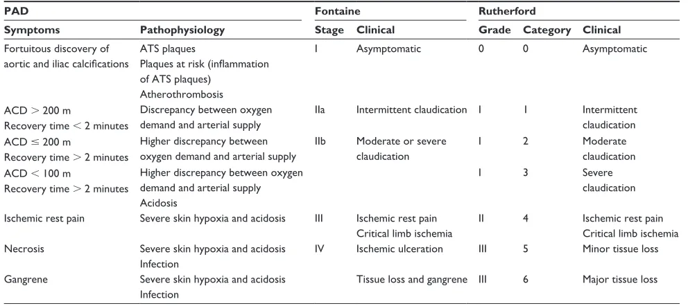
3,14–69Table 1 Two classifications of peripheral arterial disease (PAD): Fontaine and Rutherford
PAD Fontaine Rutherford
Symptoms Pathophysiology Stage Clinical Grade Category Clinical
Fortuitous discovery of aortic and iliac calcifications
ATS plaques
Plaques at risk (inflammation of ATS plaques)
Atherothrombosis
I Asymptomatic 0 0 Asymptomatic
ACD . 200 m
Recovery time , 2 minutes
Discrepancy between oxygen demand and arterial supply
IIa Intermittent claudication I 1 Intermittent
claudication ACD # 200 m
Recovery time . 2 minutes
Higher discrepancy between oxygen demand and arterial supply
IIb Moderate or severe claudication
I 2 Moderate
claudication ACD , 100 m
Recovery time . 2 minutes
Higher discrepancy between oxygen demand and arterial supply Acidosis
I 3 Severe
claudication
Ischemic rest pain Severe skin hypoxia and acidosis III Ischemic rest pain Critical limb ischemia
II 4 Ischemic rest pain
Critical limb ischemia Necrosis Severe skin hypoxia and acidosis
Infection
IV Ischemic ulceration III 5 Minor tissue loss
Gangrene Severe skin hypoxia and acidosis Infection
Tissue loss and gangrene III 6 Major tissue loss
Abbreviations: ACD, absolute claudication distance; ATS, atherosclerotic.
Dovepress
Botti et al
Stem Cells and Cloning: Advances and Applications downloaded from https://www.dovepress.com/ by 118.70.13.36 on 27-Aug-2020
What is vascular regeneration?
Vascular regeneration involves the restoration of normal
vascular function and structure and the growth of new
blood vessels. This includes a plethora of processes,
such as the distribution of blood flow via the formation
of collateral networks; the response of newly generated
vessels to hemodynamic, humoral, and local tissue factors;
the modulation of the immune response and the trafficking
of circulating cells; and the permeation of nutrients and
macromolecules through the microvasculature, which can in
turn have trophic effects on blood fluidity and hemostasis.
9Vascular regeneration is also important in a variety of
processes: during embryonic organogenesis and organ
growth in born individuals, in the course of restoration of
blood supply to ischemic tissues, and in the establishment
of blood supply to tumours.
70Neovascularization involves the growth of new structures
from preexisting vessels by migration, proliferation, and
differentiation of progenitor cells and the interplay between
growth factors and cytokines. The process of neovascularization
comprises three distinct phenomena: (1) vasculogenesis,
(2) angiogenesis, and (3) arteriogenesis.
70The essential mechanism responsible for new blood vessel
formation in adults is based on neoangiogenesis. During
angiogenesis, ECs present in vessel walls are activated in
response to various stimuli and begin to relase various growth
factors, the angiopoietins (Ang1 and Ang2) and Vascular
Endothelial Growth Factor (VEGF), which play a crucial
role in this process. While Ang1 and Ang2 participate in the
“stabilization” of the newly formed vessels, VEGF exerts its
pro-angiogenic function by binding to one of its receptors,
specifically the VEGF receptor 2 or kinase (VEGFR2 or
KDR) insert domain receptor, expressed exclusively by
ECs and their precursors. This binding triggers a cascade of
events that leads to the formation of new blood vessels and
which comprises the migration of ECs into the surrounding
tissue in response to angiogenic chemokines; proliferation
and differentiation of EPCs; and recruitment of support cells
such as pericytes for small capillaries and smooth muscle
cells for larger vessels.
The main factor inducing angiogenesis in adults is the
availability of oxygen, through the activation of
hypoxia-inducible factors.
71,72Stem cells with angiogenic potential
Stem cells are defined as cells with the capacity to self-renew
and to generate differentiated cells and are divided into two
types: embryonic and adult stem cells.
73Adult stem cells are
partially lineage-committed cells and have the capacity to
give rise to specialized cells. For this feature, adult stem cells
are so-called multipotent cells – as opposed to pluripotent
cells (ie, embryonic stem cells), which can give rise to all the
cell types in the body. Adult stem cells include three different
groups: (1) the bone marrow stem cells, (2) the circulating
pool of stem/progenitor cells (also derived from the bone
marrow), and (3) the tissue-resident stem cells.
74Bone marrow stem cells include different types of
progenitor cells, such as multipotent adult progenitor cells,
mesenchymal stem cells (MSCs), and hematopoietic stem
cells. The circulating pool of stem and progenitor cells
contains a variety of cells, but the most relevant for vascular
regeneration are the EPCs. Finally, the tissue-resident stem
cells are present in almost all tissues in a quiescent state and
can respond efficiently to different stimuli.
74Both EPCs and MSCs show promise for potential utility
in therapeutic neovascularization. MSCs are reported to
promote angiogenesis because of their capacity to stimulate
EC migration and tube formation; furthermore, MSCs support
neoangiogenesis, releasing soluble factors that contribute to
stimulate angiogenesis.
75what are the features of these cells?
MSCs are a subset of cells that express on their surface specific
molecules such as CD73, CD90, and CD105; MSCs also
express CD54/CD102, CD166, and CD49 (alpha integrin),
which regulate cell-to-cell interactions, and they do not
express any hematopoietic and/or EPC surface markers.
76MSCs can be found in many fetal and adult tissues and are
generally isolated from bone marrow, adipose tissue, umbilical
cord blood, and compact bone. Furthermore, MSCs are able
to migrate to and home to injured sites, where they act by
differentiating into specific cells and by secreting trophic
factors, which activate paracrine signaling.
77Moreover,
these cells interact with the immune system, particularly
modulating the immune response, apparently by inhibiting
tumor necrosis factor-alpha (TNFa) and interferon-gamma
(IFN-
g
) and by increasing interleukin 10 (IL-10).
78This unique
immunomodulatory property makes these cells suitable for
both autologous and heterologous transplants, since they avoid
and/or actively suppress eventual rejection of transplants.
79MSCs display a great therapeutic potential because of
their capability to differentiate into muscle, neural precursors,
cardiomyocytes, and perivascular cells. Perivascular cells
(herein referred to as pericytes) are critical cells in vascular
biology. Pericytes typically express alpha smooth muscle
actin (
α
-SMA), platelet-derived growth factor receptor beta
Dovepress Peripheral arterial disease and cell therapy
Stem Cells and Cloning: Advances and Applications downloaded from https://www.dovepress.com/ by 118.70.13.36 on 27-Aug-2020
(PDGF-
β
), and nerve/glial antigen-2 (NG-2) proteoglycan.
They are branched cells embedded within the basement
membrane of capillaries and postcapillary venules, stabilizing
the vessel wall.
80Pericytes are considered cells that control
EC proliferation and migration, and thereby also the growth
of new capillaries. In turn, ECs stimulate expansion and
activation of the pericyte precursor cell population. The balance
between ECs and pericytes is highly controlled by a series of
signaling pathway mechanisms operating in an autocrine and/
or a paracrine manner. In pathological conditions in which
angiogenic activity is impaired, pericytes and ECs could be
partly responsible for abnormalities in blood vessels.
EPCs are adult hemangioblast-derived cells characterized
by the expression of CD34, VEGF receptor 2, and CD133.
These markers are expressed by precursor cells, but not
by differentiated ones.
81In fact, as the hemangioblasts
differentiate to become ECs, they downregulate the
hematopoietic stem cell marker CD133 expression.
81EPCs
can be isolated from human peripheral or umbilical cord blood
and can also be found in bone marrow niches. EPCs have
shown in vitro all the functional properties of ECs; moreover,
EPCs have direct angiogenic action, supporting angiogenesis
through their ability to secrete paracrine mediators. In this
respect, several studies have shown that these cells release
interleukins, growth factors, and chemokines that altogether
regulate CD14-positive cells, accelerate vascular network
formation, and enhance healing processes.
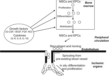
82Adult stem cells with angiogenic potential such as EPCs
and MSCs will stimulate the production of new blood vessels,
as shown in Figure 1.
Cell therapy in PAD: clinical results
Promotion of collateral vessel formation and angiogenesis
in PAD patients is an important therapeutic strategy to
minimize tissue injury associated with severe ischemia.
The Therapeutic Angiogenesis using Cell Transplantation
trial was the first report on the use of bone marrow–derived
mononuclear cells in the treatment of PAD.
14Starting from
this, the search of the literature yielded a total of 57
early-phase clinical trials for a total of 1997 enrolled patients.
The safety and feasibility of autologous cell transplantation
has been reported in 1667 treated patients (Tables 2 and 3).
Among these, a total of 303 diabetic patients with CLI and
foot ulcers underwent cell therapy. The degree of ischemia
varied throughout the groups, ranging from Rutherford
category 4/Fontaine stage III through to severe CLI
classi-fied as Rutherford category 6/Fontaine stage IV.
Only a minority of trials (n
=
13) included appropriate
controls (Table 4). In these studies, the follow-up of the
untreated or placebo group did not differ from that observed
Peripheral
circulation
Bone
marrow
Growth factors
(G-CSF, VEGF, FGF, NO)
Cytokines
(IL-6, IL-8, IL-10)
MSCs and EPCs
Proliferation
Differentiation
Mobilization
Recruitment and homing
Endothelium
Ischemic
organs
Sprouting from
pre-existing blood vessel
In situ differentiation
and proliferation
MSCs and EPCs
Figure 1 Schematic representation of neoangiogenesis promoted by circulating and bone marrow–resident stem cells.
Notes: Ischemia induces production of growth factors, cytokines, and hormones, which promotes proliferation, differentiation, and mobilization of mesenchymal stem cells (MSCs) and endothelial progenitor cells (EPCs) to form new vessels. In addition, the growth factors can stimulate EPCs sprouting from preexisting blood vessels.
Abbreviations: FGF, fibroblast growth factor; G-CSF, granulocyte colony-stimulating factor; IL, interleukin; NO, nitric oxide; VEGF, vascular endothelial growth factor. Dovepress
Botti et al
Stem Cells and Cloning: Advances and Applications downloaded from https://www.dovepress.com/ by 118.70.13.36 on 27-Aug-2020
Table 2 Clinical trials with cell therapy in peripheral arterial disease (PAD)
Published study Delivery route
Condition Patients (n)
Cell type Follow-up time Improved functional
outcomes
Tateishi-Yuyama et al14
IM PAD, DM 45 BMCs or PB-MNCs 4 and 24 weeks ABI, TcPO2, rest pain, pain-free
walking time
Esato et al15 IA PAD 8 BMCs N/D Ulceration healing
Saigawa et al16 IM PAD, DM 8 BMCs 4 weeks ABI, TcPO
2
Higashi et al17 IM PAD 7 BMCs or BM-MNCs 4 and 24 weeks ABI, TcPO
2, pain-free walking
time
Miyamoto et al18 IM CLI 12 BMCs and EPCs N/D ABI, pain-free walking time
Huang et al19 IM PAD 5 PB-MNCs 3 months ABI, LDF
Kawamura et al20 IM PAD, CLI 30 PB-MNCs N/D T°C
Lenk et al21 IA CLI 7 PB-MNCs 20 weeks ABI, TcPO
2, rest pain, pain-free
walking time
Huang et al22 IM CLI, DM 28 MPB-MNCs 3 months ABI, pain, ulcers
Ishida et al23 IM PAD 6 MPB-MNCs 4 and 24 weeks ABI, ulcers
Durdu et al24 IM PAD 28 BM-MNCs 3 and 6 months ABI, rest pain, pain-free walking
time
Koshikawa et al25 IM PAD 7 BM-MNCs 6 months ABI, pain, ulcers
Arai et al26 IM PAD 25 BMCs 1 month ABI, TcPO
2
Miyamoto et al27 IM PAD, CLI 8 BM-MNCs 4 weeks, 4 and
7 months, and 1 year
Rest pain, ulcers
Kawamura et al28 IM CLI 92 PB-MNCs 6 weeks Limb salvage, VEGF serum level
Bartsch et al29 IM and IA PAD, CLI 13 BMCs 2 and 13 months ABI, pain-free walking distance
Huang et al30 IM PAD 150 BM-MNCs or MPB-MNCs 12 weeks ABI, rest pain
Kajiguchi et al31 IM CLI 7 BM-MNCs or PB-MNCs 1 month ABI
Hernández et al32 IM CLI, DM 12 BM-MNCs 24 months ABI, SaO
2, pain-free walking time,
rest pain
Saito et al33 IM PAD 7 BM-MNCs N/D TcPO
2
Matoba et al34 IM PAD 115 BM-MNCs 3 years Pain, ulcers, pain-free walking
distance
Napoli et al35 IA PAD 18 BM-MNCs 3, 6, 12, and
18 months
ABI, ulcers, pain-free walking distance
Gu et al36 IM or IA PAD, DM 32 BM-MNCs 4 weeks ABI, TcPO
2, limb salvage
Chochola et al37 IA PAD, DM 24 BM-MNCs 1 year Limb salvage, wound healing
wester et al38 IM CLI 8 BMCs 4 and 8 months ABI, TcPO
2, ulcers
Van Tongeren et al39
IM and IA CLI 27 BMCs 6 and 12 months Pain-free walking distance,
ABI, pain reduction
De Vriese et al40 IM CLI 16 BM-MNCs 12 weeks TcPO
2, pain reduction
Cobellis et al41 IA PAD 10 BM-MNCs 12 months ABI, pain-free walking distance
Motukuru et al42 IM PAD 38 BM-MNCs 6 months Ulcer healing, limb salvage,
ABI, TcPO2
Amann et al43 IM CLI 45 BM-MNCs 3 months Limb salvage, ABI, TcPO
2,
pain-free walking distance
Amann et al44 IM PAD 51 BM-MNCs 6 months Limb salvage, ABI, TcPO
2,
pain-free walking distance
Capiod et al45 IM CLI 24 BM-MNCs or PB-MNCs N/D No clinical evaluation reported
Franz et al46 IM and IA PAD 9 BM-MNCs 2 weeks and
3 months
ABI, pain reduction, ulcers, limb salvage
Franz et al47 IM and IA PAD 20 BM-MNCs 3 months ABI, pain reduction, ulcers,
limb salvage
Zafarghandi et al48 IM CLI 50 BM-MNCs and G-CSF 4 and 24 weeks ABI, pain-free walking distance
Procházka et al49 IM CLI, DF 37 BMSCs 3 months LDF, ABI, TcPO
2, limb salvage
Procházka et al3 IM PAD 96 BMSCs 4 months ABI, limb salvage
Lara-Hernandez et al50
IM CLI 28 Mobilized EPCs 14 and 18 months
and 1 year
ABI, limb salvage
Iso et al51 IM CLI 13 BM-MSCs 4 months TcPO
2, rest pain
(Continued)
Dovepress Peripheral arterial disease and cell therapy
Stem Cells and Cloning: Advances and Applications downloaded from https://www.dovepress.com/ by 118.70.13.36 on 27-Aug-2020
in several large population studies.
2,83–87In addition,
the Edinburgh Artery Study def ined the prevalence
of asymptomatic and symptomatic PAD and related
comorbidities in the general population.
87Therefore, since
the progression of disease is well defined in CLI patients,
the lack of an untreated or a placebo group – even if
scientifically compelling – cannot diminish the significance
of the studies.
Two sources of cells were used in these trials: (1) bone
marrow aspiration (n
=
46) and (2) apheresis of peripheral
blood with or without GSF stimulation (n
=
11). The
route of cell administration was intramuscular in 39 trials,
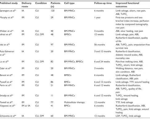
Table 2 (Continued)
Published study Delivery route
Condition Patients (n)
Cell type Follow-up time Improved functional
outcomes
Sprengers et al52 IA CLI 120 BM-MNCs 6 months Limb salvage, ulcers, rest pain,
ABI, TcPO2
Murphy et al53 IM CLI 29 BM-MNCs 1 year First-toe pressure and
toe-brachial index increase, perfusion index by computed tomography, rest pain
walter et al54 IA CLI 40 BM-MNCs 3 months ABI, ulcer healing, rest pain
Iafrati et al55 IM CLI, DM 48 BMSCs 12 weeks Limb salvage, pain, ABI,
Rutherford classification, quality of life
Idei et al56 IM CLI 97 BM-MNCs 56 months ABI, TcPO
2, pain, amputation-free
survival rate Ruiz-Salmeron
et al57
IA CLI 20 BM-MNCs 3 and 12 months Rutherford classification,
diabetic wound scales, ABI, mortality rate
Lu et al58 IM CLI, DM 82 BM-MNCs, BMSCs 6 and 24 weeks Pain-free walking time, ABI,
TcPO2, ulcers, limb salvage
Gabr et al59 IM CLI 20 BM-MNCs 3 months walking distance, rest pain,
skin condition, ABI
Benoit et al60 IM CLI 48 BMSCs 6 months Limb salvage, Rutherford
classification, ABI, pain
Powell et al61 IM CLI 86 BMCs 6 and 12 months Limb salvage, TTF, wound healing
Perin et al62 IM CLI 21 BM-MNCs 6 and 12 weeks Rutherford classification,
ABI, TcPO2, quality of life, pain
Smadja et al63 IM CLI 11 BM-MNCs 6 and 12 months TcPO
2, wound healing,
limb salvage
Powell et al64 IM CLI 77 Multicellular therapy 12 months TTF, limb salvage
Klepanec et al65 IM or IA CLI 41 BMCs 6 months Rutherford classification, ABI,
TcPO2, pain, limb salvage, wound healing
Schiavetta et al66 IA CLI, DM 60 BM-MNCs 12 months LDF, TcPO
2, limb salvage
Abbreviations: ABI, ankle-brachial index; BMCs, bone marrow cells; BMSCs, bone marrow stem cells; BM-MNCs, bone marrow–derived mononuclear cells; CLI, critical limb ischemia; DF, diabetic foot; DM, diabetes mellitus; EPCs, endothelial progenitor cells; G-CSF, granulocyte colony-stimulating factor; IA, intra-arterial injection; IM, intramuscular injection; LDF, laser Doppler flowmetry; MPB-MNCs, mobilized peripheral blood–derived mononuclear cells; N/D, not determined; PB-MNCs, peripheral blood–derived mononuclear cells; SaO2, arterial oxygen saturation; T°C, temperature expressed in degrees Celsius; TcPO2, transcutaneous oxygen tension; TTF, time to first
occurrence of treatment failure; VEGF, vascular endothelial growth factor.
Table 3 Clinical trials with intralesional administration of stem cells in foot ulcers
Published study Condition Patients (n) Cell type Follow-up time Improved functional outcomes
Vojtassák et al67 DF 1 BMSCs 29 days wound size and ulcer healing
Dash et al68 DF ulcers, PAD 24 BMSCs 3 months Wound size and pain-free walking
distance
Subrammaniyan et al69 CLI 6 BM-MNCs 6 months ABI and pain-free walking distance,
limb salvage, ulcer healing, and rest pain
Abbreviations: ABI, ankle-brachial index; BM-MNCs, bone marrow–derived mononuclear cells; BMSCs, bone marrow stem cells; CLI, critical limb ischemia; DF, diabetic foot; PAD, peripheral arterial disease.
Dovepress
Botti et al
Stem Cells and Cloning: Advances and Applications downloaded from https://www.dovepress.com/ by 118.70.13.36 on 27-Aug-2020
intra-arterial in nine trials, and combined intra-arterial plus
intramuscular in four trials (Table 2). Furthermore, two
studies compared the therapeutic effects of intramuscular or
intra-arterial delivery of bone marrow cells in patients with
lower limb ischemia, showing similar beneficial results.
36,65To prevent clot formation, harvested cells were collected
in the presence of anticoagulant.
3,14–69Intramuscular
administration is usually performed through multiple
injections at the level of limb muscles, while intra-arterial
infusion is usually performed via classic femoral access.
Three studies reported the use of intralesional administration
of bone marrow–derived stem cells in 31 diabetic patients
with foot ulcers, showing encouraging results (Table 3).
In general, bone marrow aspiration was well tolerated,
and the most frequent adverse reaction was local pain or
mild anemia. However, serious adverse reactions such as
angina with ST segment depression were observed in a small
number of patients.
55The average follow-up of these clinical studies was
8.4
±
9.55 months. Considering all studies, the reported
outcomes for therapeutic efficacy of cell therapy involved
the ankle-brachial index, TcPO
2, LDF, pain-free walking
dis-tance, ulcer healing, and amputation-free survival. In all
stud-ies, symptoms improved after the procedure, as evidenced by
clinical evaluation, relief of rest pain, and improvement by
at least one level in Rutherford and Fontaine classifications.
Furthermore, autologous cell therapy promoted
amputation-free survival with an average of 7.8 months and promoted
complete wound healing within 3 months in most patients
with ulcers prior to bone marrow stem cell transplantation, in
comparison with the natural history of PAD patients.
There-fore, autologous transplantation of bone marrow–derived
cells significantly improved both the objective and the
sub-jective endpoints.
Conclusion
Herein, the authors provide the most comprehensive review of
cell therapy trials describing the background and first results
of stem and progenitor cell therapy in patients with CLI who
are not suitable for revascularization. Both the principle,
as far as it is understood, and the methods are described.
Compelling evidence suggests that stem cell therapy may
become a useful adjunct to the current treatment options.
Because of poor prognosis and the increasing number of
patients, there is a need for new therapeutic methods.
About 1997 patients without revascularization options
were enrolled in these trials and 1667 patients were treated.
Cell therapy significantly improved functional outcomes such
as ankle-brachial index, TcPO
2or LDF, rest pain, pain-free
walking distance, ulcer healing, and limb salvage. Although
it is generally agreed that controlled trials yield more reliable
results, the authors also included noncontrolled studies,
which are the majority of published reports. The authors
believe the main reason for this majority is that the authorized
studies have chosen to treat end-stage patients, without other
therapeutic options. The procedures are generally safe and
well tolerated. Reported deaths were expected, given the
severe underlying disease, and could not be directly attributed
to cell therapy.
Challenges in this new therapeutic option still include
open questions regarding cell number, phenotype, processing,
route of optimal delivery, and frequency of application.
The number of injected cells ranged from 4
×
10
6to 10
9for bone marrow cells and from 7
×
10
7to 3
×
10
9for
peripheral blood–derived mononuclear cells, with positive
effects on blood perfusion, even when low cells were used.
Nevertheless, no correlation study between clinical response
and cell number has been performed so far, and no proven
correlation exists between the phenotype of used cells and
efficacy of neoangiogenesis. Answering these two points
is critical to understanding which and how many cells are
needed to obtain a clinical response.
The question of optimal delivery route remains open.
The rationale behind the intramuscular injection is to
generate a reservoir of cells near the ischemic area, which
can be recruited by active paracrine mechanisms. The
intra-arterial injection relies on the fact that the blood
flow transports cells up to the ischemia site; however, it
is not known how many cells are able to leave the blood
stream to reach the ischemic area. Again, no correlation
Table 4 Controlled clinical trials with cell therapy in peripheral
arterial disease
Published study
Year Patients
Total (N) Treated (n) Control (n)
Powell et al64 2012 72 48 24
Benoit et al60 2011 48 34 14
Lu et al58 2011 82 41 41
Powell et al61 2011 46 32 14
Idei et al56 2011 97 51 46
Iafrati et al55 2011 48 34 14
walter et al54 2011 40 19 21
Procházka et al3 2010 96 42 54
Sprengers et al52 2010 110 55 55
Cobellis et al41 2008 19 10 9
Bartsch et al29 2007 25 13 12
Arai et al26 2006 25 13 12
Huang et al22 2005 28 14 14
Dovepress Peripheral arterial disease and cell therapy
Stem Cells and Cloning: Advances and Applications downloaded from https://www.dovepress.com/ by 118.70.13.36 on 27-Aug-2020
study between the two routes of administration has been
performed, although the present trend is for intramuscular
administration. It has been reported that the combination of
both routes (intramuscular plus intra-arterial)
39has given
substantial improvements in clinical outcomes, but this
must be confirmed in exhaustive experiments in preclinical
models.
In summary, over the past 10 years there has been
considerable interest in stem cells, including extensive
clinical activity involving stem cells. Unfortunately, the
rationale for the clinical application of adult stem cells,
particularly in regenerative medicine, has lagged behind
initial laboratory observations. At this point, the authors
believe that additional research initiatives should be
undertaken to better identify the nature of stem cells and
that an intensive cooperation between laboratory and clinical
investigators is needed to optimize the design of cell therapy
trials and to maximize their scientific rigor. Only this will
allow the results of these investigations to develop best
clinical practices.
Additionally, although a number of stem cell therapies
exist, many treatments are performed outside international
and national regulations and many clinical trials have been
not registered on databases such as ClinicalTrials.gov
or EudraCT. Therefore, more rigorous clinical trials are
required to confirm the first hopeful results and to address
the challenging issues.
Acknowledgments
The authors are grateful to Fondazione Luigi Califano,
Fondazione Banco di Napoli, and Istituto Superiore di
Sanità. The authors thank Prof Anna Maria Molinari and
Prof Ferdinando Auricchio for helpful discussions.
Disclosure
The authors report no conflicts of interest in this work.
References
1. Hirsch AT, Haskal ZJ, Hertzer NR, et al; for American Association for Vascular Surgery/Society for Vascular Surgery; Society for Cardiovascular Angiography and Interventions; Society for Vascular Medicine and Biology; Society of Interventional Radiology; ACC/ AHA Task Force on Practice Guidelines. ACC/AHA Guidelines for the Management of Patients with Peripheral Arterial Disease (lower extremity, renal, mesenteric, and abdominal aortic): a collaborative report from the American Associations for Vascular Surgery/Society for Vascular Surgery, Society for Cardiovascular Angiography and Interventions, Society for Vascular Medicine and Biology, Society of Interventional Radiology, and the ACC/AHA Task Force on Practice Guidelines (writing committee to develop guidelines for the management of patients with peripheral arterial disease); summary of recommendations. J Vasc Interv Radiol. 2006;17(9):1383–1397.
2. Norgren L, Hiatt WR, Dormandy JA, Nehler MR, Harris KA, Fowkes FG; for TASC (Trans Atlantic Inter-Society Consensus) II Working Group. Inter-society consensus for the management of peripheral arterial disease (TASC II). J Vasc Surg. 2007;45 Suppl S:S5–S67.
3. Procházka V, Gumulec J, Jalůvka F, et al. Cell therapy, a new standard in management of chronic critical limb ischemia and foot ulcer. Cell Transplant. 2010;19(11):1413–1424.
4. Thorgeirsson TE, Geller F, Sulem P, et al. A variant associated with nicotine dependence, lung cancer and peripheral arterial disease. Nature. 2008;452(7187):638–642.
5. Wilson AM, Sadrzadeh-Rafie AH, Myers J, et al. Low lifetime recreational activity is a risk factor for peripheral arterial disease. J Vasc Surg. 2011;54(2):427–432, 432. e1–432. e4.
6. Conrad MF, Crawford RS, Hackney LA, et al. Endovascular management of patients with critical limb ischemia. J Vasc Surg. 2011; 53(4):1020–1025.
7. Saha SP, Whayne TF Jr, Mukherjee D. Current evidence for antithrombotic therapy after peripheral vascular interventions. Curr Vasc Pharmacol. Epub January 20, 2012.
8. Lessiani G, Vazzana N, Cuccurullo C, et al. Inflammation, oxidative stress and platelet activation in aspirin-treated critical limb ischaemia: beneficial effects of iloprost. Thromb Haemost. 2011;105(2): 321–328.
9. Volz KS, Miljan E, Khoo A, Cooke JP. Development of pluripotent stem cells for vascular therapy. Vascul Pharmacol. 2012;56(5–6):288–296. 10. Asahara T, Murohara T, Sullivan A, et al. Isolation of putative progenitor
endothelial cells for angiogenesis. Science. 1997;275(5302):964–967. 11. Shi Q, Rafii S, Wu MH, et al. Evidence for circulating bone
marrow-derived endothelial cells. Blood. 1998;92(2):362–367.
12. Asahara T, Masuda H, Takahashi T, et al. Bone marrow origin of endothelial progenitor cells responsible for postnatal vasculogenesis in physiological and pathological neovascularization. Circ Res. 1999; 85(3):221–228.
13. Crosby JR, Kaminski WE, Schatteman G, et al. Endothelial cells of hematopoietic origin make a significant contribution to adult blood vessel formation. Circ Res. 2000;87(9):728–730.
14. Tateishi-Yuyama E, Matsubara H, Murohara T, et al; for Therapeutic Angiogenesis using Cell Transplantation Study Investigators. Therapeutic angiogenesis for patients with limb ischaemia by autologous transplantation of bone-marrow cells: a pilot study and a randomised controlled trial. Lancet. 2002;360(9331):427–435. 15. Esato K, Hamano K, Li TS, et al. Neovascularization induced by
autologous bone marrow cell implantation in peripheral arterial disease. Cell Transplant. 2002;11(8):747–752.
16. Saigawa T, Kato K, Ozawa T, et al. Clinical application of bone marrow implantation in patients with arteriosclerosis obliterans, and the associa-tion between efficacy and the number of implanted bone marrow cells. Circ J. 2004;68(12):1189–1193.
17. Higashi Y, Kimura M, Hara K, et al. Autologous bone-marrow mononuclear cell implantation improves endothelium-dependent vasodilation in patients with limb ischemia. Circulation. 2004;109(10): 1215–1218.
18. Miyamoto M, Yasutake M, Takano H, et al. Therapeutic angiogenesis by autologous bone marrow cell implantation for refractory chronic peripheral arterial disease using assessment of neovascularization by 99 mTc-tetrofosmin (TF) perfusion scintigraphy. Cell Transplant. 2004; 13(4):429–437.
19. Huang PP, Li SZ, Han MZ, et al. Autologous transplantation of peripheral blood stem cells as an effective therapeutic approach for severe arteriosclerosis obliterans of lower extremities. Thromb Haemost. 2004;91(3):606–609.
20. Kawamura A, Horie T, Tsuda I, et al. Prevention of limb amputation in patients with limbs ulcers by autologous peripheral blood mononuclear cell implantation. Ther Apher Dial. 2005;9(1):59–63.
21. Lenk K, Adams V, Lurz P, et al. Therapeutical potential of blood-derived progenitor cells in patients with peripheral arterial occlusive disease and critical limb ischaemia. Eur Heart J. 2005;26(18):1903–1909.
Dovepress
Botti et al
Stem Cells and Cloning: Advances and Applications downloaded from https://www.dovepress.com/ by 118.70.13.36 on 27-Aug-2020
22. Huang P, Li S, Han M, Xiao Z, Yang R, Han ZC. Autologous transplantation of granulocyte colony-stimulating factor-mobilized peripheral blood mononuclear cells improves critical limb ischemia in diabetes. Diabetes Care. 2005;28(9):2155–2160.
23. Ishida A, Ohya Y, Sakuda H, et al. Autologous peripheral blood mononuclear cell implantation for patients with peripheral arterial disease improves limb ischemia. Circ J. 2005;69(10):1260–1265. 24. Durdu S, Akar AR, Arat M, Sancak T, Eren NT, Ozyurda U.
Autologous bone-marrow mononuclear cell implantation for patients with Rutherford grade II–III thromboangiitis obliterans. J Vasc Surg. 2006;44(4):732–739.
25. Koshikawa M, Shimodaira S, Yoshioka T, et al. Therapeutic angiogenesis by bone marrow implantation for critical hand ischemia in patients with peripheral arterial disease: a pilot study. Curr Med Res Opin. 2006;22(4):793–798.
26. Arai M, Misao Y, Nagai H, et al. Granulocyte colony-stimulating factor: a noninvasive regeneration therapy for treating atherosclerotic peripheral artery disease. Circ J. 2006;70(9):1093–1098.
27. Miyamoto K, Nishigami K, Nagaya N, et al. Unblinded pilot study of autologous transplantation of bone marrow mononuclear cells in patients with thromboangiitis obliterans. Circulation. 2006;114(24): 2679–2684.
28. Kawamura A, Horie T, Tsuda I, et al. Clinical study of therapeutic angiogenesis by autologous peripheral blood stem cell (PBSC) transplantation in 92 patients with critically ischemic limbs. J Artif Organs. 2006;9(4):226–233.
29. Bartsch T, Brehm M, Zeus T, Kögler G, Wernet P, Strauer BE. Transplantation of autologous mononuclear bone marrow stem cells in patients with peripheral arterial disease (the TAM-PAD study). Clin Res Cardiol. 2007;96(12):891–899.
30. Huang PP, Yang XF, Li SZ, Wen JC, Zhang Y, Han ZC. Randomised comparison of G-CSF-mobilized peripheral blood mononuclear cells versus bone marrow-mononuclear cells for the treatment of patients with lower limb arteriosclerosis obliterans. Thromb Haemost. 2007;98(6): 1335–1342.
31. Kajiguchi M, Kondo T, Izawa H, et al. Safety and efficacy of autologous progenitor cell transplantation for therapeutic angiogenesis in patients with critical limb ischemia. Circ J. 2007;71(2):196–201.
32. Hernández P, Cortina L, Artaza H, et al. Autologous bone-marrow mononuclear cell implantation in patients with severe lower limb ischaemia: a comparison of using blood cell separator and Ficoll density gradient centrifugation. Atherosclerosis. 2007;194(2):e52–e56. 33. Saito S, Nishikawa K, Obata H, Goto F. Autologous bone marrow
transplantation and hyperbaric oxygen therapy for patients with thromboangiitis obliterans. Angiology. 2007;58(4):429–434. 34. Matoba S, Tatsumi T, Murohara T, et al. Long-term clinical outcome
after intramuscular implantation of bone marrow mononuclear cells (Therapeutic Angiogenesis by Cell Transplantation [TACT] trial) in patients with chronic limb ischemia. Am Heart J. 2008;156(5): 1010–1018.
35. Napoli C, Farzati B, Sica V, et al. Beneficial effects of autologous bone marrow cell infusion and antioxidants/L-arginine in patients with chronic critical limb ischemia. Eur J Cardiovasc Prev Rehabil. 2008; 15(6):709–718.
36. Gu YQ, Zhang J, Guo LR, et al. Transplantation of autologous bone marrow mononuclear cells for patients with lower limb ischemia. Chin Med J (Engl). 2008;121(11):963–967.
37. Chochola M, Pytlík R, Kobylka P, et al. Autologous intra-arterial infusion of bone marrow mononuclear cells in patients with critical leg ischemia. Int Angiol. 2008;27(4):281–290.
38. Wester T, Jørgensen JJ, Stranden E, et al. Treatment with autologous bone marrow mononuclear cells in patients with critical lower limb ischaemia: a pilot study. Scand J Surg. 2008;97(1):56–62.
39. Van Tongeren RB, Hamming JF, Fibbe WE, et al. Intramuscular or combined intramuscular/intra-arterial administration of bone marrow mononuclear cells: a clinical trial in patients with advanced limb ischemia. J Cardiovasc Surg (Torino). 2008;49(1):51–58.
40. De Vriese AS, Billiet J, Van Droogenbroeck J, Ghekiere J, De Letter JA. Autologous transplantation of bone marrow mononuclear cells for limb ischemia in a Caucasian population with atherosclerosis obliterans. J Intern Med. 2008;263(4):395–403.
41. Cobellis G, Silvestroni A, Lillo S, et al. Long-term effects of repeated autologous transplantation of bone marrow cells in patients affected by peripheral arterial disease. Bone Marrow Transplant. 2008;42(10): 667–672.
42. Motukuru V, Suresh KR, Vivekanand V, Raj S, Girija KR. Therapeutic angiogenesis in Buerger’s disease (thromboangiitis obliterans) patients with critical limb ischemia by autologous transplantation of bone marrow mononuclear cells. J Vasc Surg. 2008;48(Suppl 6): S53–S60.
43. Amann B, Lüdemann C, Rückert R, et al. Design and rationale of a randomized, double-blind, placebo-controlled phase III study for autologous bone marrow cell transplantation in critical limb ischemia: the BONe Marrow Outcomes Trial in Critical Limb Ischemia (BONMOT-CLI). Vasa. 2008;37(4):319–325.
44. Amann B, Lüdemann C, Ratei R, Schmidt-Lucke JA. Autologous bone marrow cell transplantation increases leg perfusion and reduces amputations in patients with advanced critical limb ischemia due to peripheral artery disease. Cell Transplant. 2009;18(3):371–380. 45. Capiod JC, Tournois C, Vitry F, et al. Characterization and comparison
of bone marrow and peripheral blood mononuclear cells used for cellular therapy in critical leg ischaemia: towards a new cellular product. Vox Sang. 2009;96(3):256–265.
46. Franz RW, Parks A, Shah KJ, Hankins T, Hartman JF, Wright ML. Use of autologous bone marrow mononuclear cell implantation therapy as a limb salvage procedure in patients with severe peripheral arterial disease. J Vasc Surg. 2009;50(6):1378–1390.
47. Franz RW, Shah KJ, Johnson JD, et al. Short- to mid-term results using autologous bone-marrow mononuclear cell implantation therapy as a limb salvage procedure in patients with severe peripheral arterial disease. Vasc Endovascular Surg. 2011;45(5):398–406.
48. Zafarghandi MR, Ravari H, Aghdami N, et al. Safety and efficacy of granulocyte-colony-stimulating factor administration following autologous intramuscular implantation of bone marrow mononuclear cells: a randomized controlled trial in patients with advanced lower limb ischemia. Cytotherapy. 2010;12(6):783–791.
49. Procházka V, Gumulec J, Chmelová J, et al. Autologous bone marrow stem cell transplantation in patients with end-stage chronical critical limb ischemia and diabetic foot. Vnitr Lek. 2009;55(3):173–178. 50. Lara-Hernandez R, Lozano-Vilardell P, Blanes P, Torreguitart-Mirada N,
Galmés A, Besalduch J. Safety and efficacy of therapeutic angiogenesis as a novel treatment in patients with critical limb ischemia. Ann Vasc Surg. 2010;24(2):287–294.
51. Iso Y, Soda T, Sato T, et al. Impact of implanted bone marrow progenitor cell composition on limb salvage after cell implantation in patients with critical limb ischemia. Atherosclerosis. 2010;209(1): 167–172.
52. Sprengers RW, Moll FL, Teraa M, Verhaar MC; for JUVENTAS Study Group. Rationale and design of the JUVENTAS trial for repeated intra-arterial infusion of autologous bone marrow-derived mononuclear cells in patients with critical limb ischemia. J Vasc Surg. 2010;51(6): 1564–1568.
53. Murphy MP, Lawson JH, Rapp BM, et al. Autologous bone marrow mononuclear cell therapy is safe and promotes amputation-free survival in patients with critical limb ischemia. J Vasc Surg. 2011;53(6): 1565–1574.
54. Walter DH, Krankenberg H, Balzer JO, et al. Intraarterial administration of bone marrow mononuclear cells in patients with critical limb ischemia: a randomized start, placebo-controlled pilot trial (PROVASA). Circ Cardiovasc Interv. 2011;4(1):26–37.
55. Iafrati MD, Hallett JW, Geils G, et al. Early results and lessons learned from a multicenter, randomized, double-blind trial of bone marrow aspirate concentrate in critical limb ischemia. J Vasc Surg. 2011;54(6): 1650–1658.
Dovepress Peripheral arterial disease and cell therapy
Stem Cells and Cloning: Advances and Applications downloaded from https://www.dovepress.com/ by 118.70.13.36 on 27-Aug-2020
Stem Cells and Cloning: Advances and Applications
Publish your work in this journal
Submit your manuscript here: http://www.dovepress.com/stem-cells-and-cloning-advances-and-applications-journal
Stem Cells and Cloning: Advances and Applications is an international, peer-reviewed, open access journal. Areas of interest in stem cell research include: Embryonic stem cells; Adult stem cells; Blastocysts; Cordblood stem cells; Stem cell transformation and culture; Therapeutic cloning; Umbilical cord blood and bone marrow cells; Laboratory,
animal and human therapeutic studies; Philosophical and ethical issues related to stem cell research. This journal is indexed on CAS. The manuscript management system is completely online and includes a quick and fair peer-review system. Visit http://www.dovepress.com/ testimonials.php to read real quotes from published authors. 56. Idei N, Soga J, Hata T, et al. Autologous bone-marrow mononuclear
cell implantation reduces long-term major amputation risk in patients with critical limb ischemia: a comparison of atherosclerotic peripheral arterial disease and Buerger disease. Circ Cardiovasc Interv. 2011;4(1): 15–25.
57. Ruiz-Salmeron R, de la Cuesta-Diaz A, Constantino-Bermejo M, et al. Angiographic demonstration of neoangiogenesis after intra-arterial infusion of autologous bone marrow mononuclear cells in diabetic patients with critical limb ischemia. Cell Transplant. 2011;20(10): 1629–1639.
58. Lu D, Chen B, Liang Z, et al. Comparison of bone marrow mesenchymal stem cells with bone marrow-derived mononuclear cells for treatment of diabetic critical limb ischemia and foot ulcer: a double-blind, randomized, controlled trial. Diabetes Res Clin Pract. 2011;92(1): 26–36.
59. Gabr H, Hedayet A, Imam U, Nasser M. Limb salvage using intramuscular injection of unfractionated autologous bone marrow mononuclear cells in critical limb ischemia: a prospective pilot clinical trial. Exp Clin Transplant. 2011;9(3):197–202.
60. Benoit E, O’Donnell TF Jr, Iafrati MD, et al. The role of amputation as an outcome measure in cellular therapy for critical limb ischemia: implications for clinical trial design. J Transl Med. 2011;9:165. 61. Powell RJ, Comerota AJ, Berceli SA, et al. Interim analysis results from
the RESTORE-CLI, a randomized, double-blind multicenter phase II trial comparing expanded autologous bone marrow-derived tissue repair cells and placebo in patients with critical limb ischemia. J Vasc Surg. 2011;54(4):1032–1041.
62. Perin EC, Silva G, Gahremanpour A, et al. A randomized, controlled study of autologous therapy with bone marrow-derived aldehyde dehydrogenase bright cells in patients with critical limb ischemia. Catheter Cardiovasc Interv. 2011;78(7):1060–1067.
63. Smadja DM, Duong-van-Huyen JP, Dal Cortivo L, et al. Early endothelial progenitor cells in bone marrow are a biomarker of cell therapy success in patients with critical limb ischemia. Cytotherapy. 2012;14(2):232–239.
64. Powell RJ, Marston WA, Berceli SA, et al. Cellular therapy with ixmyelocel-T to treat critical limb ischemia: the randomized, double-blind, placebo-controlled RESTORE-CLI trial. Mol Ther. 2012;20(6): 1280–1286.
65. Klepanec A, Mistrik M, Altaner C, et al. No difference in intraarterial and intramuscular delivery of autologous bone-marrow cells in patients with advanced critical limb ischemia. Cell Transplant. Epub April 2, 2012. 66. Schiavetta A, Maione C, Botti C, et al. A phase II trial of autologous
transplantation of bone marrow stem cells for critical limb ischemia: results of NAPLES study. Stem Cells Trans Med. In press 2012. 67. Vojtassák J, Danisovic L, Kubes M, et al. Autologous biograft and
mesenchymal stem cells in treatment of the diabetic foot. Neuro Endocrinol Lett. 2006;27 Suppl 2:S134–S137.
68. Dash NR, Dash SN, Routray P, Mohapatra S, Mohapatra PC. Targeting nonhealing ulcers of lower extremity in human through autologous bone marrow-derived mesenchymal stem cells. Rejuvenation Res. 2009; 12(5):359–366.
69. Subrammaniyan R, Amalorpavanathan J, Shankar R, et al. Application of autologous bone marrow mononuclear cells in six patients with advanced chronic critical limb ischemia as a result of diabetes: our experience. Cytotherapy. 2011;13(8):993–999.
70. Carmeliet P. Angiogenesis in health and disease. Nat Med. 2003;9(6): 653–660.
71. Benoit E, O’Donnell TF Jr, Patel AN. Safety and efficacy of autologous cell therapy in critical limb ischemia: a systematic review of the literature. Cell Transplant. Epub March 28, 2012.
72. Kaelin WG Jr, Ratcliffe PJ. Oxygen sensing by metazoans: the central role of the HIF hydroxylase pathway. Mol Cell. 2008;30(4):393–402. 73. Gopall J, Huang W, Zhao Y. Prospects of adult stem cells therapy in
peripheral vascular diseases. BJMP. 2010;3(4):a345.
74. Dimmeler S, Burchfield J, Zeiher AM. Cell-based therapy of myocardial infarction. Arterioscler Thromb Vasc Biol. 2008;28(2):208–216. 75. Cobellis G, Maione C, Botti C, et al. Beneficial effects of VEGF
secreted from stromal cells in supporting endothelial cell functions: therapeutic implications for critical limb ischemia. Cell Transplant. 2010;19(11):1425–1437.
76. Volarevic V, Arsenijevic N, Lukic ML, Stojkovic M. Concise review: mesenchymal stem cell treatment of the complications of diabetes mellitus. Stem Cells. 2011;29(1):5–10.
77. Pittenger MF, Mackay AM, Beck SC, et al. Multilineage potential of adult human mesenchymal stem cells. Science. 1999;284(5411):143–147. 78. Aggarwal S, Pittenger MF. Human mesenchymal stem cells modulate
allogeneic immune cell responses. Blood. 2005;105(4):1815–1822. 79. Stagg J. Immune regulation by mesenchymal stem cells: two sides to
the coin. Tissue Antigens. 2007;69(1):1–9.
80. Ribatti D, Nico B, Crivellato E. The role of pericytes in angiogenesis. Int J Dev Biol. 2011;55(3):261–268.
81. Schatteman GC, Awad O. Hemangioblasts, angioblasts, and adult endothelial cell progenitors. Anat Rec A Discov Mol Cell Evol Biol. 2004;276(1):13–21.
82. Jarajapu YP, Grant MB. The promise of cell-based therapies for diabetic complications: challenges and solutions. Circ Res. 2010; 106(5):854–869.
83. Dormandy JA, Murray GD. The fate of the claudicant: a prospective study of 1969 claudicants. Eur J Vasc Surg. 1991;5(2):131–133. 84. Criqui MH, Langer RD, Fronek A, et al. Mortality over a period of
10 years in patients with peripheral arterial disease. N Engl J Med. 1992;326(6):381–386.
85. Hirsch AT, Criqui MH, Treat-Jacobson D, et al. Peripheral arterial disease detection, awareness, and treatment in primary care. JAMA. 2001;286(11):1317–1324.
86. Dormandy JA, Rutherford RB; for Trans Atlantic Inter-Society Consensus Working Group. Management of peripheral arterial disease (PAD). J Vasc Surg. 2000;31(1 Pt 2):S1–S296.
87. Fowkes FG, Housley E, Cawood EH, Macintyre CC, Ruckley CV, Prescott RJ. Edinburgh Artery Study: prevalence of asymptomatic and symptomatic peripheral arterial disease in the general population. Int J Epidemiol. 1991;20(2):384–392.
Dovepress
Dove
press
Botti et al
Stem Cells and Cloning: Advances and Applications downloaded from https://www.dovepress.com/ by 118.70.13.36 on 27-Aug-2020