R E S E A R C H
Open Access
A clinicopathologic study on calcifying epithelial
odontogenic tumor: with special reference to
Langerhans cell variant
Yan Chen
1, Ting-Ting Wang
2, Yan Gao
1and Tie-Jun Li
1,3,4*Abstract
Background:Calcifying epithelial odontogenic tumour (CEOT) is a rare benign odontogenic tumour, and its Langerhans cell variant is even rarer. Due to the limited number of recorded cases, the biological behaviour and histogenesis of the Langerhans cell variant of CEOT are not yet fully understood. Thus, the correlation between conventional CEOT and the Langerhans cell variant remains to be clarified.
Material (cases):Eight cases of CEOT including 2 cases of Langerhans cell variant were clinicopathologically studied and the English language literature was reviewed. Langerhans cells were detected in 2 cases of conventional CEOT and in 2 cases of Langerhans cell variant by immunohistochemistry.
Results and findings:In the 6 cases of conventional CEOT, 5 tumours involved the premolar and molar region and the anterior portion of the mandible was affected in 1 case. Four patients were followed for 2–7 years and did not show any sign of recurrence. A review of the English language literature revealed 5 cases; combined with the present 2 new cases, a total of 7 cases of Langerhans cell variant of CEOT were collected. The patients were all Asian. Six tumours occurred in the maxilla and 1 in mandible; all mainly involved the anterior region of the jaws. Five patients were followed for 2-10 years and did not show any evidence of recurrence. Langerhans cells can be seen in both the conventional and the Langerhans cell variant of CEOT; however, increased numbers of Langerhans cells are seen in the latter.
Conclusions:Although the Langerhans cell variant of CEOT is a rare entity and behaves similarly to the conventional type, it could show unique clinical and histologic features that may pose problems for differential diagnosis.
Virtual slides:http://www.diagnosticpathology.diagnomx.eu/vs/1979090740113894
Keywords:Calcifying epithelial odontogenic tumour, Langerhans cell variant, Jaws, Histogenesis, Behaviour
Introduction
Calcifying epithelial odontogenic tumour (CEOT), also known as Pindborg tumour, is a rare benign odontogenic tumour that is characterized by the presence of amyloid material that may become calcified [1,2]. It is uncom-mon, accounting for only approximately 1% of all odon-togenic tumours. Histologically, the classic form shows sheets and strands of variable size, polyhedral epithelial
cells with well-defined borders, and distinct intercellular bridges. Nuclear pleomorphism and hyperchromatism are common. Round eosinophilic homogeneous deposits or amyloid substances are frequently found and often calci-fied. A pattern of concentric calcification called Liesegang rings is common, but it is not always present [3].
Interestingly, despite the small number of documented cases of CEOT, several variants of this tumour have been reported, including the presence of clear cells [3-5], Langerhans cells [6,7], myoepithelial cells [3], bone, and cementum-like components [8]. There have also been reports of CEOT combined with adenomatoid odonto-genic tumours (AOTs) [3] or ameloblastoma [9].
* Correspondence:[email protected] 1
Department of Oral Pathology, Peking University School and Hospital of Stomatology, Beijing, China
3
Central Laboratory, Peking University School and Hospital of Stomatology, Beijing, China
Full list of author information is available at the end of the article
Asano et al. [6] and Takata et al. [7] described the Langerhans cell variant of the intraosseous CEOT in 2 Japanese patients. In both cases, the tumour chiefly consisted of scattered small islands of epithelial cells. In some nests, a few clear cells tested positive for S-100 protein, lysozome, MT 1, LN-3, and OKT 6 antibodies but not for keratin antibody. Almost no calcification of homogenous eosinophilic materials was observed. Ultrastructurally, the S-100 positive cells were identified as Langerhans cells based on the finding of rod- and racket-shaped Birbeck’s granules. Three more Asian cases were also reported later [10,11].
Due to the limited cases, the biological behaviour and histogenesis of the Langerhans cell variant of CEOT are not yet fully understood, and the relationship between normal CEOT and the Langerhans cell variant remains to be clarified. Here, we report 8 cases of CEOT, includ-ing 2 cases of the Langerhans cell variant, from our own file and review the literature with special reference to the Langerhans cell variant.
Material and methods
Cases diagnosed as CEOT or cysts with CEOT-like hyper-plasia from January 1985 to December 2010 were retrieved from the files of the Department of Oral Pathology, Peking University School of Stomatology. The study protocol was approved by the Ethical Committee for Human Experi-ments of Peking University School of Stomatology (IRB0 0001052-11041). Standard hematoxylin and eosin-stained sections were reviewed and the lesions were reclassified according to the World Health Organization Classifica-tion of Odontogenic Tumours [12].
Eight cases of CEOT were confirmed: 6 cases had typical histopathological features similar to the descrip-tion of CEOT, while the other 2 cases showed unusual
histopathological features similar to the description of the Langerhans cell variant of CEOT in the literature. After confirmation by immunohistochemical staining, the 2 cases were classified as the Langerhans cell variant of CEOT. Clinical data including age, sex, anatomic site, duration, radiographic features, clinical impression, treat-ment, and available follow-up data were recorded. Cases of the Langerhans cell variant of CEOT reported in the English language literature were reviewed.
Two cases of the Langerhans cell variant were ana-lysed by immunohistochemistry. Four-μm-thick tissue sections were deparaffinised and rehydrated. Endogen-ous peroxidase activity was blocked in 3% H2O2. For
antibodies against human S-100, CD 1a, and langerin, antigen retrieval was achieved by treating the sections with a boiling solution of tris-ethylenediaminetetraa cetic acid (pH 8.0) for 10 min in a microwave. Except for the anti-langerin antibody (Maixin; Fuzhou, Fujian, China), the primary antibodies were purchased from Zhongshan Biotechical Company (Beijing, China). The colour was developed in freshly made diaminobenzi-dine. The sections were washed briefly in running tap water and lightly stained with Mayer’s hematoxylin. Negative controls were prepared by omission of the relevant primary antibody.
To compare the distribution of Langerhans cells in the 2 cases of the Langerhans cell variant of CEOT, 2 cases of typical CEOTs (Table 1, cases 1 and 4) were also analysed by immunohistochemistry. Sections of a case of Langerhans cell histiocytosis were used as po-sitive control. The numbers of langerin-popo-sitive cells and epithelial tumour cells from 5 randomly selected areas of each section under high power magnification (× 400) in each case were scored to obtain the ratio of Langerhans cells to epithelial tumour cells.
Table 1 The clinical information of conventional CEOT
Case Age (year)/sexa
Duration (month)
Locationb Symptom Oral examination Association with impact teeth Radiographic feature Resorption of teeth root Treatment and follow-upc
1 22/M 10 Max, R6
to tubera
Swelling Swelling, no loose teeth
Yes Unilobular,
radiolucent and radiopaque
No Enucleation, 7 year
after surgery, NER
2 20/M 12 Mand, L6-8 Swelling Swelling, no
loose teeth
No Unilobular,
radiolucent
No Enucleation, 7 year
after surgery, NER
3 39/M 2 Mand, L3-5 Swelling Swelling,
loose teeth
No Multilobular,
radiolucent
No, displace of 4,5
Enucleation, lost to follow-up
4 37/F 0 Mand, L6 to
retramolar region No symptom Swelling, no loose teeth No Unilobular, radiolucent and radiopaque
No Segmental resection,
4 year after surgery, NER
5 41/F 3 Mand, L4-6 Swelling Swelling, no
loose teeth
Yes Unilocular,
radiolucent
No Curretage, 2 year
after surgery, NER
6 12/F 1 Mand, L4-R5 Swelling Swelling, no
loose teeth
Yes Unilocular,
radiolucent and radiopaque
No Currettage, no
follow-up information
a
Results
Conventional CEOT Clinical findings
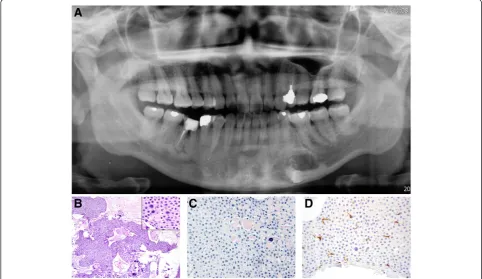
The clinical features of 6 cases of classic CEOT are sum-marized in Table 1. The patient age at first presentation was 12–41 years (median age, 29.5 years). Three pa-tients were male and 3 were female. Five tumours occurred in the mandible and 1 occurred in the maxilla. Five tumours involved the premolar and molar regions, while the anterior portion of the mandible was affected in 1 case. The first sign in 5 cases was jaw swelling, while the other case was incidentally discovered. In 5 patients, well-defined unilocular radiolucencies were noted, 3 of which had radiopaque areas and an uner-upted tooth (Figure 1A). Five patients were initially treated by enucleation or curettage and 1 patient was managed with segmental resection. Four patients were followed for 2–7 years and none of them showed any sign of recurrence.
Pathological findings
On histological examination, the tumour was com-posed of strands, nests, and masses of polyhedral epi-thelial cells with eosinophilic cytoplasm and prominent
nuclei. These cells had distinct intercellular bridges and were irregularly shaped with pleomorphic nuclei (Figure 1B). Between cells, there were round-shaped eosinophilic amyloid materials that stained positive for Congo red. The reddish homogeneous material ap-peared to undergo calcification and become concentric calcified corpuscles or calcified masses (Figure 1C). Langerin-positive cells were scattered in the epithelial mass of the tumour (Figure 1D). The ratio of langerin-positive cells to epithelial tumour cells was 1.7:100 and 0.8:100, respectively, in the 2 cases. S100 and CD1a staining showed the same pattern.
Langerhans cell variant of CEOT Case report
Case 1 A 40-year-old Chinese woman had a chief com-plaint of loosened left anterior teeth and pain that she first noted 4 years ago. The pain and loosened teeth had recently become obvious. Her past medical history was unremarkable with no evidence of systemic diseases. Intraoral examination revealed a depression over the left anterior hard palate. Loss of both labial and palatal al-veolar bone was noted. The lateral incisor was loosened.
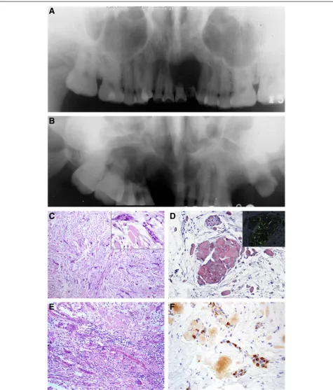
Radiographic examination revealed a well-defined uni-locular radiolucent area from the right central incisor to the first premolar (Figure 2A). There were no radiopaque foci within the lesion. Resorption of the left central and lateral incisor was detected. The clinical impression was a cyst. The lesion was curetted; during the surgery, however, the lesion was solid. The patient remained disease-free 5 years after the operation.
Case 2 A 58-year-old Chinese man was referred to our hospital for the evaluation of a swelling in the right max-illa that had been noted 3 months ago in a regular phys-ical examination. There were no obvious symptoms or signs at the lesion site reported by the patient. The pa-tient had no history of systemic disease but had suffered from allergenic rhinitis for >20 years. Intraoral examin-ation revealed a loss of alveolar bone along with the missing right central and lateral incisors. The right ca-nine was loose. There was a swelling of the plate from the right canine to the second molar area.
Radiographs revealed a multilocular radiolucent lesion without a definite sclerotic border from the left lateral incisor to the right second molar. The floor of the right maxillary sinus was not clear. No radiopaque foci were found in the lesion, while the roots of the right canine to the first molar were resorbed (Figure 2B). Ten years after a partial maxillectomy, no evidence of recurrence was found.
Pathological findings
These 2 tumours chiefly consisted of small nests and cords of epithelial cells and loose fibrous connective tissue stroma. The epithelial islands were composed of eosinophilic cytoplastic polyhedral tumour cells and a few clear cells (Figure 2C). The nuclei were relatively dense and contained a single nucleolus. Slight variation in cellular and nuclear size was seen. Intercellular brid-ges were occasionally seen between the tumour cells.
The globular masses of the homogeneous eosinophilic material tested positive for Congo red stain, were mostly located in the connective tissue, and showed apple green birefringence when subjected to polarized light (Figure 2D). There was moderate chronic inflammatory cell infiltration
in the fibrous stroma (Figure 2E). No calcification was noted. Langerhans cells positive for langerin, S100, and CD1a were seen in the epithelial islands of the tumour (Figure 2F). The ratio of Langerhans cells to epithelial tumour cells was 82.7:100 and 42.1:100, respectively, in the 2 cases.
Discussion
CEOT is an uncommon odontogenic tumour. Upon re-viewing our own file of odontogenic tumours from 1985 to 2006, we found a total of 6 cases of odontogenic tu-mours among 1,309 cases (0.46%) [13]. The male:female ratio of patients with CEOT was 1:1. The tumours were primarily in the mandible (maxilla:mandible ratio, 1:2), and approximately 82% of the cases were located in the premolar and molar regions [3]. The clinical features of the present series were in general agreement with those of earlier reports, i.e. equal sex distribution and predilec-tion for the mandible, especially the premolar and molar regions.
When first described [1], CEOT was considered a lo-cally invasive tumour, and some subsequent publications supported this concept. This view was based upon evi-dence suggestive of bone marrow involvement from ra-diographs and histological sections [2,14]. However, Vap et al [14] believed that the tumour is an expansile lesion that does not extend into the intertrabecular spaces as does the infiltrative solid ameloblastoma. Franklin and Pindborg [15] reported a recurrence rate of 14% that was primarily attributed to inadequate treatment. A few cases of malignant transformation have been reported [16,17]. It is now accepted that CEOT is a less aggressive tumour than ameloblastoma that responds well to con-servative surgery. Four cases of CEOT with follow-up data in the present series showed no signs of recurrence following surgery, which is in keeping with the notion.
As the first few reported cases of CEOT were all asso-ciated with an unerupted tooth, Pindborg [2] initially be-lieved that CEOT develops from the reduced enamel epithelium of an unerupted tooth. However, as reports of cases without an associated unerupted tooth and cases of a peripheral variant started to accumulate, it be-came evident that sources other than reduced enamel (See figure on previous page.)
epithelium should also be considered regarding CEOT histogenesis.
In the present series, 3 of 6 cases were associated with an impacted tooth. This is consistent with the literature that approximately half of CEOT cases are associated with an unerupted tooth. A few reports of a dentigerous cyst or dental follicle showed focal CEOT-like hyperpla-sia [18,19], which suggests that CEOT associated with the crowns of impacted teeth may indeed originate from the reduced enamel epithelium.
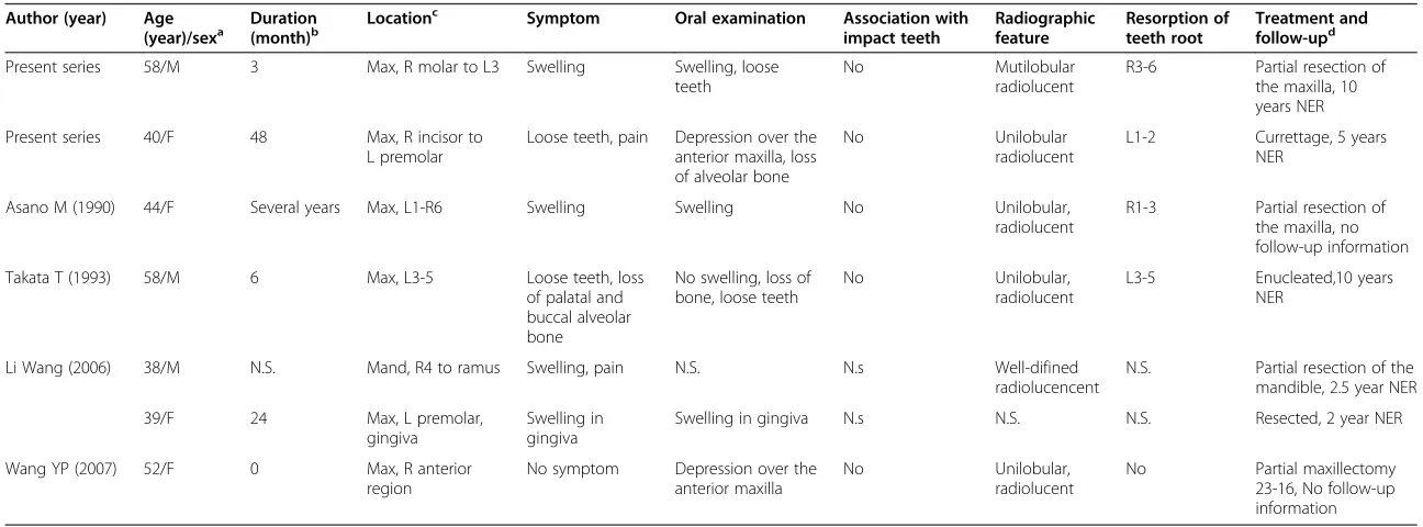
Our review of the English literature revealed 5 cases, together with our 2 new cases, for a total of 7 cases of the Langerhans cell variant of CEOT (Table 2). All of the pa-tients were Asian. The patient age range at first presenta-tion was 38–58 years (median, 44 years). Three patients were male and 4 were female. Six tumours occurred in the maxilla, while 1 occurred in the mandible. Five of the 6 maxillary tumours involved the anterior region. This fea-ture is in contrast to the findings of conventional CEOT. Jaw swelling was noted in 4 cases. Two patients com-plained of loosened teeth and 1 patient showed no obvious symptoms. During an intraoral examination, bony depres-sion of the tumour area was noted in 3 cases, a unique fea-ture not seen in conventional CEOT.
Of the 6 cases with radiographic records, well-defined unilocular radiolucency was noted in 5 cases and multi-locular radiolucency was noted in one. The radiopaque area was not evident in any cases. Root resorption was detected in 4 cases. Three patients were initially treated by enucleation or curettage and the other 4 cases under-went partial jaw resection. Five patients were followed for 2–10 years and no recurrence was recorded. Thus, the Langerhans cell variant of CEOT may show similar behaviours to those of conventional CEOT.
Histologically, the Langerhans cell variant of CEOT is unusual. Although Congo red-positive homogenous amyl-oid material could be seen, there was no calcification. The tumour islands were composed of small numbers of epi-thelial cells with clear cells suggestive of Langerhans cells. Langerin is the protein expressed in Langerhans cells that is localized in the Birbeck granules present within the cytoplasm. Langerin staining, accompanied by S100 and CD1a staining, confirmed the presence of Langerhans cells in the tumour.
With regard to histogenesis, none of the 7 cases sum-marized here were associated with an unerupted tooth; thus, the Langerhans cell variant of CEOT is less likely to arise from a reduced enamel epithelium. The tumour was mainly located within the jaw bone with apparent root resorption of the apical part of the involved tooth. Other tissue origins like the rests of Malassez should be considered.
Another interesting finding of the present study was that Langerhans cells were detected not only in the Langerhans
cell variant of CEOT but also in the epithelial nests of the conventional type. Similar reports have been presented by other groups [20]. Langerhans cells are bone marrow-derived cells that migrate into the oral epithelium and serve as antigen-presenting cells. Because both oral and odontogenic epithelia originate from the same oral ecto-derm, it is possible that Langerhans cells may also migrate into the epithelial nests of odontogenic tumours. Thus, Langerhans cells may not exclusively exist in the Langer-hans cell variant of CEOT. However, the number of Lang-erhans cell increased dramatically in the LangLang-erhans cell variant of CEOT. The increased number of Langerhans cells may be associated with inflammation. Interestingly, 6 of the 7 cases of Langerhans cell variant here had a mod-erate number of lymphocytes and plasma cell infiltration. Asono [6] postulated that Langerhans cells may play an important role in antigen presentation or regression of CEOT.
Although Langerhans cells increases are due to im-munogenic reactions and do not exist exclusively in this kind of CEOT, we continue to use the term“Langerhans cell variant of CEOT”to describe it since a limited num-ber of cases have been reported. At this point, the use of another name may not be helpful for accumulating data about this kind of CEOT with special clinical and histo-pathological characteristics. A more suitable name may be adopted after more cases have been reported.
Differentiating conventional CEOT from the Langer-hans cell variant is not difficult. The conventional type affects the premola and molar region as an asymptom-atic slow-growing expansile mass and may feature an unerupted tooth. In contrast, lesions of the Langerhans cell variant have a predisposition for the anterior maxilla and a possibly overlying bone depression. Histopatho-logically, in the conventional type, the tumour consists of islands and sheets of polyhedral epithelial cells, eo-sinophilic homogenous amyloid substance, and calcified tissue. In the Langerhans cell variant, very small islands and cords of neoplastic cells with abundant amyloid sub-stance and no calcification are characteristic features.
In conclusion, the Langerhans cell variant of CEOT is rare but consistent. To date, it has only been reported in Asian people, mainly in the anterior portion of the max-illa. This jaw tumour could present features of bone de-pression with loss of teeth and alveolar bone. The lesion comprises small epithelial islands and amorphous eo-sinophilic globules of amyloid materials with no appar-ent calcification. These features are somewhat differappar-ent from those of conventional CEOT and should alert the pathologists to the differential diagnosis.
Consent
Table 2 The clinical information of reported cases of Langerhans cell variant of CEOT
Author (year) Age (year)/sexa
Duration (month)b
Locationc Symptom Oral examination Association with impact teeth
Radiographic feature
Resorption of teeth root
Treatment and follow-upd
Present series 58/M 3 Max, R molar to L3 Swelling Swelling, loose
teeth
No Mutilobular
radiolucent
R3-6 Partial resection of the maxilla, 10 years NER
Present series 40/F 48 Max, R incisor to
L premolar
Loose teeth, pain Depression over the anterior maxilla, loss of alveolar bone
No Unilobular
radiolucent
L1-2 Currettage, 5 years NER
Asano M (1990) 44/F Several years Max, L1-R6 Swelling Swelling No Unilobular,
radiolucent
R1-3 Partial resection of the maxilla, no follow-up information
Takata T (1993) 58/M 6 Max, L3-5 Loose teeth, loss
of palatal and buccal alveolar bone
No swelling, loss of bone, loose teeth
No Unilobular,
radiolucent
L3-5 Enucleated,10 years
NER
Li Wang (2006) 38/M N.S. Mand, R4 to ramus Swelling, pain N.S. N.s Well-difined
radiolucencent
N.S. Partial resection of the mandible, 2.5 year NER
39/F 24 Max, L premolar,
gingiva
Swelling in gingiva
Swelling in gingiva N.s N.S. N.S. Resected, 2 year NER
Wang YP (2007) 52/F 0 Max, R anterior
region
No symptom Depression over the anterior maxilla
No Unilobular,
radiolucent
No Partial maxillectomy
23-16, No follow-up information
a
M = male, F = female;b
N.S. = not stated;c
max = maxilla, mand = mandible, R = right, L = left;d
NER = no evidence for recurrence.
Patholog
y
2014,
9
:37
Page
7
o
f
8
cpathology.o
rg/content/9/1
Competing interest
The authors declare that they have no competing interests.
Authors’contributions
Study concepts and study design: TJL, Data acquisition: YC, TTW, Quality control of data and algorithms: TJL, Data analysis and interpretation: YC, TTW, Manuscript preparation: YC, TTW, Manuscript editing: TJL, Manuscript review: TJL. YC and TTW: co-first author, TJL: corresponding author. All authors read and approved the final manuscript.
Authors’information
Yan Chen and Ting-Ting Wang: co-first author.
Acknowledgements
This work was supported by Research Grants from the National Natural Science Foundation of China (81141092, 81030018 and 81302349). The authors also gratefully acknowledge the patients for their cooperation.
Author details
1Department of Oral Pathology, Peking University School and Hospital of
Stomatology, Beijing, China.2Department of Oral Medicine, Hebei United University, School and Hospital of Stomatology, Tangshan, Hebei, China. 3Central Laboratory, Peking University School and Hospital of Stomatology,
Beijing, China.4National Engineering Laboratory for Digital and Material Technology of Stomatology, Beijing, China.
Received: 21 November 2013 Accepted: 5 January 2014 Published: 20 February 2014
References
1. Pindborg JJ:Calcifying epithelial odontogenic tumors.Acta Pathol Microbiol Scand Suppl1955,111:71.
2. Pindborg JJ:A calcifying epithelial odontogenic tumor.Cancer1958, 11(4):838–843.
3. Philipsen HP, Reichart PA:Calcifying epithelial odontogenic tumour: biological profile based on 181 cases from the literature.Oral Oncol2000, 36(1):17–26.
4. Anavi Y, Kaplan I, Citir M, Calderon S:Clear-cell variant of calcifying epithelial odontogenic tumor: clinical and radiographic characteristics.
Oral Surg Oral Med Oral Pathol Oral Radiol Endod2003,95(3):332–339. 5. Germanier Y, Bornstein MM, Stauffer E, Buser D:Calcifying epithelial
odontogenic (pindborg) tumor of the mandible with clear cell component treated by conservative surgery: report of a case.J Oral Maxillofac Surg2005,63(9):1377–1382.
6. Asano M, Takahashi T, Kusama K, Iwase T, Hori M, Yamanoi H, Tanaka H, Moro I:A variant of calcifying epithelial odontogenic tumor with Langerhans cells.J Oral Pathol Med1990,19(9):430–434.
7. Takata T, Ogawa I, Miyauchi M, Ijuhin N, Nikai H, Fujita M:Non-calcifying Pindborg tumor with Langerhans cells.J Oral Pathol Med1993, 22(8):378–383.
8. Slootweg PJ:Bone and cementum as stromal features in Pindborg tumor.J Oral Pathol Med1991,20(2):93–95.
9. Seim P, Regezi JA, O’Ryan F:Hybrid ameloblastoma and calcifying epithelial odontogenic tumor: case report.J Oral Maxillofac Surg2005, 63(6):852–855.
10. Wang L, Wang SZ, XMC:Langerhans cells containing calcifying epithelial odontogenic tumor: report of two cases and review of the literature.
Oral Oncology EXTRA2006,42:144–146.
11. Wang YP, Lee JJ, Wang JT, Liu BY, Yu CH, Kuo RC, Chiang CP:Non-calcifying variant of calcifying epithelial odontogenic tumor with Langerhans cells.
J Oral Pathol Med2007,36(7):436–439.
12. Barnes L, EJ RP, Sidransky:World Health Organization Classification of Tumours. Pathology & Genetics Head and Neck Tumours.Lyon: IARC Pres; 2005.
13. Luo HY, Li TJ:Odontogenic tumors: a study of 1309 cases in a Chinese population.Oral Oncol2009,45(8):706–711.
14. Vap DR, Dahlin DC, Turlington EG:Pindborg tumor: the so-called calcifying epithelial odontogenic tumor.Cancer1970,25(3):629–636.
15. Franklin CD, Pindborg JJ:The calcifying epithelial odontogenic tumor. A review and analysis of 113 cases.Oral Surg Oral Med Oral Pathol1976, 42(6):753–765.
16. Demian N, Harris RJ, Abramovitch K, Wilson JW, Vigneswaran N:Malignant transformation of calcifying epithelial odontogenic tumor is associated with the loss of p53 transcriptional activity: a case report with review of the literature.J Oral Maxillofac Surg,68(8):1964–1973.
17. Kawano K, Ono K, Yada N, Takahashi Y, Kashima K, Yokoyama S, Yanagisawa S: Malignant calcifying epithelial odontogenic tumor of the mandible: report of a case with pulmonary metastasis showing remarkable response to platinum derivatives.Oral Surg Oral Med Oral Pathol Oral Radiol Endod2007, 104(1):76–81.
18. Gopalakrishnan R, Simonton S, Rohrer MD, Koutlas IG:Cystic variant of calcifying epithelial odontogenic tumor.Oral Surg Oral Med Oral Pathol Oral Radiol Endod2006,102(6):773–777.
19. Ficarra G, Hansen LS, Stiesmeyer EH:Intramural calcifying epithelial odontogenic tumor.Int J Oral Maxillofac Surg1987,16(2):217–221. 20. Poomsawat S, Punyasingh J:Calcifying epithelial odontogenic tumor: an
immunohistochemical case study.J Mol Histol2007,38(1):103–109.
doi:10.1186/1746-1596-9-37
Cite this article as:Chenet al.:A clinicopathologic study on calcifying epithelial odontogenic tumor: with special reference to Langerhans cell variant.Diagnostic Pathology20149:37.
Submit your next manuscript to BioMed Central and take full advantage of:
• Convenient online submission
• Thorough peer review
• No space constraints or color figure charges
• Immediate publication on acceptance
• Inclusion in PubMed, CAS, Scopus and Google Scholar
• Research which is freely available for redistribution