R E V I E W
Open Access
Chimeric antigen receptors for adoptive T
cell therapy in acute myeloid leukemia
Mingxue Fan
1, Minghao Li
1, Lipeng Gao
1, Sicong Geng
2, Jing Wang
1, Yiting Wang
1, Zhiqiang Yan
1*and Lei Yu
1*Abstract
Currently, conventional therapies for acute myeloid leukemia (AML) have high failure and relapse rates. Thus, developing new strategies is crucial for improving the treatment of AML. With the clinical success of anti-CD19 chimeric antigen receptor (CAR) T cell therapies against B-lineage malignancies, many studies have attempted to translate the success of CAR T cell therapy to other malignancies, including AML. This review summarizes the current advances in CAR T cell therapy against AML, including preclinical studies and clinical trials, and discusses the potential AML-associated surface markers that could be used for further CAR technology. Finally, we describe strategies that might address the current issues of employing CAR T cell therapy in AML.
Keywords:Chimeric antigen receptors, Acute myeloid leukemia, Immunotherapy
Background
Acute myeloid leukemia (AML) is a cancer of the myeloid line of blood cells that is characterized by the clonal ex-pansion of abnormal myeloid progenitors in the bone marrow and peripheral blood, which interferes with the normal production of blood cells. AML is a rare disease, and its incidence increases with an aging population, as this disease is most commonly found in adults [1]. In the past 5 years, the cure rate was 35–40% for AML patients under 60 years old and 5–15% for patients older than 60. The elderly, who are unable to withstand intensive chemotherapy, have an average survival of 5–10 months [2]. Despite improving our understanding of AML, the disease still has poor outcomes due to high disease- and treatment-related mortality.
Forty years ago, the combined injection of cytarabine and anthracycline was introduced as the first standard treatment for AML [3, 4]. Since then, many chemotherapy regimens have improved outcomes for some AML patients [5]. However, the effectiveness of traditional chemotherapy may have hit a ceiling for treating AML, especially for older patients and those who either tend to relapse or have
intermediate- or high-risk factors associated with AML [6]. In addition, allogeneic hematopoietic stem cell transplant-ation (allo-HSCT) has been the most successful immuno-therapy for AML over the past decade, especially with the advances made in using alternative donors [7–9]. Unfortu-nately, older and less fit patients are poor candidates for allogeneic HSCT due to significant toxicity and a high relapse rate [10]. The limited success and high toxicity of the currently available strategies indicate an urgent need for new therapeutics. It is possible that the infusion of allo-geneic chimeric antigen receptor (CAR) T cells could enhance the efficacy of allogeneic HSCT [11]. This possi-bility is supported by recent evidence that a child with acute lymphoblastic leukemia (ALL) at the Children’s Hospital of Philadelphia relapsed after a cord blood trans-plant and then received infusions of CTL019 CAR T cells, resulting in a remission of leukemia without graft-versus-host disease (GVHD) [12]. In addition, another recent study showed that the treatment of allogeneic CAR T cells is beneficial for patients with relapsed B cell malignancies after allo-HSCT with low toxicities and complications [13]. Therefore, the CAR-expressing T cell technology, which has been successfully implemented in treating acute lymphoblastic leukemia (ALL), has been considered a promising immunological approach for the treatment of AML [12, 14–19]. This new type of targeted immunother-apy merges the exquisite targeting specificity of monoclo-nal antibodies with the potent cytotoxicity and long-term * Correspondence:zqyan@sat.ecnu.edu.cn;yulei@nbic.ecnu.edu.cn
1Institute of Biomedical Engineering and Technology, Shanghai
Engineering Research Center of Molecular Therapeutics and New Drug Development, School of Chemistry and Molecular Engineering, East China Normal University, NO. 3663 Zhongshan Road, Shanghai 200062, People’s Republic of China
Full list of author information is available at the end of the article
persistence provided by cytotoxic T cells. CAR is an artificial antigen receptor that mediates antibody-targeted recognition. The binding between CAR and its antigen on tumor cells triggers a signal transduction cascade through signaling domains and then activates T cells to kill the target directly or through other compo-nents of the immune system (Fig. 1) [20]. At the begin-ning of the in vitro expansion stage, CAR can be transferred to the patient’s selected T cells using either viral vectors or non-viral approaches [21]. The viral vectors include retroviruses (including lentivirus), adenovirus and adeno-associated virus. Among them,
γ-retroviral and lentiviral vectors have been the most useful carriers for long-term gene expression because of their ability to integrate into the host genome and their low intrinsic immunogenicity [22, 23]. In contrast toγ-retroviral vectors, lentiviral vectors can deliver lar-ger DNA sequences and integrate into non-dividing cells, which are less susceptible to silencing by host restriction factors [24]. Lentiviral vectors are more commonly used in clinical trials because of their safer integration site pro-file [25]. Non-viral systems, including nude DNA, mRNA, liposomes, etc., are very effective in gene delivery because of their higher efficiency, non-infectiousness, unlimited carrier capacity, controlled chemical constitution and
generous production. For instance, mRNA electroporation in clinical trials induced the transient expression of CAR for approximately one week and prevented the potential toxicity of CRS [11].
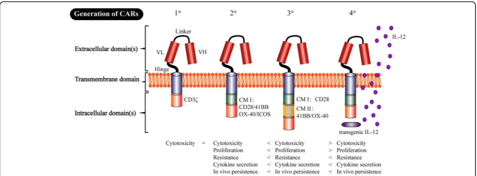
The adoptive cell therapy of CAR-expressing T cells is a new but promising approach in the field of cancer im-munotherapy. The development of CAR T cells can be divided into four generations based on the different characteristics of intracellular domains (Fig. 2). The CAR prototype consists of an extracellular domain that serves as the targeting moiety (which is usually a single chain variable fragment (scFv) formed from a monoclo-nal antibody, mAb), a transmembrane domain, and an intracellular signaling domain(s) [26]. CARs with the typical structure of“scFv-spacer-CD3z”design are called
“1st-generation CARs”. In contrast to the T cell receptor (TCR), “1st-generation CAR” recognizes the targets in-dependent of major histocompatibility complex (MHC) restriction, therefore making it highly specific for various surface antigens on tumor cells [27]. However, further research has gradually shown that“1st-generation CARs” exhibit the problems of deficient secreted cytokines, inadequate proliferation, and low persistence in vivo. To overcome these weaknesses, CD3ζ as well as co-stimulatory signaling domains, such as 41BB and CD28,
were incorporated into the intracellular domain to form the so-called“2nd-generation CAR”[28–30]. The added co-stimulatory signal can help complete the activation of T cells and avoid apoptosis by promoting the IL-2 syn-thesis. The CD28ζ CAR T cells primarily caused consti-tutive stimulation, proliferation and growth. In contrast, the 41BBζCAR T cells induced early exhaustion, thereby limiting the antitumour efficacy [21]. Correspondingly, the CD3ζ plus two co-stimulatory signaling domains, 41BB- and CD28, were introduced into the intracellular domain of “3rd-generation CARs” to augment cytokine production and cancer-killing ability [31, 32]. Unfortu-nately, the latest studies have shown that the“ 3rd-gener-ation CARs” did not produce more desirable outcomes compared with the“2nd-generation CARs”. Because the stronger stimulation may produce potential side effects, such as cytokine release syndrome (CRS), further studies should be performed to explore the safety of“ 3rd-gener-ation CARs”. Notably, recent studies have indicated that CAR reached its limit when targeting tumors with a remarkable phenotypic heterogeneity. Subsequently, the
“4th-generation CARs”, i.e., so-called TRUCK T cells, were proposed, which are formed by an additional modi-fication with an inducible expression cassette for a trans-genic protein. For example, a cytokine such as IL-12 can be released by CAR T cells to modulate the T cell response, which can help keep the local therapeutic con-centration and avoid systemic toxicity by releasing a variety of therapeutic proteins [27]. We anticipate that the future new generations of CAR will overcome some of the corresponding problems of CAR T cell therapy.
Translating the success of CAR T cell therapy to other malignancies that have an unmet medical need, such as AML, is currently underway. For instance, CAR T cell therapy has been developed in patients with AML, as many detailed studies have been published. This review summarizes the recent application of CAR T cell therapy in AML and focuses on AML-associated cell surface an-tigens that could be potential target candidates for CAR T cell therapy. Finally, we discuss the common issues of CAR T cell therapy in AML and summarize the strat-egies of building CAR T cells with improved safety and availability.
The application of CAR-modified T cells in AML
Despite the enormous challenge of developing CAR T cells for multiple diseases, several potential CAR T cell targets have been actively explored in preclinical studies and clinical trials over the past decade (Fig. 3). From the evolution of CAR T cell therapy for treating AML, it may be observed that we have gradually shifted the focus of our research on creating safer and more effective CAR-modified T cells for the past two years using gen-omic editing.
Lewis Y antigen
One of the remarkable advantages of CAR T cell therapy is the ability to recognize a broad variety of targets such as non-protein antigens. The Lewis Y antigen (LeY) is an example of this situation; LeY is an oligosaccharide that is overexpressed on many epithelial cancers and hematological malignancies (including AML) [33, 34]
Fig. 2The four generations of CAR production. The extracellular domain of CAR includes a single chain variable fragment (scFv) (H (heavy) and L (light) chain) that is spliced by a linker. A hinge (e.g., hinge region of human immunoglobulin D molecule) ensures flexibility and connects to the transmembrane domain (TM). The TM is routinely the constant region of the human G immunoglobulin, whereas the intracellular domain includes only the CD3ζsignaling domain known as the“1st-generation CAR”. Subsequently, to augment T cell persistence and proliferation [28], CD3ζas well as the costimulatory endo-domains 41BB- or CD28-signaling domains were incorporated into the“2nd-generation CAR”. The intracellular domain includes CD3ζplus two costimulatory domains 41BB- and CD28-signaling domains that were included in the
but has limited expression on normal healthy tissues [35, 36]. The LeY - CAR T cell trial was the first CART therapy clinical trial targeting AML (ClinicalTrials.gov number, NCT01716364), evaluating the effect of an au-tologous second-generation anti-LeY CAR T cell therapy in 4 patients with relapsed AML. Following fludarabine preconditioning, the patients were administered up to 1.3 × 10⁹ of total T cells (14–38% CAR T cells). The results showed that two patients achieved protracted re-mission, one patient achieved cytogenetic rere-mission, and the fourth patient with active leukemia presented a re-duction in peripheral blood (PB) blasts. Incredibly, no grade 3 or 4 toxicity was observed. The most notable finding from this study was the lack of toxicity and the durable in vivo persistence after infusion [37]. In addition, LeY is the first antigen that was successfully
implemented in CAR T cell therapy to target AML. Retroviral transduction of anti-LeY-CD28ζ into CAR T cells has exhibited potent cytotoxicity against LeY+ epi-thelial tumor cell lines in vitro and animal models in vivo without affecting normal tissues [38].
CD44v6
The hyaluronate receptor CD44 is a type I transmem-brane glycoprotein commonly used as a marker to iden-tify cancer stem/initiating cells. CD44 variant domain 6 (CD44v6) is a CD44 variant isoform expressed in AML [39] and multiple myeloma (MM) [40], correlating with a poor prognosis. Importantly, CD44v6 is absent in hematopoietic stem cells (HSCs) and expressed at low levels on normal cells, which may provide a therapeutic window. The Italy San Raffaele Scientific Institute
designed a second-generation CD28-CD3ζCAR and de-rived the scFv from a mutated sequence of the human-ized CD44v6-specific mAb (bivatuzumab). This CAR exerted a significantly positive effect in targeting cancer cells in vitro and in vivo. However, these anti-CD44v6-CD28ζ CAR T cells caused an unexpected and dose-limiting toxicity (DLT), monocytopenia. Subsequently, this group focused their attention on co-expressing clinical-grade suicide genes [41, 42] to control these ad-verse events [43].
NKG2D ligand
Natural killer group 2D (NKG2D) ligands contain six members of the UL16-binding protein, or the retinoic acid early transcript (ULBP/RAET) family and two members of the MHC class I-related chain (MIC) family [44], all of which are either absent or minimally expressed on healthy tissues but widely expressed on numerous malignancies (including ovarian cancer [45] and AML [46]). Several different variants of NKG2D-di-rected CAR have been developed and tested for their cytotoxicity and the ability of achieving complete remissions [47]. From April 2015 to July 2016, a phase I (ClinicalTrials.gov number, NCT02203825) dose-escalation study was performed to establish the feasibil-ity and safety of NKG2D-DAP10-CD3ζ CAR T cells (CM-CS1 T cells) in treating AML and was completed ahead of schedule. A total of 11 subjects were infused with 1 × 10⁶to 3 × 10⁹(8 cohorts) of CM-CS1 T cells based on a 3 + 3 design. The results showed that 9 sub-jects treated in the first 3 cohorts completed their 28-day evaluation period without any DLTs. It is worth men-tioning that there was no case of cell-related neurotoxicity, cytokine release syndrome (CRS), autoimmunity or CAR T cell-related death during treatment [48].
Folate receptorβ
The folate receptor β(FRβ) is a member of the folate-binding protein receptors family, which is primarily expressed on myeloid-lineage hematopoietic cells and frequently up-regulated in AML blasts (~70%) [49, 50]. Preclinical models using anti-FRβ-CD28ζ CAR T cells presented potent and targeted killing of leukemia cells while preserving healthy CD34+ cells. Interest-ingly, the investigators also used all-trans retinoic acid (ATRA), an FDA-approved drug for subclass M3 AML [51, 52], to up-regulate the target antigen, which led to an improved anti-leukemia activity [53]. This general concept of increasing antigen expression on diseased tissue to improve the potency of the CAR T cell agent is very likely to be further explored in follow-up studies.
CD38
CD38, also known as cyclic ADP ribose hydrolase, is a glycoprotein expressed on the surface of many immune cells. Previous studies have shown that CD38 is expressed on the majority of AML blasts but not healthy human hematopoietic stem cells (HSCs) [54, 55]. Ac-cordingly, one research group has focused on CD38 as a candidate therapeutic target and developed an anti-CD38-41BBζ CAR. Remarkably, studies involving this CAR revealed another example of ATRA-enhanced cyto-toxicity on AML cells regarding enhanced CD38 expres-sion [56]. Therefore, these results may provide a new paradigm for pharmacologically inducible immunother-apy that combines ATRA and CAR T cell therimmunother-apy to treat AML.
FLT-3
Fms-like tyrosine kinase 3 (FLT-3), also known as CD135, is a cytokine receptor belonging to the class III receptor tyrosine kinases. The FLT3 gene is one of the most commonly mutated genes in AML, with internal tandem duplications of FLT3 (FLT3-ITD) as the most frequent mutation (25%) associated with AML. In a re-cent study, researchers generated anti-FLT3-41BBζCAR T cells, which demonstrated potent anti-AML activity in vitro and in vivo. Notably, compared with anti-CD33 CAR T cells, anti-FLT3 CAR T cells indicated a lower hematological toxicity [57].
CD7
CD7 is an NK and T cell marker that is highly expressed in 30% of AML cases. Its expression is associated with a worse prognosis and chemoresistance [58, 59]. CD7-directed CAR T cells have been created and exhibited potent cytotoxicity against T-ALL and AML cell lines as well as against primary AML blasts, but there was no observed toxicity against normal myeloid progenitors [60]. This finding indicates that CD7 is a potential target for AML that should be further explored in future studies.
CD33
AML, which resulted in a transient response [67]. How-ever, as CD33 is expressed in healthy myeloid cells and other tissues [69–71], the toxicity that occurs following CD33-directed CAR T cell infusion must be well con-trolled before further evaluation in clinical trials. One re-search group proposed a novel solution to this problem by removing CD33 from normal hematopoietic stem progenitor cells (HSPCs) using genomic editing during CD33-mediated CAR T cell treatment of AML, as CD33 is not essential to hematopoietic differentiation, and a lack of CD33 in myeloid progeny does not cause any vis-ible functional changes [72]. Overall, recent studies were committed to reducing the toxicity of CD33-specific CAR T cells and proposed many strategies, which will be further described in detail below.
CD123
As the transmembrane alpha chain of the interleukin-3 receptor, CD123 is widely expressed in the majority of AML blasts but presents low expression levels on nor-mal hematopoietic cells [73–77]. Both anti-CD123-CD28ζ CAR and anti-CD123-41BBζ CAR T cells have demonstrated potent leukemia killing ability in vitro and in vivo but produced incongruous results regarding their myeloablative effect on healthy CD123+cells [78, 79]. In addition, two phase I trials (ClinicalTrials.gov number,
NCT02159495, NCT02623582) for CD123-directed
CAR T cell therapy are currently underway to validate the effect and safety profiles. Subsequently, one group
generated a novel
anti-CD123-CD28-CD137-CD27-CD3ζ-iCasp9 CAR (4SCAR123) that exhibited potent cytotoxicity against AML in vitro and then infused 4SCAR123 into a 47-year-old male patient with AML-M2. The patient exhibited a rapid response consistent with a controllable CRS and achieved partial remission within 20 days without any off-target cytotoxicities [80]. One significant concern is that CD123-directed CAR T cells could irreversibly increase the myeloablative impact on normal hematopoiesis. Some strategies have been proposed to develop safer CD123-directed CAR T cells, one of which involves using the irreversible myeloabla-tion of CD123-directed CAR T cells in conjuncmyeloabla-tion with allogenic HSCT, such as the chemotherapy precondi-tioning prior allo-HSCT, to reduce the risk of AML relapse and pave the way to further explore CAR T cell combination therapies [78].
CLEC12A
CLEC12A (also known as CLL1) has been previously described as selectively overexpressed in leukemia stem cells (LSCs). One group confirmed that CLEC12A is heterogeneously expressed on AML blasts and overex-pressed on AML LSCs. Lentivirally transduced anti-CLEC12A-41BBζ CAR T cells can successfully target
CLEC12A+ cells, which are resistant to chemotherapy. Hence, anti-CLEC12A CAR T cells can potentially be used as a consolidation regimen after induction chemo-therapy to eradicate LSC and minimal residual disease (MRD) in AML [81].
AML-related surface antigens as candidates for CAR therapies
Due to its potent and durable anti-tumor activity, CAR T cell therapy has been recently regarded as a promising curative therapy against B-lineage malignancies. The rea-son for these positive results is that CD19 is an ideal tar-get for B-cell malignancies [65]. As is well known, new tumor-related antigens may arise following somatic mu-tations in the dividing tumor cells, which can serve as valuable therapeutic targets. These antigens are classi-fied as tumor-specific antigens and mutation-causing over-expression antigens [82]. CD19 is a unique tumor-specific antigen expressed on the tumor cells of B-lineage malignancies but not on normal cells. Unfor-tunately, truly AML-specific surface antigens have not been identified to date. Most of the antigens currently studied are mutation-causing over-expression antigens, which result in fatal “on-target/off-tumor toxicity” of CAR T cell treatments because of the expression of these antigens on normal tissue. Therefore, one pre-requisite for developing clinically effective CAR therap-ies is the confirmation of specific AML-associated surface targets. Theoretically, these antigens should meet the following specific requirements [83]: 1) a con-firmed AML surface antigen; 2) expressed on as few normal tissues as possible; 3) expressed in an ad-equately large percentage of AML patients; 4) homoge-nously expressed on the tumor cells of a given patient; and 5) exerts an essential function in the pathophysi-ology and/or bipathophysi-ology of AML [84].
In addition to the above-mentioned targets used in CAR T cell therapy to treat AML, several other surface molecules, which are listed in Table 1, have been identi-fied and may be useful for directing the future explor-ation of CAR T cells in AML based on their distribution in normal tissue and specific involvement in potential toxicity [84].
CD123 is a typical LSC target in AML, and it has been reported that CD123-CAR T cells may be a promising tool as a chemotherapy-free myeloablative conditioning regimen for HSCT, which is particularly critical to avoid relapse [79]. As shown in Table 1, CD47 is overex-pressed on LSCs and can be detected in almost all AML samples, and its expression is often associated with worse outcomes [86]. AML LSCs escape macrophage phagocytosis by the recognition between CD47 on the LSCs and extracellular region of signal regulatory pro-tein alpha (SIRPα) on the macrophages [87]. By contrast, CD47 is faintly expressed in most normal tissues [84]. These findings make CD47 an ideal marker of AML LSCs. T-cell immunoglobulin mucin-3 (TIM-3) is an-other ideal marker of AML LSCs and is highly expressed in LSCs in most types of AML (except for M3) but is
not expressed in normal LSCs [88]. TIM-3 plays an im-portant role in the viability, proliferation, and differenti-ation of AML LSCs [89], as well as in the exhaustion of CD8+ T cells. Several recent studies have shown that AML relapse after CAR T cell therapy is directly associ-ated with the significant up-regulation of TIM-3 recep-tors on T cells. TIM-3 pathways are also involved in the exhaustion of CAR T cells and the dysfunction of AML [90, 91]. This pathway is worth further exploration as a potential target in the clinical setting.
The challenges and corresponding strategies of CAR T cell therapy in treating AML
CAR-redirected T cells are an emerging powerful tool for treating patients with cancer, with an especially high rate of long-term complete remission achieved by CAR
Table 1Cell surface antigens expressed on AML compared with HSC
Antigen AML
expression
Function Normal tissue expression Comment Reference
CD44 100%
(samples)
Mediates cell adhesion and can transduce signals
Ubiquitously expressed with many alternatively spliced isoforms
Cancer stem cell marker on several solid tumors
[123]
CD45RA >90% (samples)
Regulates a variety of cellular processes including cell growth, differentiation, mitotic cycle, and oncogenic transformation
naive T cells;
CD34+CD38+normal progenitors A specific marker for leukemiastem cell subpopulations in AML [124]
CLL-1 (CLEC12A, DCAL-2, MICL)
≈95% (samples)
ND Restricted to hematopoietic
cells of myeloid lineage
Expression may identify minimal residual disease and predict relapse
[81,125, 126]
CD96 66%
(samples)
May have a function in NK cell adhesion and/or activation
Resting and activated T cells and NK cells, possibly intestinal epithelium
Expressed on only 5% of CD34+CD38−CD90+cells in bone marrow
[127]
CD47 100%
(samples)
Binds SIRPa and inhibits phagocytosis
Widely expressed at low levels Differential expression facilitated prospective separation of residual normal HSC from LSC
[128,129]
CD32 34%
(samples)
Fc-g receptor 2 (FCGR2) Restricted to hematopoietic cells Not expressed on functional HSC [130]
CD25 25%
(samples)
High-affinity IL-2 receptor (IL2RA)
Restricted to hematopoietic cells Not expressed on functional HSC [130]
TIM-3 (HAVCR2)
most AML types (except for M3)
An important regulator of Th1 cell immunity and tolerance induction
Not expressed in CD34+CD38− normal HSCs or the majority of CD34+CD38+normal progenitors
An immune checkpoint, also a Th1-specific cell surface protein that regulates macrophage activation
[131–133]
NDnot detected,NKnatural killer,HSChematopoietic stem cell,LSCleukemia stem cell,SIRPasignal regulatory protein-a
Table 2AML-related surface molecules as potential targets for CAR therapies
Antigen Antibody clone Efficacy in treatment model Effect on normal References
CD47 B6H12
(mouse IgG1)
Treatment initiated 8–12 weeks post transplantation: decrease AML in 3/3 samples (8/8 mice) with clearance of the bone marrow in 3/8 mice
No effect on in vitro phagocytosis of CD34+ normal bone marrow progenitors
[128]
Hu5F9 (Human IgG4)
Completely eradicated human AML in vivo, leading to long-term disease-free survival of patient-derived xenografts
Safely administered intravenously at doses by toxicokinetic studies in non-human primates
[134]
TIM3 ATIK2a
(human IgG2b)
Effective in killing TIM-3 expressing cell lines by its CDC and ADCC activities;
In vivo xenogeneic transplantation efficiently eradicated AML LSCs
No effect on cord blood or bone marrow engrafted mice
[133]
T cell treatments in relapsed/refractory CD19+ ALL pa-tients [17, 19, 92]. Over the past few years, several groups have concertedly focused on translating CAR T cell therapy to AML, and they have demonstrated that CAR T cells can eradicate AML in both preclinical and clinical trials. Thus, the efficacy of anti-AML CAR T cells appears to be equivalent to that of anti-ALL CAR T cells. Nevertheless, critical questions remain in this field. Here, we will outline the challenges of CAR T cell ther-apies when applied to AML, and focus on discussing the available and potentially feasible strategies to optimize the efficacy and safety of CAR T cell therapy (Fig. 4).
Cytokine release syndrome
When CAR T cells exert a clinical effect, persistence and proliferation are required; however, these activities may also cause significant toxicity. The most common and harmful toxicity is cytokine release syndrome (CRS), a rapid and evident inflammatory systemic re-sponse caused by dramatic increases in many inflam-matory cytokines (e.g., soluble IL-2R, IL-6 levels, ferritin, C-reactive protein (CRP), etc.) that occur with the in vivo activation and exponential proliferation of CAR T cells. [93]
As previously reported by Wang et al., one AML pa-tient treated with approximately 4 × 10⁸anti-CD33 CAR T cells experienced CRS [67]. Another group submitted an abstract that described a single patient treated with anti-CD123 CAR T cells who showing severe CRS in the absence of overt off-target cytotoxicity [94].
Many studies have indicated that IL-6 is a central me-diator of CRS-related toxicity [93]. Furthermore, several clinical studies have proved that the combined adminis-tration of tocilizumab, an anti-IL-6R antagonist, and sys-temic corticosteroids showed successful and rapid relief of CRS following CAR T cell infusions [12]. The clinical treatment algorithm for CRS has been well reviewed; please refer to reference 95 [95].
Strategies of further optimizing the treatment algo-rithms for CRS are currently under investigation (Clini-calTrials.gov number, NCT02906371), and gene-editing technology could be applied to CAR T cells to avoid CRS-related toxicities. For example, either gene silen-cing or the CRISPR/Cas9 system can be used to disturb IL-6 and other CRS-related cytokines in T cells prior to transduction with CARs. Additionally, T cells could simultaneously express a corresponding scFv specific to the IL-6 receptor such as tocilizumab as well as CARs
in order to block the IL-6 receptors actively avoiding CRS (Fig. 5h).
In all, the mechanisms by which CAR T cells cause CRS are varied and poorly understood. How to effect-ively control the CRS toxicity of CAR T cells is one of the most important challenges for improving the field of CAR T cell therapies overall.
On-target/off-tumor toxicity
Because on-target/off-tumor toxicity results from the ex-pression of tumor-associated antigens (TAAs) on normal tissue, minimizing the risk of toxicity is critical in the suc-cessful implementation of CAR T cell therapy. The first step in this process is to select more specific AML-associated surface targets, as mentioned above. However, it is highly difficult to identify surface antigens that are uniquely expressed on malignant myeloid tumors. There are many reports regarding insignificant myelosuppression caused by CAR T cells in preclinical models of AML. In addition, one AML patient enrolled in NCT01864902
experienced moderate hepatotoxicity and a transient reduction in marrow blasts following infusion with CD33 CAR T cells [67]. Another clinical trial with anti-LeY CARs in AML did not reveal any major off-target toxicities [37].
In consideration of the serious consequences of the
“on-target/off-tumor”toxicities reported in other clinical cases [96, 97], we should prepare corresponding strat-egies to address the “on-target/off-tumor” effects that may arise at any time.
mRNA electroporation
The expression of CARs using mRNA electroporation of T cells ensures the gradual loss of surface CAR expres-sion as T cells divide, which may be a useful strategy for determining the potential toxicity of novel constructs. One group transiently expressed an mRNA CAR con-struct targeting CD33 to avoid prolonged toxicity [65], whereas another clinical study is currently ongoing in which T cells expressing anti-CD123 CARs via mRNA
Fig. 5Different types of“Dual targeting”approaches.aThe CD3ζand costimulatory domains are separated in individual molecules targeting two diverse tumor antigens, an event known as trans-signaling CARs. These proteins will be activated when both antigens are identified [115–117].bThe
electroporation were infused into patients with AML (ClinicalTrials.gov number, NCT02623582) to evaluate efficacy and safety.
Suicide gene applications
A suicide gene is a genetically encoded molecule that allows for the selective destruction of adoptively transferred cells. The addition of a suicide gene to cellular therapeutic prod-ucts can lead to the selective ablation of gene-modified cells, which can mitigate or prevent collateral damage to contiguous cells and/or tissues [32]. This approach may be useful in abrogating the on-target and off-tumor toxicities of CAR-directed T cells. The inducible Caspase9 (iC9) sui-cide gene comprises a drug-binding domain cloned in frame with human Caspase9. Upon the exogenous adminis-tration of a non-therapeutic small molecule chemical in-ducer of dimerization (CID), iC9 dimerizes and induces apoptosis of the transduced cells within hours. CD44v6-, CD33-, and CD123-directed CAR T cells all contain an iC9 suicide gene as a tool for controlling the adverse events, which has been tested in preclinical research [37, 68, 80].
“Kill switch”—EGFRt
A“kill switch”is based on a tag derived from the epider-mal growth factor receptor (EGFRt) that retains the epi-tope recognized by the commercially available FDA-approved mAb cetuximab [98]. Anti-CD33- and anti-CD123-CD28ζ-EGFRt cells have been designed that can be eliminated by cetuximab if either CRS or any on-target/off-tumor toxicities are observed [99–101].
Dual-targeting strategies
When off-tumor toxicity is observed, these above strat-egies could enhance the ability of either ameliorating or abrogating these deleterious effects. Therefore, the inclu-sion of up-front safeguards is in an urgent need to pre-vent off-target toxicity in healthy tissues. Specific novel strategies are described in Fig. 5a-d, but future studies are required to expand on these ideas.
Relapse
Despite the scarcity of clinical cases regarding AML re-lapse after CAR T cell therapy, several preclinical studies have been performed to explore the reasons for the re-lapse. The corresponding strategies to address this issue have also been proposed.
Reduced efficacy and LSCs
Relapse is primarily caused by the lack of effectiveness of CAR T cells, which can be attributed to two factors: the immunosuppressive microenvironment and LSCs. To address the first issue, one approach is the use of so-called “TRUCK cells”, which can induce IL-12 release and activate innate immune cells to the targeted tumor
and thus eliminate cancer cells not recognized by CAR T cells [27]. This strategy can enhance the efficacy of CAR T cell therapy, thereby eliminating cancer cells and preventing tumor relapse caused by the residual cancer cells. To address the second issue regarding LSCs, the best solution is to identify the optimal markers for AML LSCs applied to CAR, which we have discussed in detail above.
Immune checkpoint
Inhibitory receptors/pathways, such as the PD-1 and TIM-3 pathways, induce the dysfunction and exhaustion of CAR T cells in AML and are also the mechanism of immune escape. Recently, several studies have indicated that there is a significantly higher expression of PD-1 and TIM-3 on T cells in relapsed AML samples com-pared with that seen in remittent or healthy donors [91, 102, 103]. Gene-editing technology could allow for the permanent disruption of negative signaling pathways [104]. Combined approaches using blocking antibodies may also interrupt this interaction, thus leading to the increased CAR T cell-induced cytotoxicity [103]. The lat-est technology is the use of switch receptors that incorp-orate a segment of the PD-1 receptor into the CAR construct (Fig. 5e), thereby inducing PD-L1 expression within the tumor microenvironment (TME) to augment the cytokine secretion, proliferation and granzyme ex-pression of CAR T cells, improving tumor therapy [105].
Antigen escape
robust anti-tumor activity. Currently, our group is evaluat-ing CD33/CD123 dual-targeted CARs to prevent antigen escape-caused relapse and may evaluate them as promising myeloablative tools for HSCT in a follow-up study.
Conclusion
In the past few years, the progress of CAR-engineered T cells has rapidly developed and made great achieve-ments. Nevertheless, there still exist certain limitations in this field that should not be ignored. One of the most concerning issues is that there is no convincing evidence of an AML-specific cell surface antigen that can be safely used to maximize the usefulness of CAR T cells. Admirably, many research groups are still confident and have developed numerous strategies to improve the current status of CAR T cells as a therapeutic in the AML field, such as gene-editing technology, antibodies, and combination therapies, most of which have been presented in this review. If these strategies could be suc-cessfully employed in clinical trials, the ability of CAR-expressing T cells in treating AML would be immeasur-able. In addition, we hope that this review provides use-ful information regarding the overall progress of CAR T cell therapy in the AML and injects new ideas into fu-ture research. In conclusion, the adoptive transfer of CAR-engineered T cells represents a valuable and at-tractive therapeutic strategy that has the potential to provide new prospects for cancer immunotherapy.
Abbreviations
ADCC:Antibody-dependent cell-mediated cytotoxicity; ALL: Acute lymphoblastic leukemia; allo-HSCT: Hematopoietic stem cell transplantation; AML: Acute myeloid leukemia; ATRA: All-trans retinoic acid; CAR: Chimeric antigen receptor; CDC: Complement dependent cytotoxicity; CIK: Cytokine induced killer; CRS: Cytokine release syndrome; EBV-CTL: Human Epstein Barr Virus-cytotoxic lymphocyte; EGFR: Epidermal growth factor receptor; FLT-3: Fms-like tyrosine kinase 3; FRβ: Folate receptorβ; GVHD: Graft-versus-host disease; IL12: Interleukin-12; iMC: Inducible MyD88/CD40; LAG3: Lymphocyte activating 3; LSC: Leukemia stem cell; mAb: Monoclonal antibody; MHC: Major histocompatibility complex; mRNA: Messenger ribonucleic acid; NKG2D: Natural killer group 2D; PD1: Programmed death 1; scFv: Single chain variable fragment; SIRPa: Signal regulatory protein-a; TAAs: Tumor-associated antigens (TAAs); TCR: T cell receptor; TIM-3: T-cell immunoglobulin mucin-3; TME: Tumor microenvironment (TME)
Acknowledgements
The authors are deeply thankful for their parents’encouragement. We also thank Professor Yan and Minghao Li for their generous and constant support. Finally, Mingxue Fan would like to acknowledge Sicong Geng and“MayDay” (Ashin, Monster, Stone, Masa, Ming); if we had never met, I would not have had the courage to follow my dreams.
Funding
This work was supported by the National Basic Research Program of China (2013CB932500), the National Natural Science Foundation of China (60976004), and the“985”grants of East China Normal University (ECNU).
Availability of data and materials
The material supporting the conclusion of this review has been included within the article.
Authors’contributions
Mingxue Fan designed the study. All authors read and approved the final manuscript.
Ethics approval and consent to participate This is not applicable for this review.
Consent for publication This is not applicable for this review.
Competing interests
The authors declare that they have no competing interests.
Publisher’s Note
Springer Nature remains neutral with regard to jurisdictional claims in published maps and institutional affiliations.
Author details 1
Institute of Biomedical Engineering and Technology, Shanghai Engineering Research Center of Molecular Therapeutics and New Drug Development, School of Chemistry and Molecular Engineering, East China Normal University, NO. 3663 Zhongshan Road, Shanghai 200062, People’s Republic of China.2China Novartis Institutes for Biomedical Research Co., Ltd., GDD/TRD/Chemical and Pharmaceutical Profiling, 5F, Building 3, Novartis Campus 4218 Jinke Rd, Zhangjiang Hi-Tech Park Pudong District, Shanghai 201203, China.
Received: 7 June 2017 Accepted: 17 August 2017
References
1. Eaves CJ, Humphries RK. Acute myeloid leukemia and the Wnt pathway. N Engl J Med. 2010;362:2326–7.
2. Döhner H, Weisdorf DJ, Bloomfield CD. Acute Myeloid Leukemia. N Engl JMed. 2015;373:1136–52.
3. Yates J, Glidewell O, Wiernik P, Cooper MR, Steinberg D, Dosik H. Cytosine arabinoside with daunorubicin or adriamycin for therapy of acute myelocytic leukemia: a CALGB study. Blood. 1982;60:454–62.
4. Yates JW, Wallace J Jr, Ellison RR, Holland JF. Cytosine arabinoside (NSC-63878) and daunorubicin (NSC-83142) therapy in acute nonlymphocytic leukemia. Cancer Chemother Rep. 1973;57:485–8.
5. Medinger M, Lengerke C, Passweg J. Novel therapeutic options in Acute Myeloid Leukemia. Leukemia Research Reports. 2016;6:39–49. 6. Kantarjian H, O'Brien S. Questions regarding frontline therapy of acute
myeloid leukemia. Cancer. 2010;116:4896–901.
7. Passweg JR, Baldomero H, Bader P, Bonini C, Cesaro S, Dreger P. Hematopoietic stem cell transplantation in Europe 2014: more than 40 000 transplants annually. Bone Marrow Transplant. 2016;51:786–92.
8. Versluis J, Hazenberg CL, Passweg JR, van Putten WL, Maertens J, Biemond BJ. Post-remission treatment with allogeneic stem cell transplantation in patients aged 60 years and older with acute myeloid leukaemia: a time-dependent analysis. Lancet Haematol. 2015;2:e427.
9. Gupta V, Tallman MS, Weisdorf DJ. Allogeneic hematopoietic cell transplantation for adults with acute myeloid leukemia: myths, controversies, and unknowns. Blood. 2011;117:2307–18. 10. Duval M, Klein JP, He W, Cahn JY. Cairo:Hematopoietic Stem-Cell
Transplantation for Acute Leukemia in Relapse or Primary Induction Failure. J Clin Oncol. 2010;28:3730–8.
11. Barrett DM, Singh N, Porter DL, Grupp SA, June CH. Chimeric antigen receptor therapy for cancer. Annu Rev Med. 2014;65:333–47.
12. Grupp SA, Kalos M, Barrett D, Aplenc R, Porter DL, Rheingold SR. Chimeric Antigen Receptor–Modified T Cells for Acute Lymphoid Leukemia. N Engl J Med. 2013;368:1509.
13. Liu J, Zhong JF, Zhang X, Zhang C. Allogeneic CD19-CAR-T cell infusion after allogeneic hematopoietic stem cell transplantation in B cell malignancies. J Hematol Oncol. 2017;10:35.
15. Cruz CR, Micklethwaite KP, Savoldo B, Ramos CA, Lam S, Ku S. Infusion of donor-derived CD19-redirected virus-specific T cells for B-cell malignancies relapsed after allogeneic stem cell transplant: a phase 1 study. Blood. 2013;122:2965–73. 16. Kalos M, Levine BL, Porter DL, Katz S, Grupp SA, Bagg A. T Cells with Chimeric Antigen Receptors Have Potent Antitumor Effects and Can Establish Memory in Patients with Advanced Leukemia. Sci Transl Med. 2011;3:95ra73.
17. Davila ML, Riviere I, Wang X, Bartido S, Park J, Curran K. Efficacy and toxicity management of 19-28z CAR T cell therapy in B cell acute lymphoblastic leukemia. Sci Transl Med. 2014;6:224ra225.
18. Kochenderfer JN, Dudley ME, Kassim SH, Somerville RPT, Carpenter RO. Chemotherapy-Refractory Diffuse Large Cell Lymphoma and Indolent B-Cell Malignancies Can Be Effectively Treated With Autologous T B-Cells Expressing an Anti-CD19 Chimeric Antigen Receptor. J Clin Oncol Off J Am Soc Clin Oncol. 2014;33:540.
19. Maude SL, Frey N, Shaw PA, Aplenc R, Barrett DM, Bunin NJ. Chimeric Antigen Receptor T Cells for Sustained Remissions in Leukemia—NEJM. N Engl J Med. 2014;371:1507.
20. Davila ML, Bouhassira DC, Park JH, Curran KJ, Smith EL, Pegram HJ. Chimeric antigen receptors for the adoptive T cell therapy of hematologic malignancies. Int J Hematol. 2014;99:361–71.
21. Zhang C, Liu J, Zhong JF, Zhang X. Engineering CAR-T cells. Biomark Res. 2017;5:22.
22. Miller AD, Rosman GJ. Improved retroviral vectors for gene transfer and expression. BioTechniques. 1989;7:989–90.
23. Naldini L, Blomer U, Gallay P, Ory D, Mulligan R. In vivo gene delivery and stable transduction of nondividing cells by a lentiviral vector. Science-AAAS-Weekly Paper Edition. 1996;272(5259):263-67.
24. Ellis J. Silencing and variegation of gammaretrovirus and lentivirus vectors. Hum Gene Ther. 2005;16:1241.
25. Mcgarrity GJ, Hoyah G, Winemiller A, Andre K, Stein D, Blick G. Patient monitoring and follow-up in lentiviral clinical trials. Journal of Gene Medicine. 2013;15:78–82.
26. Eshhar Z. The T-Body Approach: Redirecting T Cells with Antibody Specificity. Handb Exp Pharmacol. 2008;181:329–42.
27. Chmielewski M, Abken H. TRUCKs: the fourth generation of CARs. Expert Opin Biol Ther. 2015;15:1145.
28. Milone MC, Fish JD, Carpenito C, Carroll RG, Binder GK, Teachey D. Chimeric receptors containing CD137 signal transduction domains mediate enhanced survival of T cells and increased antileukemic efficacy in vivo. Molecular Therapy the Journal of the American Society of Gene Therapy. 2009;17:1453.
29. Bridgeman JS, Hawkins RE, Hombach AA, Abken H, Gilham DE. Building better chimeric antigen receptors for adoptive T cell therapy. Current Gene Therapy. 2010;10:77–90.
30. Song DG, Ye Q, Poussin M, Harms GM, Figini M, Powell DJ Jr. CD27 costimulation augments the survival and antitumor activity of redirected human T cells in vivo. Blood. 2012;119(3):696–706.
31. Sadelain M, Brentjens R, Rivière I. The basic principles of chimeric antigen receptor design. Cancer Discovery. 2013;3:388.
32. Minagawa K, Zhou X, Mineishi S, Di SA. Seatbelts in CAR therapy: How Safe Are CARS? Pharmaceuticals. 2015;8:230–49.
33. Sakamoto J, Furukawa K, Cordoncardo C, Yin BW, Rettig WJ, Oettgen HF. Expression of Lewisa, Lewisb, X, and Y blood group antigens in human colonic tumors and normal tissue and in human tumor-derived cell lines. Cancer Res. 1986;46:1553–61.
34. Zhang S. Selection of tumor antigens as targets for immune attack using immunohistochemistry: II. Blood group-related antigens. Int J Cancer. 1997;73:50. 35. Kobayashi K, Sakamoto J, Kito T, Yamamura Y, Koshikawa T, Fujita M. Lewis
blood group-related antigen expression in normal gastric epithelium, intestinal metaplasia, gastric adenoma, and gastric carcinoma. Am J Gastroenterol. 1993;88:919–24.
36. Yuriev E, Farrugia W, Scott AM, Ramsland PA. Three-dimensional structures of carbohydrate determinants of Lewis system antigens: Implications for effective antibody targeting of cancer. Immunol Cell Biol. 2006;83:709–17. 37. Ritchie DS, Neeson PJ, Khot A, Peinert S, Tai T, Tainton K. Persistence and
efficacy of second generation CAR T cell against the LeY antigen in acute myeloid leukemia. Mol Ther. 2013;21:2122.
38. Peinert S, Prince HM, Guru PM, Kershaw MH, Smyth MJ, Trapani JA. Gene-modified T cells as immunotherapy for multiple myeloma and acute myeloid leukemia expressing the Lewis Y antigen. Gene Ther. 2010;17:678.
39. Legras S, Günthert U, Stauder R, Curt F, Oliferenko S, Kluin-Nelemans HC. A strong expression of CD44-6v correlates with shorter survival of patients with acute myeloid leukemia. Blood. 1998;91:3401.
40. Liebisch P, Eppinger S, Schöpflin C, Stehle G, Munzert G, Döhner H. CD44v6, a target for novel antibody treatment approaches, is frequently expressed in multiple myeloma and associated with deletion of chromosome arm 13q. Haematol. 2005;90(4):489–93.
41. Ciceri F, Bonini C, Stanghellini MT, Bondanza A, Traversari C, Salomoni M. Infusion of suicide-gene-engineered donor lymphocytes after family haploidentical haemopoietic stem-cell transplantation for leukaemia (the TK007 trial): a non-randomised phase I-II study. Lancet Oncol. 2009;10:489–500. 42. Di SA, Tey SK, Dotti G, Fujita Y, Kennedy-Nasser A, Martinez C. Inducible apoptosis
as a safety switch for adoptive cell therapy. N Engl J Med. 2011;365:1673–83. 43. Casucci M, Nicolis di Robilant B, Falcone L, Camisa B, Norelli M, Genovese P.
CD44v6-targeted T cells mediate potent antitumor effects against acute myeloid leukemia and multiple myeloma. Blood. 2013;122:3461–72. 44. Nausch N, Cerwenka A. NKG2D ligands in tumor immunity. Oncogene.
2008;27:5944–58.
45. Barber A, Zhang T, Demars LR, Conejo-Garcia J, Roby KF, Sentman CL. Chimeric NKG2D receptor-bearing T cells as immunotherapy for ovarian cancer. Cancer Res. 2007;67:5003–8.
46. Sinha C, Ju B, Su SO, Goktug A, Lin W, Seth A. Modulation of NKG2D Ligands Expression in Acute Myeloid Leukemia. Biol Blood Marrow Transplant. 2016;22:S211.
47. Sentman CL, Meehan KR. NKG2D CARs as cell therapy for cancer. Cancer J. 2014;20:156–9.
48. Nikiforow S, Werner L, Murad J, Jacobs M, Johnston L, Patches S. Safety Data from a First-in-Human Phase 1 Trial of NKG2D Chimeric Antigen Receptor-T Cells in AML/MDS and Multiple Myeloma. Am Soc Hematol. 2016;128(22): 4052.
49. Ross JF, Wang H, Behm FG, Mathew P, Wu M, Booth R. Folate receptor type beta is a neutrophilic lineage marker and is differentially expressed in myeloid leukemia. Cancer. 1999;85:348–57.
50. Pan XQ, Zheng X, Shi G, Wang H, Ratnam M, Lee RJ. Strategy for the treatment of acute myelogenous leukemia based on folate receptor beta-targeted liposomal doxorubicin combined with receptor induction using all-trans retinoic acid. Blood. 2002;100:594–602.
51. Wang H, Zheng X, Behm FG, Ratnam M. Differentiation-independent retinoid induction of folate receptor typeβ, a potential tumor target in myeloid leukemia. Blood. 2000;96:3529–36.
52. Hao H, Qi H, Ratnam M. Modulation of the folate receptor type beta gene by coordinate actions of retinoic acid receptors at activator Sp1/ets and repressor AP-1 sites. Blood. 2003;101:4551–60.
53. Lynn RC, Poussin M, Kalota A, Feng Y, Low PS, Dimitrov DS. Targeting of folate receptorβon acute myeloid leukemia blasts with chimeric antigen receptor-expressing T cells. Blood. 2015;125:3466.
54. Terstappen LW, Safford M, Unterhalt M, Könemann S, Zurlutter K, Piechotka K. Flow cytometric characterization of acute myeloid leukemia: IV. Comparison to the differentiation pathway of normal hematopoietic progenitor cells. Leukemia. 1992;6:993–1000.
55. Konopleva M, Rissling I, Andreeff M. CD38 in Hematopoietic Malignancies. Chem Immunol. 2000;75:189.
56. Tetsumi Yoshida KM, Yoshifumi Takei All-trans retinoic acid enhances cytotoxic effect of T cells with an anti-CD38 chimeric antigen receptor in acute myeloid leukemia. Clin Transl Immunol. 2016;73.
57. Chien CD, Sauter CT, Ishii K, Nguyen SM, Shen F, Tasian SK. Preclinical Development of FLT3-Redirected Chimeric Antigen Receptor T Cell Immunotherapy for Acute Myeloid Leukemia. Am Soc Hematol. 2016; 128(220):1072.
58. Chang H, Salma F, Yi QL, Patterson B, Brien B, Minden MD. Prognostic relevance of immunophenotyping in 379 patients with acute myeloid leukemia. Leuk Res. 2004;28:43.
59. Satoh C, Tamura H, Yamashita T, Tsuji T, Dan K, Ogata K. Aggressive characteristics of myeloblasts expressing CD7 in myelodysplastic syndromes. Leuk Res. 2009;33:326.
60. Silva D, Tashiro H, Srinivasan M, Brenner MK, Mamonkin M. CD7 CAR for the Treatment of Acute Myeloid and Lymphoid Leukemia. Am Soc Hematol. 2016;128(22):455.
62. Walter RB, Gooley TA, Vh VDV, Loken MR, van Dongen JJ, Flowers DA. CD33 expression and P-glycoprotein-mediated drug efflux inversely correlate and predict clinical outcome in patients with acute myeloid leukemia treated with gemtuzumab ozogamicin monotherapy. Blood. 2007;109:4168. 63. Marin V, Pizzitola I, Agostoni V, Attianese GM, Finney H, Lawson A.
Cytokine-induced killer cells for cell therapy of acute myeloid leukemia: improvement of their immune activity by expression of CD33-specific chimeric receptors. Haematologica. 2010;95:2144.
64. Dutour A, Marin V, Pizzitola I, Valsesia-Wittmann S, Lee D, Yvon E. In Vitro and In Vivo Antitumor Effect of Anti-CD33 Chimeric Receptor-Expressing EBV-CTL against CD33 Acute Myeloid Leukemia. Adv Hematol. 2012;2012:683065. 65. Kenderian SS, Ruella M, Shestova O, Klichinsky M, Aikawa V, Morrissette JJ.
CD33-specific chimeric antigen receptor T cells exhibit potent preclinical activity against human acute myeloid leukemia. Leukemia. 2015;29:1637. 66. O'Hear C, Heiber JF, Schubert I, Fey G, Geiger TL. Anti-CD33 chimeric antigen
receptor targeting of acute myeloid leukemia. Haematologica. 2015;100:336–44. 67. Wang QS, Wang Y, Lv HY, Han QW, Fan H, Guo B. Treatment of
CD33-directed chimeric antigen receptor-modified T cells in one patient with relapsed and refractory acute myeloid leukemia. Mol Ther J Am Soc Gene Ther. 2015;23:184–91.
68. Minagawa K, Jamil MO, Al-Obaidi M, Pereboeva L, Salzman D, Erba HP. In Vitro Pre-Clinical Validation of Suicide Gene Modified Anti-CD33 Redirected Chimeric Antigen Receptor T-Cells for Acute Myeloid Leukemia. PLoS One. 2016;11:e0166891.
69. Dinndorf PA, Andrews RG, Benjamin D, Ridgway D, Wolff L, Bernstein ID. Expression of normal myeloid-associated antigens by acute leukemia cells. Blood. 1986;67:1048–53.
70. Schwonzen M, Diehl V, Dellanna M, Staib P. Immunophenotyping of surface antigens in acute myeloid leukemia by flow cytometry after red blood cell lysis. Leuk Res. 2007;31:113–6.
71. Hoyer JD, Grogg KL, Hanson CA, Gamez JD, Dogan A. CD33 detection by immunohistochemistry in paraffin-embedded tissues: a new antibody shows excellent specificity and sensitivity for cells of myelomonocytic lineage. Am J Clin Pathol. 2008;129:316–23.
72. Kim MY, Kenderian SS, Schreeder D, Klichinsky M, Kozlowski M. Engineering Resistance to Antigen-Specific Immunotherapy in Normal Hematopoietic Stem Cells By Gene Editing to Enable Targeting of Acute Myeloid Leukemia. Am Soc Heamtol. 2016;128(22):1000.
73. Muñoz L, Nomdedéu JF, López O, Carnicer MJ, Bellido M, Aventín A. Interleukin-3 receptor alpha chain (CD123) is widely expressed in hematologic malignancies. Haematologica. 2001;86:1261–9.
74. Jin L LE, Ramshaw HS, et al: Monoclonal antibody-mediated targeting of CD123, IL-3 receptor alpha chain, eliminates human acute myeloid leukemic stem cells.Cell Stem Cell 2009, 5(1):31-42.
75. Jordan CT. Targeting myeloid leukemia stem cells. Sci Transl Med. 2010;2: 31ps21.
76. Song DG, Ye Q, Carpenito C, Poussin M, Wang LP, Ji C. In vivo persistence, tumor localization, and antitumor activity of CAR-engineered T cells is enhanced by costimulatory signaling through CD137 (4-1BB). Cancer Res. 2011;71:4617–27.
77. Rongvaux A, Takizawa H, Strowig T, Willinger T, Eynon EE, Flavell RA. Human Hemato-Lymphoid System Mice: Current Use and Future Potential for Medicine. Annu Rev Immunol. 2013;31:635–74.
78. Mardiros A, Dos SC, Mcdonald T, Brown CE, Wang X, Budde LE. T cells expressing CD123-specific chimeric antigen receptors exhibit specific cytolytic effector functions and antitumor effects against human acute myeloid leukemia. Blood. 2013;122:3138–48.
79. Gill S, Tasian SK, Ruella M, Shestova O, Li Y, Porter DL. Preclinical targeting of human acute myeloid leukemia and myeloablation using chimeric antigen receptor-modified T cells. Blood. 2014;123:2343.
80. Luo Y, Chang LJ, Hu Y, Dong L, Wei G, Huang H. First-in-Man CD123-Specific Chimeric Antigen Receptor-Modified T Cells for the Treatment of Refractory Acute Myeloid Leukemia. Am Soc Hematol. 2015;126(23):3778.
81. Kenderian SS, Ruella M, Shestova O, Klichinsky M, Kim MY, Soderquist C. Leukemia Stem Cells Are Characterized By CLEC12A Expression and Chemotherapy Refractoriness That Can be Overcome By Targeting with Chimeric Antigen Receptor T Cells. Am Soc Hematol. 2016;128(22):766. 82. Pitcovski J, Shahar E, Aizenshtein E, Gorodetsky R. Melanoma antigens and
related immunological markers. Crit Rev Oncol Hematol. 2017:36–49. 83. Atanackovic D, Radhakrishnan SV, Bhardwaj N, Luetkens T. Chimeric Antigen
Receptor (CAR) therapy for multiple myeloma. Br J Haematol. 2016;172:685–98.
84. Majeti R. Monoclonal antibody therapy directed against human acute myeloid leukemia stem cells. Oncogene. 2010;30:1009.
85. Stahl M, Kim TK, Zeidan AM. Update on acute myeloid leukemia stem cells: New discoveries and therapeutic opportunities. World J Stem Cells. 2016;8:316–31. 86. Majeti R, Chao MP, Alizadeh AA, Pang WW, Jaiswal S, Gibbs KD Jr. CD47 Is
an Adverse Prognostic Factor and Therapeutic Antibody Target on Human Acute Myeloid Leukemia Stem Cells. Cell. 2009;138:286–99.
87. Jaiswal S, Jamieson CH, Pang WW, Park CY, Chao MP, Majeti R. CD47 is upregulated on circulating hematopoietic stem cells and leukemia cells to avoid phagocytosis. Cell. 2009;138:271–85.
88. Akashi K: TIM-3 Is a Novel Therapeutic Target for Eradicating Acute Myelogenous Leukemia Stem Cells. In Innovative Medicine. Springer. 2015; 07–315.
89. Lee CJ, Kane LP. Expression and function of TIM-3, a potential therapeutic target in acute myeloid leukemia. Am Soc Clin Oncol. 2014;32(15 suppl): 7095.
90. Kenderian SS, Ruella M, Shestova O, Klichinsky M, Kim MY, Porter DL: Identification of PD1 and TIM3 as checkpoints that limit chimeric antigen receptor T cell efficacy in leukemia. Am Soc Hematol. 2015.
91. Zhou Q, Munger ME, Veenstra RG, Weigel BJ, Hirashima M, Munn DH. Coexpression of Tim-3 and PD-1 identifies a CD8+ T-cell exhaustion phenotype in mice with disseminated acute myelogenous leukemia. Blood. 2011;117:4501.
92. Lee DW, Kochenderfer JN, Stetler-Stevenson M, Cui YK, Delbrook C, Feldman SA. T cells expressing CD19 chimeric antigen receptors for acute
lymphoblastic leukaemia in children and young adults: a phase 1 dose-escalation trial. Lancet. 2015;385:517–28.
93. Lee DW, Gardner R, Porter DL, Louis CU, Ahmed N, Jensen M. Current concepts in the diagnosis and management of cytokine release syndrome. Blood. 2014;124:188.
94. Luo Y, Chang L-J, Hu Y, Dong L, Wei G, Huang H: First-in-man CD123-specific chimeric antigen receptor-modified T cells for the treatment of refractory acute myeloid leukemia. Am Soc Hematol. 2015.
95. Lee DW, Gardner R, Porter DL, Louis CU, Ahmed N, Jensen M. Current concepts in the diagnosis and management of cytokine release syndrome. Blood. 2014;124:188–95.
96. Lamers CHJ, Sleijfer S, Vulto AG, Kruit WHJ, Kliffen M, Debets R. Treatment of metastatic renal cell carcinoma with autologous T-lymphocytes genetically retargeted against carbonic anhydrase IX: first clinical experience. J Clin Oncol Off J Am Soc Clin Oncol. 2006;24:e20.
97. Morgan RA, Yang JC, Kitano M, Dudley ME, Laurencot CM, Rosenberg SA. Case Report of a Serious Adverse Event Following the Administration of T Cells Transduced With a Chimeric Antigen Receptor Recognizing ERBB2. Mol Ther J Am Soc Gene Ther. 2010;18:843.
98. Wang X, Chang WC, Wong CW, Colcher D, Sherman M, Ostberg JR. A transgene-encoded cell surface polypeptide for selection, in vivo tracking, and ablation of engineered cells. Blood. 2011;118:1255–63.
99. Song D, Swartz MH, Biesecker SG, Borda F, Shah RR, Emtage P: Chimeric Antigen Receptor-Modified T Cells for the Treatment of Acute Myeloid Leukemia Expressing CD33. Am Soc Hematol. 2016.
100. Mardiros A, Forman SJ, Budde LE. T cells expressing CD123 chimeric antigen receptors for treatment of acute myeloid leukemia. Curr Opin Hematol. 2015;22:484–8.
101. Song D, Swartz MH, Biesecker SG, Borda F, Shah RR, Emtage P. Chimeric Antigen Receptor-Modified T Cells for the Treatment of Acute Myeloid Leukemia Expressing CD33. Am Soc Hematol. 2016;128(22):4058. 102. Schnorfeil FM, Lichtenegger FS, Emmerig K, Schlueter M, Neitz JS, Draenert
R. T cells are functionally not impaired in AML: increased PD-1 expression is only seen at time of relapse and correlates with a shift towards the memory T cell compartment. J Hematol Oncol. 2015;8:93.
103. Kenderian SS, Ruella M, Shestova O, Klichinsky M, Kim MY, Porter DL, June CH, Gill SI. Identification of PD1 and TIM3 as checkpoints that limit chimeric antigen receptor T cell efficacy in leukemia. Am Soc Hematol. 2015;126(23):852. 104. Su S, Hu B, Shao J, Shen B, Du J, Du Y. CRISPR-Cas9 mediated efficient
PD-1 disruption on human primary T cells from cancer patients. Sci Rep. 2016;6:20070.
105. Liu X, Ranganathan R, Jiang S, Fang C, Sun J, Kim S. A Chimeric Switch-Receptor Targeting PD1 Augments the Efficacy of Second-Generation CAR T Cells in Advanced Solid Tumors. Cancer Res. 2016;76:1578–90.
107. Sotillo E, Barrett DM, Black KL, Bagashev A, Oldridge D. Convergence of Acquired Mutations and Alternative Splicing of CD19 Enables Resistance to CART-19 Immunotherapy. Cancer Disc. 2015;5(12):1282–95.
108. Hegde M, Corder A, Chow KK, Mukherjee M, Ashoori A, Kew Y. Combinational targeting offsets antigen escape and enhances effector functions of adoptively transferred T cells in glioblastoma. Mol Ther. 2013; 21:2087–101.
109. Ruella M, Barrett DM, Kenderian SS, Shestova O, Hofmann TJ, Perazzelli J. Dual CD19 and CD123 targeting prevents antigen-loss relapses after CD19-directed immunotherapies. J Clin Investig. 2016;126:3814.
110. Hegde M, Mukherjee M, Grada Z, Pignata A, Landi D, Navai SA. Tandem CAR T cells targeting HER2 and IL13Rα2 mitigate tumor antigen escape. J Clin Investig. 2016;126:3036.
111. Zah E, Lin MY, Silvabenedict A, Jensen MC, Chen YY. T cells expressing CD19/CD20 bi-specific chimeric antigen receptors prevent antigen escape by malignant B cells. Cancer Immunol Res. 2016;4:498.
112. Grada Z, Hegde M, Byrd T, Shaffer DR, Ghazi A, Brawley VS. TanCAR: A Novel Bispecific Chimeric Antigen Receptor for Cancer Immunotherapy. Mol Ther Nucl Acids. 2013;2:e105.
113. Spear P, Wu MR, Sentman ML, Sentman CL. NKG2D ligands as therapeutic targets. Cancer Immun. 2013;13:8.
114. Foster AE, Duong M, Lu A, Chang P, Mahendravada A, Shinners N: Inducible MyD88/CD40 (iMC) Costimulation Provides Ligand-Dependent Tumor Eradication By CD123-Specific Chimeric Antigen Receptor T Cells. Am Soc Hematol. 2016;128(22):4551.
115. Wilkie S, van Schalkwyk MC, Hobbs S, Davies DM, Sj VDS, Pereira AC. Dual targeting of ErbB2 and MUC1 in breast cancer using chimeric antigen receptors engineered to provide complementary signaling. J Clin Immunol. 2012;32:1059–70.
116. Kloss CC, Condomines M, Cartellieri M, Bachmann M, Sadelain M.
Combinatorial antigen recognition with balanced signaling promotes selective tumor eradication by engineered T cells. Nat Biotechnol. 2013;31:71–5. 117. Lanitis E, Poussin M, Klattenhoff AW, Song D, Sandaltzopoulos R, June CH.
Chimeric antigen receptor T cells with dissociated signaling domains exhibit focused anti-tumor activity with reduced potential for toxicity in vivo. Cancer Immunol Res. 2013;1:43–53.
118. Wu CY, Roybal KT, Puchner EM, Onuffer J, Lim WA. Remote control of therapeutic T cells through a small molecule-gated chimeric receptor. Science. 2015;350:aab4077.
119. Roybal KT, Rupp LJ, Morsut L, Walker WJ, Mcnally KA, Park JS. Precision Tumor Recognition by T Cells With Combinatorial Antigen-Sensing Circuits. Cell. 2016;164:770.
120. Fedorov VD, Themeli M, Sadelain M. PD-1- and CTLA-4-based inhibitory chimeric antigen receptors (iCARs) divert off-target immunotherapy responses. Sci Transl Med. 2013;5:215ra172.
121. Sharyn T, Stauss HJ, Morris EC. Molecular immunology lessons from therapeutic T-cell receptor gene transfer. Immunology. 2010;129:170–7. 122. Liu X, Ranganathan R, Jiang S, Fang C, Sun J, Kim S. A Chimeric
Switch-Receptor Targeting PD1 Augments the Efficacy of Second-Generation CAR T Cells in Advanced Solid Tumors. Cancer Res. 2016;76:1578.
123. Jordan CT, Guzman ML, Noble M. Cancer stem cells. N Engl J Med. 2006; 355:1253–61.
124. Kersten B, Valkering M, Wouters R, Amerongen R, Hanekamp D, Kwidama Z. CD45RA, a specific marker for leukaemia stem cell sub-populations in acute myeloid leukaemia. Br J Haematol. 2016;173:219–35.
125. Bakker AB, van den Oudenrijn S, Bakker AQ, Feller N, Van MM BJA. C-type lectin-like molecule-1: a novel myeloid cell surface marker associated with acute myeloid leukemia. Cancer Res. 2004;64:8443–50.
126. Van RA, van Dongen GA, Kelder A, Rombouts EJ, Feller N, Moshaver B. The novel AML stem cell associated antigen CLL-1 aids in discrimination between normal and leukemic stem cells. Blood. 2007;110:2659–66. 127. Hosen N, Park CY, Tatsumi N, Oji Y, Sugiyama H, Gramatzki M. CD96 is a
leukemic stem cell-specific marker in human acute myeloid leukemia. Proc Natl Acad Sci U S A. 2007;104:11008.
128. Majeti R, Chao MP, Alizadeh AA, Pang WW, Jaiswal S, Gibbs KD Jr. CD47 is an adverse prognostic factor and therapeutic antibody target on human acute myeloid leukemia stem cells. Cell. 2009;138:286–99.
129. Galli S, Zlobec I, Schürch C, Perren A, Ochsenbein AF, Banz Y. CD47 protein expression in acute myeloid leukemia: A tissue microarray-based analysis. Leuk Res. 2015;39:749–56.
130. Yoriko Saito HK, Hijikata A, Tomizawa-Murasawa M, Tanaka S, Takagi S. Identification of Therapeutic Targets for Quiescent, Chemotherapy-Resistant Human Leukemia Stem Cells. Sci Transl Med. 2010;2:17ra19.
131. Akashi K. TIM-3 Is a Novel Therapeutic Target for Eradicating Acute Myelogenous Leukemia Stem Cells. Japan: Springer; 2015. 132. Kikushige Y, Miyamoto T. Identification of TIM-3 as a Leukemic Stem Cell
Surface Molecule in Primary Acute Myeloid Leukemia. Oncology. 2015;89:28–32. 133. Kikushige Y, Shima T, Takayanagi S, Urata S, Miyamoto T, Iwasaki H. TIM-3 is
a promising target to selectively kill acute myeloid leukemia stem cells. Cell Stem Cell. 2010;7:708–17.
134. Majeti R. Abstract IA13: Preclinical development of a humanized anti-CD47 antibody targeting AML stem cells. Clin Cancer Res. 2015;21
• We accept pre-submission inquiries
• Our selector tool helps you to find the most relevant journal
• We provide round the clock customer support
• Convenient online submission
• Thorough peer review
• Inclusion in PubMed and all major indexing services • Maximum visibility for your research
Submit your manuscript at www.biomedcentral.com/submit


![Fig. 5 Different types ofwill be connected [118].comprise two different linked scFvs to allow for targeting of two different antigens using a single construct [112].reduce CRS and was inspired by thedrives the inducible expression of the CAR target to a se](https://thumb-us.123doks.com/thumbv2/123dok_us/716184.1568040/9.595.59.540.340.613/different-different-targeting-different-construct-thedrives-inducible-expression.webp)
