R E S E A R C H A R T I C L E
Open Access
Dengue-specific serotype related to clinical
severity during the 2012/2013 epidemic in
centre of Brazil
Benigno A. M. Rocha
1,3*, Adriana O. Guilarde
1, Angela F. L. T. Argolo
1, Marianna Peres Tassara
1,
Lucimeire A. da Silveira
1, Isabela C. Junqueira
1, Marília D. Turchi
1, Valéria C. R. Féres
2and Celina M. T. Martelli
1Abstract
Multilingual abstracts:Please see Additional file 1 for translations of the abstract into the five official working languages of the United Nations.
Background:Currently, in Brazil, there is a co-circulation of the four dengue (DENV-1 to DENV-4) serotypes. This study aimed to assess whether different serotypes and antibody response patterns were associated with the severity of the disease during a dengue outbreak, which occurred in 2012/2013 in centre of Brazil.
Methods:We conducted a prospective study with 452 patients with laboratory confirmed dengue in central Brazil, from January 2012 to July 2013. The clinical outcome was the severity of cases: dengue, dengue with warning signs, and severe dengue. The patients were evaluated at three different moments. Blood sampling for laboratory testing and confirmatory tests for dengue infection were performed. We performed a multinomial analysis considering the three categories of the dependent variable, as outlined above. The odds ratios (ORs) were calculated. A multinomial logistic regression model was applied for variables with aP-value <0.20. Statistical analysis was performed with STATA 12.0 software.
Results:Four hundred fifty-two patients (452/632, 71.5%) were diagnosed with dengue. The dengue virus (DENV) serotypes were identified in 243 cases. DENV-4 was detected in 135 patients (55.6%), DENV-1 in 91 (37.4%), DENV-3 in 13 (5.3%), and DENV-2 in 4 (1.6%). Patients with the DENV-1 serotype were more prone to present with several clinical and laboratory features as compared with DENV-4 patients, including spontaneous bleeding (P= 0.03), intense abdominal pain (P= 0.004), neurological symptoms (P= 0.09), and thrombocytopenia (P= 0.01). Secondary infection was more predominant among DENV-4 cases (80.0%) compared with DENV-1 cases (62.3%) (P= 0.03). The univariate analysis showed that females (OR= 2.12; 95%CI: 1.44–3.13;P< 0.01) had a higher risk of having dengue with warning signs. The multinomial analysis showed that severe dengue cases with secondary infection had an adjustedORof 2.80 (95%CI: 0.78–10.00;P= 0.113) as compared with dengue fever with primary infection when adjusted for age and sex.
Conclusion:The current data show that 5.8% of patients recruited for treatment in healthcare centres and hospitals during the study period had severe dengue. DENV-4 was the predominant serotype, followed by DENV-1, in a large outbreak of dengue in central Brazil. Our findings contribute to the understanding of clinical differences and immune status related to the serotypes DENV-1 and DENV-4 in central of Brazil.
Keywords:Dengue, Secondary infection, Severe dengue, Dengue type 4, Brazil
* Correspondence:benigno.rocha@gmail.com
1Institute of Tropical Pathology and Public Health, Federal University of Goiás,
Goiânia, Brazil
3School of Nursing, State University of Goiás, Ceres, Brazil
Full list of author information is available at the end of the article
Background
The etiologic agents of dengue fever and dengue hemorrhagic fever (DF/DHF) are four serotypes: dengue virus (DENV)-1, DENV-2, DENV-3, and DENV-4, part of the dengue complex of the genusFlavivirus[1]. Dengue is a vector-borne viral disease considered to be a global public health issue due to an increasing incidence and its potential to cause epidemics and/or continuous viral circulation in most urban areas in tropical and subtrop-ical regions of the world. In 2010, approximately 390 million dengue-infected individuals and 20,000 deaths were estimated worldwide [2–4].
In Brazil, dengue has been reported yearly since 1986 being widespread from the Atlantic coastal area to other Brazilian macroregions. In 2000, 60% of the dengue cases reported in South America occurred in Brazil [5]. The Brazilian Surveillance System registered at least four dengue epidemics in 2002, 2008, 2010, and 2013, with a predominance with a predominance of the serotypes DENV-3, DENV-2, DENV-1 and DENV-4 in each year, respectively. Currently there is a co-circulation of the four dengue serotypes after DENV-4 was reintroduced in 2010 [6–9].
Dengue presents with a range of symptoms, ranging from asymptomatic through to mild infection to severe illness with life-threatening outcomes. According to the disease progression, there are three clinical phases: the initial febrile phase from 1 to 3 days after the onset of symptoms, followed by the critical phase (4–7 days), and recovery or death. The majority of symptomatic cases progress to DF, considered to be the mild form of the disease.
Clinical classification of dengue has been a matter of extensive discussion in literature [10–15]. The current classification reflects the severity of the clinical features namely DF, dengue with warning signs (DwS), and se-vere dengue (SD). It has been adopted by the World Health Organization (WHO) and the Brazilian Ministry of Health to guide clinical management [2, 16, 17]. The potential for increasing vascular permeability is the hall-mark of severe disease progression [2, 18–20]. Other specific organ involvement such as skin, eye, musculo-skeletal system, gastrointestinal tract, liver, kidney and genitourinary tract, heart, and respiratory system are part of the dengue clinical presentation [21–23].
As the four serotypes are considered antigenically related but distinct, the previous immune status of the infected in-dividuals plays an important role in disease progression [24]. In fact, several potential individual risk factors are im-plicated in dengue severity such as age, gender, immune status related to previous heterologous DENV infection, and co-morbidities, among others [24–28]. Most of the current literature is from Southeast Asia, where the DENV has been circulating for longer time (several decades). In
this sense, there is a greater opportunity for research due to the distinct epidemiologic scenarios related to virus circulation and the immunity of the population in many endemic regions [29–32].
In a previous study, we explored the effects of viremic levels of type of infection, primary and second-ary, in relation to the severity of the disease in the adult population during a DENV-3 epidemic in early 2000 in central of Brazil [21]. Here, we present a clin-ical cohort of dengue patients recruited during a DENV-4 outbreak in 2013, which had the largest re-ported number of incident cases (2233 suspected cases per 100,000 inhabitants) at state level (Goiás, central Brazil) [33, 34]. This was the first time that a simultan-eous co-circulation of the four dengue serotypes was detected regionally. This scenario represents an oppor-tunity to explore the immune status of the population, serotypes, and other potential risk factors related to se-vere disease progression.
The aim of the current study was to assess whether different serotypes and antibody response patterns were associated with the severity of the disease during a den-gue outbreak in 2012/2013 in central of Brazil.
Methods
Study design and setting
We recruited 632 clinically suspected dengue cases, out which 452 (71.5%) were laboratory confirmed dengue cases. We conducted a prospective study of these laboratory con-firmed dengue patients recruited at three healthcare units and four hospitals in the city of Goiânia (1.4 million inhabi-tants; Instituto Brasileiro de Geografia e Estatística, 2013), central Brazil, from January 2012 through to July 2013. We recruited patients who attended dengue reference centres established by the Secretariat of Health to deal with the re-ferral of patients during the dengue outbreak in a timely manner. All recruitment sites had clinical expertise and op-erational capability to provide day healthcare monitoring and intravenous fluid replacement for suspected dengue cases (see Additional file 2).
Eligibility criteria and follow-up procedures
Inclusion criteria were laboratory confirmed dengue cases. The virologic and serologic tests for dengue diagnosis were: NS1 antigen positive test and/or detection of se-rotypes by multiplex polymerase chain reaction (PCR) and/or immunoglobulin M (IgM) serologic positive re-sult by antibody capture enzyme-linked immunosorbent assay (MAC-ELISA).
Exclusion criteria were clinically suspected dengue cases with communication impairment, residents living outside city boundaries, and those with restrictions to comply with follow-up procedures. We also excluded outpatients who did not remain in health facilities for clinical manage-ment or monitoring.
Independently of this study protocol, local clinicians were responsible for all management decisions relating to routine health attendance, following the official guide-lines [16].
Clinical outcome
The clinical outcome was the severity of dengue cases defined as DF, DwS, or SD [2]. Two infectious disease doctors classified the dengue cases at the end of the follow-up period.
Screening procedures at baseline
We screened patients with clinically suspected dengue in-dependently of their age. We examined outpatients who were receiving intravenous fluid replacement and hospital-ized patients. After informed consent was given, trained re-searchers obtained demographic information and clinical history, and performed medical examinations using stand-ard case report forms. In addition, we collected an initial blood sample for diagnostic laboratory confirmation(t1).
Follow-up
Follow-up visits were scheduled: during the early conva-lescent phase ≥7 days after the onset of symptoms(t2); and late convalescent phase at 30–45 days after the onset of symptoms(t3). In addition to the clinical examination, we collected blood samples for dengue monitoring, adher-ing to the same intervals.
For outpatients, the duration of follow-up was the time lag between the first and last blood collections (t2 ort3) performed during the convalescent phase of the disease. The duration of follow-up for hospitalized patients was defined as the period from the first medical visit to either the discharge date or death.
Data collection
We collected sociodemographic characteristics, such as age, sex, socioeconomic status, education, previous dengue episodes, and key warning signs of illness (e.g.,
hypotension, intense abdominal pain, and significant bleeding), from the patients.
Dengue classification
We used the current dengue classification guidelines rec-ommended by the Dengue Control Program (Brazilian Ministry of Health, 2009), with the recommended WHO classification [2], as:
DF: The disease may manifest as a nonspecific febrile syndrome including the presence of acute febrile illness and two of the following symptoms: headache, retro-orbital pain, myalgia, arthralgia, rash, or hemorrhagic manifestations.
DwS: The patient may present with persistent and severe abdominal pain, persistent vomiting, fluid accumulation, mucosal bleeding, altered mental status, hepatomegaly, and progressive increase in hematocrit.
SD: Defined by one or more of the following: (i) shock from plasma leakage, fluid accumulation with respiratory distress, or both, (ii) severe bleeding as evaluated by a clinician, or (iii) severe organ
involvement; liver: aspartate aminotransferase (AST) or alanine aminotransferase (ALT) > 1000; central nervous system: impaired consciousness; and injury heart and other organs.
Definition of variables
The first seven days after the onset of symptoms denote the acute phase of illness. We defined illness“day 1” as the day of the onset of symptoms. Patients receiving day care were those patients who stayed in the hospital for intravenous fluid replacement for 24 h. Ambulatory pa-tients were papa-tients who attended ambulatory care units and required clinical monitoring and/or intravenous fluid replacement for <24 h.
Laboratory procedures
Blood samples (10 ml) were collected at the initial clinical visit and at follow-up visits. Samples were prepared and sera were cryopreserved according to biosafety guidelines in freezers at −20 °C for serological tests and −80 °C for molecular tests, at the research centre (Laboratory of Mo-lecular Biology and Immunology of Infectious Diseases) of the Institute of Tropical Pathology and Public Health, Federal University of Goiás, central Brazil.
University of Goiás, independently of the laboratory rou-tine procedures followed at the healthcare units.
Serological tests
Acute and convalescent-paired sera were tested using the Dengue IgM Capture ELISA (Panbio®, Brisbane, Australia) and Dengue IgG Indirect ELISA (Panbio®, Brisbane, Australia) commercial kits. A NS1 antigen test was performed at baseline (Platelia™, Bio-Rad, California, USA). All tests were conducted according to the manu-facturers’instructions.
Molecular tests
The serotypes were identified by protocol, which includes viral RNA extraction using the QiAamp® Viral RNA Mini Kit (QIAGEN Inc., Germantown, MD, EUA). The comple-mentary DNA (cDNA) was obtained by reverse transcrip-tion (RT) at 40 °C for 60 min with 10μl of viral RNA and a mix containing 10 mL random primer and 50 U/μl re-verse transcriptase (Applied Biosystems™ High-Capacity cDNA Reverse Transcription Kit, Applied Biosystems, MA, USA).
The viral typing was performed by cDNA amplification using consensus primers (D1 and D2) for the four sero-types of DENV, the complementary sequences of the genes encoding the C and prM proteins. For the amplification of viral RNA the thermocycler SwiftTM Maxi Thermal Cycler (ESCO Technologies, OR, USA) was used. Then, the semi-nested specific primers TS1, TS2, TS3, and TS4 were used to detect each serotype of DENV, from 1 to 4 Lanciotti et
al. [36]. The bands were visualized on 1.2% agarose gel using a transilluminator (UV Transilluminator, MUV Major Science, CA US).
A primary infection was defined by detecting IgM antibodies and/or nucleic acid and/or antigen NS1 in acute serum samples and seroconversion of IgG in the convalescent phase. A secondary infection was defined by detecting IgG antibodies in acute serum samples of patients with laboratory confirmed dengue.
Data analysis
Initially, we applied descriptive statistics to assess the distribution of the variables in order to characterize the study population. The chi-square test andt-test were ap-plied in order to verify statistical differences between categorical or continuous variables, respectively. We per-formed a multinomial analysis taking into account the three categories of the dependent variables (DF, DwS, SD) to calculate the association between the outcomes and the independent variables, considering DF as the reference. The odds ratios (ORs), with their respective 95% confi-dence intervals (95% CIs), and the P-values (chi-square test) were calculated. Variables that presented with a P< 0.20 in association with the outcome were eligible for the multivariate analysis. We applied a multinomial logis-tic regression model to adjust ORs for sex and age as a continuous variable. The software used for the multi-nomial analysis was STATA version 12.0 (StataCorp LLC, TE, USA).
Ethical considerations
This study was approved by the Ethics Committee on Research of the Aggeu Magalhães Research Center (FIOCRUZ-PE) (No. 24/11) and the review board of each institution. Patients gave informed consent or when less than 18 years old this was given by the par-ents or guardians.
Results
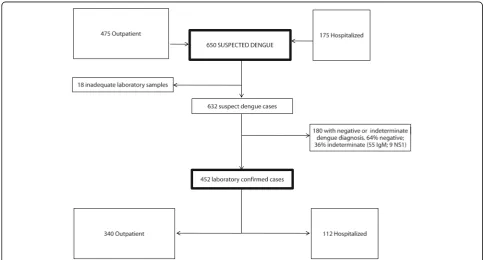
Of the 632 symptomatic dengue cases clinically and labor-atorially screened at baseline, 452 (71.5%) were diagnosed as dengue confirmed by specific serology and/or viral de-tection (see Fig. 1). Table 1 presents the clinical and epi-demiological data according to case ascertainment. In both groups (dengue confirmed and symptomatic cases), the majority of patients (~ 80%) were adult and approxi-mately half were females. Approxiapproxi-mately 75% of the pa-tients were recruited in ambulatory settings and reported previous medical visits during the acute phase of the dis-ease. Adult population comorbidity was similar between the groups.
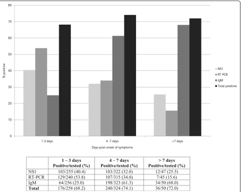
At baseline, the RT-PCR (53.8%) and NS1 (40.4%) tests yielded higher positive results compared with IgM ser-ology (25%) during days 1–3 after the onset of symptoms. In contrast, higher frequencies of IgM positive results (68%) were detected seven days after the onset of symp-toms versus RT-PCR (15.6%) and/or NS1 (25.5%) results (see Fig. 2).
Table 2 presents the main clinical and laboratory find-ings of confirmed dengue patients stratified by age group. At baseline, the most frequent symptoms reported were fever (100%), headache (~ 80%), and prostration (~ 90%), and these were similar among the age groups. Approxi-mately 65% of patients presented with a cutaneous rash and approximately half reported vomiting at baseline. During the course of the illness, higher frequencies of signs and symptoms were observed among children, such as spontaneous bleeding (47.6% versus 26.6% among adults). In addition, intensive abdominal pain, effusion, and ascites were predominant among children as com-pared to adults, and a statistically significant difference was observed between the age groups. Children had a
Table 1Clinical and epidemiological characteristics of 632 suspected dengue cases according to laboratory confirmation, recruited in Central Brazil, 2012 and 2013
Characteristics Laboratory confirmedadengue cases (%) Unconfirmed dengue cases (%) Pb
Suspected cases dengue 452 (71.5) 180 (28.5) –
Mean age (dp) 35.7 (17.5) 34.8 (16.6) 0.546
Age group (years)
≤1 2 (0.4) 3 (1.7) 0.288
2–15 48 (10.6) 15 (8.3) 0.371
16–39 233 (51.5) 100 (55.6) 0.320
40–59 118 (26.1) 44 (24.4) 0.629
≥60 51 (11.3) 18 (10.0) 0.619
Gender
Female 235 (52.0) 88 (48.9) 0.481
Male 217 (48.0) 92 (51.1) 0.481
Health care settingc
Ambulatory 340 (75.2) 126 (70.0) 0.178
Hospital 112 (24.8) 54 (30.0) 0.178
Previous medical visit 142 (31.4) 64 (31.9) 0.316
Comorbidityd 115 (25.4) 48 (29.4) 0.751
Hypertension 82 (18.1) 30 (16.7) 0.661
Diabetes 25 (5.5) 11 (6.1) 0.776
Asthma 14 (3.1) 8 (4.4) 0.404
Chronic renal failure 8 (1.8) 2 (1.1) 0.549
Othersc 15 (3.3) 11 (7.2) 0.669
Deaths 1 (0.2) 1 (0.6) 0.487
a
laboratory confirmed dengue cases were positive by at least one of the test (RT-PCR and/or NS1 and/or IgM). b
χ2 test
cconsidering 540 adult patient (≥ 18 years). d
higher frequency of primary infection (39.4% versus 22% among the adult population), which was a statistically sig-nificant difference.
Of the 452 patients surveyed, they were classified as Dengue Fever 188 (41.6%), Dengue with Alert Signs 238 (52.6%) and Severe Dengue 26 (5.8%). The DENV sero-types were identified in 243 (53.8%) out of 452 patients, tested by RT-PCR. DENV-4 was the predominant serotype, detected in 135 (55.6%) patients, followed by DENV-1 in 91 (37.4%) patients. DENV-3 and DENV-2 were de-tected in 13 (5.3%) and 4 (1.6%) patients, respectively (see Table 3).
Among the 26 cases of SD, nine had the DENV sero-types identified and all were infected with DENV-1; comprising five males and four females, aged seven to 57 years (median: 43 years). Descriptive analyses by sero-type showed similar age and gender distributions of the patients infected with serotypes DENV-1 and DENV-4; the majority of patients were adults and approximately
half were females. Each serotype included a large range of clinical forms and biological variations (data not shown).
Patients with a detectable DENV-1 serotype were more prone to present with several clinical and laboratory fea-tures as compared with DENV-4 patients, including spon-taneous bleeding (DENV-1: 33.0% versus DENV-4: 20.0%; P =0.03); intense abdominal pain (DENV-1: 29.7% versus DENV-4: 14.1%; P = 0.004); neurological symptoms (DENV-1: 6.7% versus DENV-4: 2.2%; P = 0.09); and thrombocytopenia (DENV-1: 33.7% versus DENV-4: 18.2%;P =0.01). The immune status measured by primary or secondary infections were available for 202 patients (DENV-1 or DENV-4). Secondary infection was more pre-dominant among DENV-4 cases (80.0%) compared with DENV-1 cases (62.3%), a statistically significant difference between the serotypes (P =0.03).
Table 4 presents the results of the multinomial analysis (the association between the antibody response pattern,
serotype, and severity of disease taking DF as a refer-ence). The univariate analysis showed that females were at a higher risk of having DwS (OR = 2.12; 95% CI: 1.44–3.13;P< 0.01) in comparison with DF patients. Fe-males classified as having SD did not differ from the ref-erence. Adult patients (≥ 15 years old) had an OR of 0.53 (95% CI: 0.26–1.08; P = 0.082) compared to chil-dren with DF. Comorbidity was not associated with the
severity of the disease. Patients classified as having DwS and that mounted secondary infection were found to be not at risk (OR = 1.03; 95% CI: 0.66–1.60; P = 0.890) compared with DF patients with primary infection. In addition, SD cases with secondary infection had an OR of 2.63 (95%CI: 0.74–9.30;P =0.134). Results of DENV-1 and DENV-4 were presented for DwS versus FD, as few cases of DENV-2 and DENV-3 were detected in this
Table 2Clinical and laboratory findings of confirmed dengue patients estratified by age-group
Features < 15 years
N= 42 (%) ≥
15 years
N= 410 (%) P
a
Signs and symptoms reported or observed during recruitment
Age, years - median (min-max) 10.1 (0–14) 38.4 (15–83) –
Female sex 25 (59.5) 210 (51.2) 0.305
Days of illness - median (min-max) 4 (2–8) 5 (1–13) 0.535g
Headache 36 (85.7) 335 (81.7) 0.519
Prostration 41 (97.6) 373 (91.0) 0.139
Rash 29 (69.0) 269 (65.6) 0.654
Vomit 23 (54.8) 175 (42.7) 0.133
Signs and symptoms reported or observed during the course of the disease
Spontaneous bleeding 20 (47.6) 109 (26.6) 0.004
Gastrointestinal bleeding 3 (7.1) 22 (5.4) 0.631
Neurological alterations 1 (2.4) 24 (5.9) 0.346
Breathing difficulties 0 51 (12.4) 0.015
Icterus 1 (2.4) 17 (4.1) 0.576
Intense abdominal pain 12 (28.6) 66 (16.1) 0.042
Hepatomegaly 3 (11.1) 24 (5.9) 0.737
Effusions and Ascites 4 (9.5) 8 (2.0) 0.004
Main laboratory results during the course of the disease
Hemoconcentrationb 12 (29.3) 77 (19.6) 0.146
Leukopenia (< 4000 cel/ml)c 20 (50.0) 230 (58.4) 0.223
Thrombocytopenia (< 100.000 cel/ml)d 9 (22.0) 123 (31.1) 0.307
AST (> 1000)e 0 0 –
ALT (> 1000)f 0 0 –
Clinical classification
Dengue Fever 12 (28.6) 176 (42.9) 0.075
Dengue with Warning Signs 27 (64.3) 211 (51.5) 0.103
Severe Dengue 3 (7.1) 23 (5.6) 0.307
Antibody response patternh
Primary 13 (39.4) 67 (22.7) 0.034
Secondary 20 (60.6) 228 (77.3) 0.034
Hemoconcentration (children >44%, adult >48%) a
χ2 test b
432 results available c
435 results available d
433 results available e
416 results available f
415 results available g
Mann-Whitney Test h
study. The multinomial analysis showed that SD cases with secondary infection had an adjusted OR of 2.80 (95% CI: 0.78–10.00; P = 0.113) compared with DF pa-tients with primary infection, when adjusted for age and sex (see Table 5).
Discussion
Our results show differences in the clinical features of dengue patients infected during a large DENV-4 out-break in central of Brazil. Children presented higher fre-quencies of several warning signs of disease severity
Table 3Clinical and epidemiological characteristics of 452 laboratory confirmed dengue cases, Central Brazil, 2012 and 2013
Parameters Dengue Fever
N= 188 (%)
Dengue with Warning Signs
N= 238 (%)
Severe Dengue
N= 26 (%) Gender
Female 78 (41.5) 143 (60.1) 14 (53.8)
Age group (years)
< 1 1 (0.5) 1 (0.5) 0
2 a 14 11 (5.8) 26 (10.9) 3 (11.5)
15 a 39 108 (57.5) 123 (51.6) 10 (38.5)
40 a 59 44 (23.4) 63 (26.5) 11 (42.3)
≥60 24 (12.8) 25 (10.5) 2 (7.7)
Comorbidity 46 (24.5) 60 (25.2) 9 (34.6)
Antibody response pattern
Primary 38 (27.3) 40 (23.0) 2 (13.3)
Secondary 101 (72.7) 134 (77.0) 13 (86.7)
Serotypesa
DENV-1 35 (18.6) 47 (19.7) 9 (34.6)
DENV-2 2 (1.1) 2 (0.8) 0
DENV-3 3 (1.6) 10 (4.2) 0
DENV-4 63 (33.5) 72 (30.3) 0
Undetectable 75 (39.9) 98 (41.2) 15 (57.7)
Not done 10 (5.3) 9 (3.8) 2 (7.7)
a
Serotypes determined by reverse-transcription polymerase chain reaction (RT-PCR)
Table 4Multinomial analysis of the association between antibody response pattern, serotype and severity of dengue disease as outcome
Parameters Dengue Fever Dengue with Warning Signs Severe Dengue
N= 188 (%) N= 238 (%) OR(95%CI) P-value N= 26 (%) OR(95%CI) P-value
Gender
Female 78 (41.5) 143 (60.1) 2.12 (1.44–3.13) 0.000 14 (53.8) 1.64 (0.72–3.75) 0.236
Age group (years)
< 15 12 (6.4) 27 (11.3) Reference – 3 (11.5) Reference –
≥15 176 (93.6) 211 (88.7) 0.53 (0.26–1.08) 0.082 23 (88.5) 0.52 (0.14–1.99) 0.342
Comorbidity 46 (24.5) 60 (25.2) 1.04 (0.67–1.62) 0.860 9 (34.6) 1.63 (0.68–3.92) 0.271
Antibody response patterna
Primary 53 (28.2) 62 (26.1) Reference – 3 (11.5) Reference –
Secondary 121 (64.4) 146 (61.3) 1.03 (0.66–1.60) 0.890 18 (69.2) 2.63 (0.74–9.30) 0.134
Serotypeb
DENV-1 35 (19.7) 47 (19.7) Reference – 9 (34.6) NA –
DENV-4 63 (35.4) 72 (30.3) 0.85 (0.49–1.48) 0.568 0 NA –
Reference for the multinomial analysis: dengue fever NANot applicable
a
49 patients not evaluated b
such as spontaneous bleeding, intensive abdominal pain, and neurological symptoms when compared to adults. Secondary infections were more prone to occur in the adult population, however, more than 60% of the chil-dren and almost 80% of the adult patients in this study were found to have a previous dengue infection, highlighting the high DENV circulation in the region.
Few studies have compared clinical features and la-boratory abnormalities in the pediatric age group and adults in Brazil [37, 38]. A study conducted in the same region in 2005 showed that secondary infection was not a predictor of severe clinical manifestation in adults in-fected with the DENV-3 serotype [21].
In a prospective clinical study conducted in the same city in 2000, we found that mild cases of dengue were predominant among adults [21]. In this study, we classi-fied the majority of the pediatric and adult patients as having DwS, followed by DF, and a few cases of SD. This distribution reflects the clinical characteristics of dengue patients treated at reference day clinics and hospitals during the 2012/2013 epidemic. It does not resemble the entire cohort of dengue patients, as most of the cases were milder cases classified as DF, as according to the surveillance system (SINAN, 2012/2013).
It is important to note that all patients classified as hav-ing SD were infected with DENV-1 and none were in-fected with DENV-4. However, we are aware that our sample size relating to the severe form of the disease is too small to draw a conclusion. Interestingly, the historical data outlined by Hastead in the early decades of dengue epidemics regarding differences in clinical manifestations of DENV-1 and DENV-4 serotypes in Southeast Asia similarly describe DENV-1 as being more prone to cause severe cases as compared with DENV-4 [39].
In our study, we found a high percentage of undetect-able viremia among severe cases. Dengue patients may
progress to severe disease during the defervescence period, which is the period of hospital admission and re-cruitment that is beyond the viremic period [21]. We are aware that this could represent a potential selection bias in the recruitment of severe cases.
One of the strengths of this study was the recruitment of dengue patients approximately two years after the introduction of the DENV-4 serotype in central Brazil. In this context, it is likely that the majority of the popu-lation in the study area was naïve to DENV-4, which ex-plains the current outbreak with the predominance of the DENV-4 circulation. In fact, DENV-4 had been pre-viously isolated in nine patients in the neighbouring state of Mato Grosso do Sul in 2012. The authors had warned about the potential for outbreaks due to the introduction of the DENV-4 serotype in a susceptible population to this serotype in central Brazil [40].
Another strength of our study is that we recruited pa-tients in several ambulatories and hospital settings within the region. However, our study population in-cluded only patients living in one city and the results may not be generalizable to rural areas or other regions of the country.
Comparison of clinical manifestations, antibody response patterns, and severity of the disease were restricted to the DENV-4 and DENV-1 serotypes, as few patients had de-tectable DENV-2 or DENV-3 serotypes in this study. These findings are concordant with the official laboratory system in charge of DENV surveillance regionally. It is interesting that the DENV-2 serotype had not been predominantly registered by the viral surveillance system in the last two decades in the study area [35]. We are aware that accord-ing to the period when the blood samples for serological tests (IgM or IgG) were collected, this may yield negative or positive results, which could lead to the misclassification of primary and secondary infections.
Conclusions
In summary, the present study shows the incidence of SD among pediatric and adult patients in the first regis-tered DENV-4 outbreak in central Brazil. To our know-ledge, this is the first prospective clinical study to compare DENV-1 and DENV-4 patients in relation to antibody re-sponse patterns and severity of the disease. Our findings contribute to the understanding of clinical differences and immune status related to the serotypes DENV-1 and DENV-4 in central Brazil.
Additional file
Additional file 1:Multilingual abstracts in the five official working languages of the United Nations. (PDF 644 kb)
Additional file 2:Association between clinical or laboratory markers and dengue serotypes, Central Brazil, 2012 and 2013. (DOCX 13 kb)
Table 5Multinomial analysis adjusted by age and sex of the association between antibody response pattern, serotype and severity of dengue disease as outcome
Parameters Dengue with Warning Signs Severe Dengue
ORAdj(95%CI) P-value ORAdj(95%CI) P-value
Comorbidity 1.20 (0.72–2.00) 0.479 1.74 (0.64–4.73) 0.279 Antibody response pattern
Primary Reference – Reference –
Secondary 1.20 (0.76–1.89) 0.433 2.80 (0.78–10.0) 0.113 Missing 1.93 (0.92–4.09) 0.083 6.23 (1.32–29.4) 0.021 Serotypea
DENV-1 Reference – NA –
DENV-4 0.80 (0.45–1.41) 0.444 NA –
Not evaluated 0.95 (0.56–1.62) 0.856 NA – a
Abbreviations
ALT:Alanine aminotransferase; AST: Aspartate aminotransferase; cDNA: 23 complementary DNA; cDNA: Deoxyribonucleic acid;CI: Confidence interval; CNS: Central nervous system; DENV: Dengue virus; DENV: Virus dengue; DF: Dengue fever; DF: Fever dengue; DHF: Hemorrhagic dengue fever; DwC: Dengue with warning signs; ELISA: Enzyme-linked immunosorbent assay; IBGE: Intituto Brasileiro de Geografia e Estatística; Ig: Immunoglobulin; IgG: Immunoglobulin G; IgM: Immunoglobulin M; NS1: Non-structural protein-1; OR: Odds ratio; PCR: Polymerase chain reaction; RNA: Ribonucleic acid; RT: Reverse transcription; RT-PCR: Reverse transcription-polymerase chain reaction; SD: Severe dengue; STATA: Data analysis and statistical software; WHO: World Health Organization
Acknowledgements
We would like to thank the State Health Department of Goiás, the Municipal Health Department of Goiânia for its collaboration, and the cooperation of the employees of the health units participating in the study.
We thank the director and staff of the participating hospitals and the Secretariat of Health. We also thank the patients for their collaboration and generosity.
Funding
This study was funded by the National Council for Scientific and Technological Development (Conselho Nacional de Desenvolvimento Cientifico e Tecnologico, CNPq); and the Foundation for the State of Goiás Research (FAPEG).
Availability of data and materials
The datasets collected and/or analyzed during the current study are available from the corresponding author upon reasonable request.
Authors’contributions
BAMR conducted the molecular biology tests, participated in the data analysis, and wrote the paper. AOG held the patients’clinical classification (FD, DwS e SD) and contributed to the writing of the paper. AFLTA participated in the data collection and assisted in the laboratory exams. MPT participated in the data collection and conducted the patients’clinical classification. LAS coordinated the fieldwork and assisted with the laboratory exams. ICJ participated in the data collection and performed the laboratory exams. MDT assisted in the patients’clinical classification and contributed to the writing of the paper. VCRF coordinated the study at the regional level, and assisted in the data collection and the molecular biology tests. CMTM contributed to the study design and the writing of the paper. All authors approve the paper for publication.
Authors’information
BAMR has a PhD in Tropical Medicine and Public Health with an emphasis on epidemiology from the Institute of Tropical Pathology and Public Health of the Federal University of Goiás. He is also a public health professor at the State University of Goiás. Visit this link to see his CV: http://lattes.cnpq.br/ 7049130317115406.
Competing interests
The authors declare that they have no competing interests.
Consent for publication Not applicable.
Ethics approval and consent to participate
This study was approved by the Ethics Committee on Research of the Aggeu Magalhães Research Center (FIOCRUZ-PE) (No. 24/11) and the review boards of each institution. Patients gave informed consent or when less than 18 years old this was given by the parents or guardians.
Trial registration number
This study is not a clinical trial. It is a prospective observational study and therefore did not need to be registered.
Author details
1Institute of Tropical Pathology and Public Health, Federal University of Goiás,
Goiânia, Brazil.2Faculty of Pharmacy, Federal University of Goiás, Goiânia,
Brazil.3School of Nursing, State University of Goiás, Ceres, Brazil.
Received: 14 July 2016 Accepted: 18 June 2017
References
1. Gubler DJ. Dengue viruses: their evolutins, history and emergence as a global public health problem. In: Duane J. Gubler, Eng Eong Ooi, Subhash Vasudevan JF, editor. Dengue and dengue hemorrhagic fever. 2nd ed. London; 2014. p. 1–29.
2. World Health Organization. Dengue: guidelines for diagnosis, treatment, prevention, and control. New Edition. Geneva: World Health Organization; 2009.
3. Guzman MG, Halstead SB, Artsob H, Buchy P, Farrar J, Gubler DJ, et al. Dengue: a continuing global threat. Nat. Rev. Microbiol. 2010;8:S7–16. 4. Bhatt S, Gething PW, Brady OJ, Messina JP, Farlow AW, Moyes CL, et al. The
global distribution and burden of dengue. Nature. 2013;496:504–7. 5. Brathwaite Dick O, San Martín JL, Montoya RH, del Diego J, Zambrano B,
Dayan GH. The history of dengue outbreaks in the Americas. Am J Trop Med Hyg. 2012;87:584–93.
6. Siqueira JB, Martelli CMT, Coelho GE, da R Simplicio AC, Hatch DL. Dengue and dengue hemorrhagic fever, Brazil, 1981-2002. Emerg Infect Dis. 2005;11:48–53.
7. Teixeira MG, Siqueira JB, Ferreira GLC, Bricks L, Joint G. Epidemiological trends of dengue disease in Brazil (2000-2010): a systematic literature search and analysis. PLoS Negl Trop Dis. 2013;7:e2520.
8. Number of Reporter Cases of Dengue and Severe Dengue (SD) in the Americas, by country: Figures for 2014 (to week noted each country). In: Annual cases reported of dengue. PAHO. 2015. http://www2.paho.org/hq/ index.php?option=com_topics&view=readall&cid=3273&Itemid=&lang=pt. Accessed 18 Jul 2015.
9. Temporao JG, Penna GO, Carmo EH, Coelho GE, do Socorro Silva Azevedo R, Teixeira Nunes MR, et al. Dengue virus serotype 4, Roraima State, Brazil. Emerg Infect Dis. 2011;17:938–40.
10. Gutiérrez G, Gresh L, Pérez MÁ, Elizondo D, Avilés W, Kuan G, et al. Evaluation of the diagnostic utility of the traditional and revised WHO dengue case definitions. PLoS Negl Trop Dis. 2013;7:e2385.
11. Jayaratne SD, Atukorale V, Gomes L, Chang T, Wijesinghe T, Fernando S, et al. Evaluation of the WHO revised criteria for classification of clinical disease severity in acute adult dengue infection. BMC Res Notes. 2012;5:645. 12. Bandyopadhyay S, Lum LCS, Kroeger A. Classifying dengue: a review of the
difficulties in using the WHO case classification for dengue haemorrhagic fever. Tropical Med Int Health. 2006;11:1238–55.
13. Prasad D, Kumar C, Jain A, Kumar R. Accuracy and applicability of the revised WHO classification (2009) of dengue in children seen at a tertiary healthcare facility in northern India. Infection. 2013;41:775–82. 14. Deen JL, Harris E, Wills B, Balmaseda A, Hammond SN, Rocha C, et al. The
WHO dengue classification and case definitions: time for a reassessment. Lancet. 2006;368:170–3.
15. Alexander N, Balmaseda A, Coelho ICB, Dimaano E, Hien TT, Hung NT, et al. Multicentre prospective study on dengue classification in four South-east Asian and three Latin American countries. Tropical Med Int Health. 2011;16:936–48.
16. BRASIL. Dengue diagnóstico e manejo clínico adulto e criança. 4oed.
Ministério da Saúde; Brasília. 2013. (in Portuguese).
17. BRASIL. Nova classificação de caso de Dengue - OMS. 1° ed. Ministério da saúde; Brasília. 2014. (in Portuguese).
18. dos RFC BM, de A Luna EJ, Braga Júnior LL, de RVB O, LTM R, do S da Silva M, et al. Risk factors associated with death in Brazilian children with severe dengue: a case-control study. Clinics. 2014;69:55–60.
19. Bhaskar E, Sowmya G, Moorthy S, Sundar V. Prevalence, patterns, and factors associated with bleeding tendencies in dengue. J Infect Dev Ctries. 2015;9:105–10.
20. Trung D the, Wills B. Clinical features of dengue. In: Duane J. Gubler, Eng Eong Ooi, Subhash Vasudevan JF, editor. Dengue and dengue hemorrhagic fever. 2nd ed. London; 2014. p. 115–44.
21. Guilarde AO, Turchi MD, Siqueira JB, Feres VCR, Rocha B, Levi JE, et al. Dengue and dengue hemorrhagic fever among adults: clinical outcomes related to viremia, serotypes, and antibody response. J Infect Dis. 2008;197:817–24.
factors for severe dengue viral infection. Ooi EE, editor. PLoS One. 2014;9:–e114499. Public Library of Science
23. Solomon T, Dung NM, Vaughn DW, Kneen R, Thao LT, Raengsakulrach B, et al. Neurological manifestations of dengue infection. Lancet. 2000;355:1053–9. 24. Pawitan JA. Dengue virus infection: predictors for severe dengue. Acta Med
Indones. 2011;43:129–35.
25. Guzmán MG, Kourí G, Valdés L, Bravo J, Vázquez S, Halstead SB. Enhanced severity of secondary dengue-2 infections: death rates in 1981 and 1997 Cuban outbreaks. Rev Panam Salud Publica. 2002;11:223–7.
26. Kalayanarooj S, Nimmannitya S. Is dengue severity related to nutritional status?. Southeast Asian J Trop Med Public Health. 2005. p. 378–84. 27. Lee M-S, Hwang K-P, Chen T-C, Lu P-L, Chen T-P. Clinical characteristics of
dengue and dengue hemorrhagic fever in a medical center of southern Taiwan during the 2002 epidemic. J Microbiol Immunol Infect. 2006;39:121– 9.
28. Martins AC, Pereira TM, Oliart-Guzmán H, Delfino BM, Mantovani SAS, Braña AM, et al. Seroprevalence and seroconversion of dengue and implications for clinical diagnosis in amazonian children. Interdiscip Perspect Infect Dis. 2014;2014:703875.
29. Halstead S. Pathogenesis of dengue: challenges to molecular biology. Science. 1988;239:476–81.
30. Thomas L, Verlaeten O, Cabié A, Kaidomar S, Moravie V, Martial J, et al. Influence of the dengue serotype, previous dengue infection, and plasma viral load on clinical presentation and outcome during a dengue-2 and dengue-4 co-epidemic. Am J Trop Med Hyg. 2008;78(6):990-8. 31. Rosen L. The Emperor’s new clothes revisited, or reflections on the pathogenesis
of dengue hemorrhagic fever. Am J Trop Med Hyg. 1977;26:337–43. 32. Kouri GP, Guzmán MG, Bravo JR. Why dengue haemorrhagic fever in Cuba?
2. An integral analysis. Trans R Soc Trop Med Hyg. 1987;81:821–3. 33. Boletim Semanal de Dengue - Goiás 2014: Semana Epidemiológica 1 a 53
(14/12/2013 a 03/01/2015). In: Boletim Semanal de Dengue. Secr. Estadual de Saúde de Goiás; 2015. https://extranet.saude.go.gov.br/public/dengue. html. Accessed 5 Aug 2015. (in Portuguese).
34. Boletim Semanal de Dengue - Goiás 2013. In: Boletim Semanal de Dengue. Secr. Estadual Saúde; 2013. http://www.saude.go.gov.br/index.
php?idEditoria=4208. Accessed 9 Aug 2015. (in Portuguese).
35. Feres VCR, Martelli CMT, Turchi MD, Junior JBS, Nogueira RMR, Rocha BAM, et al. Laboratory surveillance of dengue virus in Central Brazil, 1994-2003. J Clin Virol. 2006;37:179–83.
36. Lanciotti RS, Calisher CH, Gubler DJ, Chang GJ, Vorndam AV. Rapid detection and typing of dengue viruses from clinical samples by using reverse transcriptase-polymerase chain reaction. J Clin Microbiol. 1992;30:545–51. 37. Brito CAA de. Dengue em Recife, Pernambuco: padrões clínicos,
epidemiológicos, laboratoriais e faotres de risco associados à forma grave da doença. https://www.arca.fiocruz.br/handle/icict/3908. (2007). Acessede 9 Jul 2014. (in Portuguese).
38. Escosteguy CC, Pereira AGL, Medronho RDA, Rodrigues CS, Chagas KKFD. Diferenças, segundo faixa etária, do perfil clínico-epidemiológico dos casos de dengue grave atendidos no Hospital Federal dos Servidores do Estado, Rio de Janeiro-RJ, Brasil, durante a epidemia de 2008. Epidemiol e Serviços Saúde. 2013;22:67–76. (in Portuguese)
39. Nishiura H, Halstead SB. Natural history of dengue virus (DENV)-1 and DENV-4 infections: reanalysis of classic studies. J Infect Dis. 2007;195:1007–13. 40. Bertolacci-Rocha LG, da Cunha RV, de Castro Lichs GG, Dal Fabbro MMFJ,
Motta-Castro ARC. Introduction of the dengue virus type 4 in the State of Mato Grosso do Sul. Brazil Cad Saude Publica. 2014;30:1789–92.
• We accept pre-submission inquiries
• Our selector tool helps you to find the most relevant journal
• We provide round the clock customer support
• Convenient online submission
• Thorough peer review
• Inclusion in PubMed and all major indexing services
• Maximum visibility for your research
Submit your manuscript at www.biomedcentral.com/submit