Open Access
Case report
A large left ventricular pseudoaneurysm in Behçet's disease: a case
report
Seyed Mojtaba Marashi*
1, Payam Eghtesadi-Araghi
2and
Mohammad Hussein Mandegar
3Address: 1Assistant Professor of Anesthesiology, Department of Cardiac Surgery, Dr. Shariati Hospital Complex, Tehran Univ. of Medical Sciences,
Tehran 14197 Iran, 2Anesthesiologist, President of Parsteb Pajouheshyar Medical Sciences Research Institute (NGO), Department No. 5, 37th
(Eastern), First Golzar St., Ashrafi Esfahani Bulv., Ponak Sq., Tehran 1476783476, Iran and 3Associate Professor of Cardiac Surgery, Department
of Cardiac Surgery, Dr. Shariati Hospital Complex, Tehran Univ. of Medical Sciences, Tehran 14197 Iran
Email: Seyed Mojtaba Marashi* - marashi@doctor.com; Payam Eghtesadi-Araghi - payam_eghtesadi@yahoo.com; Mohammad Hussein Mandegar - mhmandegar@yahoo.com
* Corresponding author
Behçet's diseasecardiacleft ventricularpseudoaneurysm
Abstract
Background: Behçet's disease is a collagen-vascular disease most commonly seen in Asia and Mediterranean area. Different organs and systems including cardiovascular system could be involved. Pseudoaneurysm is the most common form of arterial involvement in Behçet's disease; however, cardiac pseudoaneurysm is rare.
Case Presentation: A rare case of 13 years old boy with a 4-year history of Behçet's disease with development of a huge left ventricular pseudoaneurysm is reported who had been admitted because of cough, chills, fever, and chest pain. Findings obtained on echocardiography, magnetic resonance imaging, chest computed tomography and coronary angiography confirmed a left ventricular pseudoaneurysm. There was no complication for next 24 months follow up period after surgical treatment.
Conclusion: Considering its fatality and nonspecific manifestations, one should consider cardiac pseudoaneurysms as a potential risk in any patient with Behçet's disease.
Background
In 1937, Hulusi Behçet, a Turkish dermatologist, first described a chronic autoimmune disease bearing his name with characteristic orogenital aphtous ulceration and uveitis [1]. The etiology of Behçet's disease, which is most commonly seen in Asia and Mediterranean area, is still unknown. The primary pathology is a vasculitis affect-ing skin, joints, pulmonary, gastrointestinal, urinary, and
nervous systems [2]. Its vascular complications are most frequently manifested as thromboembolism in veins and pseudoaneurysm in arteries [3]. Although pseudoaneu-rysms are the most common form of arterial involvement in Behçet's disease, we could only find one case reported
by Rolland et al. [4] with Behçet's disease and cardiac
pseudoaneurysm. Occasional cases of cardiac pseudoan-eurysms have been reported in association with
Published: 14 June 2005
BMC Surgery 2005, 5:13 doi:10.1186/1471-2482-5-13
Received: 20 November 2004 Accepted: 14 June 2005
This article is available from: http://www.biomedcentral.com/1471-2482/5/13
© 2005 Marashi et al; licensee BioMed Central Ltd.
rheumatoid arthritis [5] and Kawasaki's disease [6]; how-ever large cardiac pseudoaneurysms are mostly complica-tions of cardiac surgery, myocardial infarction, endocarditis, and chest trauma [7].
In this report we present a patient with Behçet's disease and a huge left ventricular pseudoaneurysm.
Case presentation
A 13 years old boy with Behçet's disease was referred to our hospital with chills, fever, cough, and chest pain of one month duration in June 2001. The diagnosis of Behçet's disease was established 4 years prior to this admission, that had presented with oral aphtae, orchitis, right eye uveitis leading to blindness, recurrent pseudofol-liculitis, knee arthritis, and lower extremity deep vein thrombosis, all attributable to this autoimmune disorder. He had been treated by Prednisolone (15 mg/day) and Methotrexate (7.5 mg/week) for last 7 month.
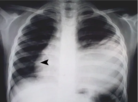
Physical examination revealed III/VI to-and-fro murmur along the left sternal border and an S3 gallop. The poste-rior-anterior chest X-ray (Fig. 1), computed tomography scan with intravenous contrast media at the level of T5–T8 (Fig. 2), magnetic resonance imaging (MRI) (Fig. 3), cor-onary angiography and echocardiography all revealed a 10.1 × 14.8 cm left ventricular pseudoaneurysm in the anterior wall of left ventricle. There was no narrowing sug-gesting coronary artery disease in his coronary
angiogra-phy. In two-dimensional echocardiography, the typical features of pseudoaneurysm was noted including a rela-tively narrow neck in comparison with the diameter of the aneurysm, sharp discontinuity of the endocardium at the site at which the aneurysm communicates with the left ventricle, no noticeable valvular dysfunction, and left ven-tricular wall motion abnormalities. The orifice to radius ratio was not measurable due to the large size of pseudoaneurysm.
At surgery, the ostium (arrowhead in Fig. 4) was measured 2.5 × 3.0 cm. The above mentioned results warranted a surgery with median sternotomy approach. After pericar-dotomy, a pulsatile mass appeared at the tip of left ventri-cle with a fistula to the heart. The pseudoaneurysm and large amounts of thrombus within it were resected, and the defect in the left ventricular wall was repaired by Teflon Plegeted Prolen 4/0. The patient made an unevent-ful recovery. The pathologic examination revealed a fibrous pseudoaneurysm including areas of old hemor-rhage and thrombosis and chronic inflammation. There was no complication for next 24 months follow up period after the operation.
Conclusion
We have presented an unusual patient with Behçet's dis-ease and a large (10.1 × 14.8 cm) left ventricular pseu-doaneurysm. Behçet's disease is a systemic disorder with
The PA chest X-ray: left-sided pleural effusion and a large mass in anterolateral part of left lung which had overshad-owed the left border of the heart
Figure 1
The PA chest X-ray: left-sided pleural effusion and a large mass in anterolateral part of left lung which had overshad-owed the left border of the heart. Arrowhead indicates shift of the heart to the right side.
Chest computed tomography scan with contrast at the level of T7 showing the large pseudoaneurysm
Figure 2
mucocutaneous, ophthalmic, neurological, cardiovascu-lar, pulmonary, gastrointestinal, urogenital and muscu-loskeletal involvement. Its vascular manifestations are thrombophlebitis and, less frequently, arterial lesions such as pseudoaneurysms, occlusions and stenoses [8].
About 8% of the patients with Behçet's disease have severe vascular complications such as arterial pseudoaneurysms and occlusions [1]. Pseudoaneurysms are the most com-mon form of arterial involvement in Behçet's disease [3].
Cardiac involvement is rare in Behçet's disease [9] and engages only about 6% of patients [4]. Cardiac involve-ment in this disorder is a diffuse process that involves both cardiac structure and vascular elements. Higher inci-dences of interatrial septum aneurysm (6% to 31%), mitral valve prolapse (3% to 25%), mitral regurgitation (6% to 40%), and aneurysmal dilatations of valsalva sinus and ascending aorta were observed in the Behçet's disease patients than in the normal subjects [10]. Pericarditis, myocarditis, endocardial fibrosis, conduction defects, and aortic regurgitation were also observed [11]. Although left ventricular aneurysms with Behçet's disease have occa-sionally been reported, we found only one case of cardiac pseudoaneurysm in these patients reported in literature.
Rolland et al. [4] reported a 29 years old patient with
Behçet's syndrome and a false left ventricular aneurysm and coronary artery aneurysm, which were repaired under cardiopulmonary bypass with no postoperative complications.
Cardiac pseudoaneurysm is defined as a rupture of the myocardium that is contained by pericardial adhesions or the epicardial wall. This phenomenon can be explained by myocardial fragility induced by ischemia due to vascu-litis process of Behçet's disease [4]. Because there was no evidence suggesting coronary artery disease in his coro-nary angiography of our patient, it could be assumed that this pseudoaneurysm was resulted from rupture of the left ventricle due to angiitis and the myocardial fragility induced by ischemia.
In contrast to a true ventricular aneurysm, in which the wall is composed of myocardial scar tissue, the wall of a pseudoaneurysm is composed of thick fibrous tissue and pericardium [12]. In our case, pseudoaneurysm was con-sisted of profuse fibrous tissues and thromboses in vari-ous stages.
Cardiac pseudoaneurysms have the potential to leak or rupture and can be the source of peripheral emboli [10]. Different reports have discussed that such a contained rupture has a greater propensity for rupture than a true aneurysm, whose wall contains myocardium. Rupture of a left ventricular pseudoaneurysm is usually fatal; hence appropriate recognition and treatment (early surgery) even for asymptomatic patients is strongly recommended [7].
The diagnosis of pseudoaneurysm is not straightforward and is rarely suggested by clinical signs and symptoms [7].
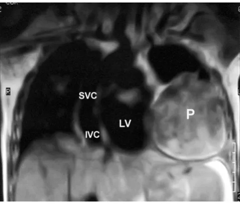
The MRI indicated a mass with inhomogeneous signals imply-ing the presence of blood and clots in addition to calcification
Figure 3
The MRI indicated a mass with inhomogeneous signals imply-ing the presence of blood and clots in addition to calcifica-tion. (SVC = Superior Vena Cava; IVC = Inferior Vena Cava; LV = Left Ventricle; P = Pseudoaneurysm)
Chest computed tomography scan with contrast at T7 level
Figure 4
In our patient the pseudoaneurysm presented with non-specific symptoms and signs. Thus, such a diagnosis was highly unlikely before getting the results of imaging. Vari-ous imaging methods have been used to diagnose pseu-doaneurysm, including two-dimensional and contrast echocardiography [13], computed tomography, magnetic resonance imaging, and left ventricular angiography. Each has its advantages and disadvantages but echocardiogra-phy has become the most common examination used for first diagnosis because it can evaluate other associations such as valvular regurgitation, thrombus formation, and ventricular function, are often important supplements to clinical management [7].
Like our patient, chest radiography sometimes shows a localized bulge on the cardiac silhouette. On computed tomography, pseudoaneurysms are characterized by an abrupt disappearance of the myocardial wall at the border of the pseudoaneurysm. Magnetic resonance imaging shows the low signal of the pericardium, which consti-tutes the only wall of the pseudoaneurysm [7].
Surgical repair is usually recommended when a left ven-tricular pseudoaneurysm is detected [14,15]. In this case surgical intervention was mandatory, partly due to the young age of the patient. However, in cases of post-infarc-tion left ventricular pseudoaneurysm, which is one of the most common causes, routine surgical repair regardless of other clinical characteristics of the patient remains as a matter of discussion. Some authors believe that the neces-sity of surgical repair in these cases should be individual-ized for each patient [7,16].
In patients with post-infarction left ventricular pseudoan-eurysm, surgical repair of pseudoaneurysm was associated with an acceptable surgical mortality rate [7] and the long term outcome appears relatively benign [13] and late death was related primarily to the underlying disease or cardiac dysfunction [7]. There is no data on the long term prognosis of patients with cardiac pseudoaneurysms in Behçet's disease; however, long-term survival could not be expected given the diffuse involvement of cardiac struc-ture and vascular elements [10].
Considering its fatality and nonspecific manifestations, one should consider cardiac pseudoaneurysms as a poten-tial risk in any patient with Behçet's disease. Thanks to early diagnosis and surgery, our patient was treated suc-cessfully and had no complications in a follow-up period of 24 months.
Abbreviations
SVC = Superior Vena Cava
IVC = Inferior Vena Cava
LV = Left Ventricle
Competing interests
The author(s) declare that they have no competing interests.
Authors' contributions
SMM: Data collection, participated in the design of the study, critical review of the manuscript.
PE: Conceived the study and wrote the manuscript, partic-ipated in the design of the study.
MHM: Supervisor and conductor of treatment team and critical reviewer of the manuscript.
All authors read and approved the final manuscript.
Acknowledgements
Authors wish to thank Dr. Mohammad Kazazi, Dr. Abbas Salehi-Omran, Dr. Vafi Salmasi, Dr. Zahra Alizadeh, Dr. Golnar Mortezaie and Dr. Alireza Moayeri for their invaluable help.
References
1. Cakir O, Eren N, Ulka R, Nazaroghi H: Bilateral subclavian arte-rial aneurysm and ruptured abdominal aorta pseudoaneu-rysm in Behçet's disease. Ann Vasc Surg 2002, 16(4):516-20. 2. Alkim H, Gurkaynak G, Sezgin O, Oguz D, Saritas U, Sahin B:
Chronic pancreatitis and aortic pseudoaneurysm in Behçet's disease. Am J Gastroenterol 2001, 96(2):591-3.
3. Silistreli E, Karabay O, Erdel C, Serbest O, Guzeloglu M, Catalyurek H, Acikel U: Behçet's disease: treatment of popliteal pseu-doaneurysm by an endovascular stent graft implantation.
Ann Vasc Surg 2004, 18(1):118-20.
4. Rolland JM, Bical O, Laradi A, Robinault J, Benzidia R, Vanetti A, Her-reman G: False aneurysm of the left ventricle and coronary aneurysms in Behçet's disease. Arch Mal Coeur Vaiss 1993,
86(9):1383-5.
5. Rittoo D, Sutherland GR: Posterior left ventricular pseudoaneu-rysm after aortic valve replacement in a patient with Rheu-matoid Arthritis: diagnosis by transesophageal echocardiography. J Am Soc Echocardiogr 1994, 7(4):429-33. 6. Pillay D: Post infarction left ventricular pseudoaneurysm in a
patient with Kawasaki's disease. Heart 2001, 86(6):692. 7. Yeo TC, Malouf JF, Oh JK, Seward JB: Clinical profile and
out-come in 52 patients with cardiac pseudoaneurysm. Ann Intern Med 1998, 128(4):299-305.
8. Ko GY, Byun JY, Chai BG, Cho SH: The vascular manifestations of Behçet's disease: angiographic and CT findings. Br J Radiol 2000, 73(876):1270-4.
9. Srairi JE, Ghannam R, Maouni A, Akalay O, Benomar S, el Haitem N, Mesbahi R, Benomar M: Coronary aneurysm in Behçet's dis-ease: report of a case. Arch Mal Coeur Vaiss 1998, 91(12):1509-12. 10. Gurgun C, Ercan E, Ceyhan C, Yavuzgil O, Zoghi M, Aksu K, Cinan CS, Turkoglu C: Cardiovascular involvement in Behçet's disease. Jpn Heart J 2002, 43(4):389-98.
11. Di Eusanio G, Mazzola A, Gregorini R, Esposito G, Di Nardo W, Di Manici G, Villani C: Left ventricularaneurysm secondary to Behcet's disease. Ann Thorac Surg 1991, 51(1):131-2.
12. Burger A, Sherman HB: Images in clinical medicine: Left ven-tricular pseudoaneurysm. N Engl J Med 2001, 344(25):1910. 13. Moreno R, Zamorano JL, Almeria C, Rodrigo JL, Villate A, Serra V,
Alvarez L, Aubele A, Sanchez-Harguindey L: Usefulness of contrast agents in the diagnosis of left ventricular pseudoaneurysm after acute myocardial infarction. Eur J Echocardiogr 2002,
Publish with BioMed Central and every scientist can read your work free of charge "BioMed Central will be the most significant development for disseminating the results of biomedical researc h in our lifetime."
Sir Paul Nurse, Cancer Research UK
Your research papers will be:
available free of charge to the entire biomedical community
peer reviewed and published immediately upon acceptance
cited in PubMed and archived on PubMed Central
yours — you keep the copyright
Submit your manuscript here:
http://www.biomedcentral.com/info/publishing_adv.asp
BioMedcentral 14. Yeo TC, Malouf JF, Reeder GS, Oh JK: Clinical characteristics and
outcome in postinfarction pseudoaneurysm. Am J Cardiol 1999,
84(5):592-5.
15. Csapo K, Voith L, Szuk T, Edes I, Kereiakes DJ: Postinfarction left ventricular pseudoaneurysm. Clin Cardiol 1997, 20(10):898-903. 16. Moreno R, Gordillo E, Zamorano J, Almeria C, Garcia-Rubira JC, Fernandez-Ortiz A, Macaya C: Long term outcome of patients with postinfarction left ventricular pseudoaneurysm. Heart 2003, 89(10):1144-6.
Pre-publication history
The pre-publication history for this paper can be accessed here: