R E S E A R C H A R T I C L E
Open Access
Oral soft tissue disorders are associated
with gastroesophageal reflux disease:
retrospective study
Masaaki Watanabe
1, Eiji Nakatani
2, Hiroo Yoshikawa
1, Takahiro Kanno
1, Yoshiki Nariai
3, Aya Yoshino
1,
Michael Vieth
4, Yoshikazu Kinoshita
5and Joji Sekine
1*Abstract
Background:Dental erosion (DE), one of oral hard tissue diseases, is one of the extraoesophageal symptoms defined as the Montreal Definition and Classification of gastroesophageal reflux disease (GERD). However, no study evaluated the relationship between GERD and oral soft tissues. We hypothesized that oral soft tissue disorders (OSTDs) would be related to GERD. The study aimed to investigate the association OSTDs and GERD.
Methods:GERD patients (105 cases), older and younger controls (25 cases each) were retrospectively examined for oral symptoms, salivary flow volume (Saxon test), swallowing function (repetitive saliva swallowing test [RSST]), teeth (decayed, missing, and filled [DMF] indices), and soft tissues (as evaluation of OSTDs, gingivitis; papillary, marginal, and attached [PMA] gingival indexes, simplified oral hygiene indices [OHI-S], and inflammatory oral mucosal regions). Clinical histories, which included body mass index [BMI], the existence of alcohol and tobacco use, and bruxism, were also investigated. APvalue of <0.05 was defined as statistically significant.
Results:GERD patients, older and younger controls participated and aged 66.4 ± 13.0, 68.3 ± 8.2 and 28.7 ± 2.6 years old, respectively. The most common oral symptom in the GERD patients was oral dryness. Salivary flow volume and swallowing function in the GERD patients were significantly lower than in either of the controls (all P < 0.05). Inflammatory oral mucosal regions were found only in the GERD patients. The DMF indices, as a measure of dental caries, in the GERD patients were higher than in the younger controls (P < 0.001), but lower than in
the older controls (P = 0.033). The PMA gingival indexes, as a measurement for gingival inflammation, and
OHI-S, as a measure for oral hygiene, in the GERD patients were significantly higher than in either of the controls (all P < 0.05). Though no significant differences in BMI, the existence of alcohol and tobacco use were found, bruxism, as an exacerbation factor of periodontal disease, in the GERD patients was significantly more frequent than in either control group (P = 0.041).
Conclusions: OSTDs were associated with GERD, which was similar to the association between DE and GERD.
Keywords: Dental erosion, Gastroesophageal reflux disease, Gingivitis, Inflammatory oral mucosal regions, Oral soft tissue disorders, Salivary flow volume, Swallowing function
* Correspondence:georges@med.shimane-u.ac.jp
1Department of Oral and Maxillofacial Surgery, Shimane University Faculty of Medicine, 89-1 Enya-cho, Izumo, Shimane 693-8501, Japan
Full list of author information is available at the end of the article
Background
Salivary flow volume and swallowing function in gastro-esophageal reflux disease (GERD) were revealed to be significantly reduced in our preliminary study [1]. Under normal circumstances, saliva and swallowing function have roles in preventing the oral cavity from acid reflux through neutralization and clearing effects [1–3]. Ac-cording to the Montreal Definition and Classification of GERD, dental erosion (DE) is a major oral symptom caused by acid reflux in patients with GERD [4]. DE is defined as the irreversible loss of dental hard tissue by a chemical process that does not involve bacteria [5, 6]. In our previous study, we found that DE, as an extraoeso-phageal symptom of GERD, was caused by acid reflux, which resulted in reduced salivary flow volume and swallowing function [1, 4]. Salivary flow and swallowing function may decrease along with oral health condition related to aging [7, 8]. Our preliminary study showed the salivary flow volume and swallowing function in GERD patients were reduced even though considering the effect of aging [1].
The oral cavity consists of teeth, periodontal tissue including gingiva and other oral mucosal regions in-cluding the tongue, buccal mucosa, palate, vestibular and oral floor [9–12]. The oral cavity disorders in-clude dental caries, periodontal diseases including gingivitis and periodontitis, and inflammatory oral mucosal regions such as redness, erosion, and ulcer [5, 6, 9–12]. Periodontal disease also as a part results from reduced salivary flow volume [13]. Although we have already revealed the relationships between DE and GERD in our previous preliminary study, complete edentulous patients are included in the GERD patients [1]. Understandably, DE is not in the edentulous GERD pa-tients. We focused on oral soft tissues to detect the edentu-lous GERD patients from oral findings. Then, oral soft tissue disorders (OSTDs) were defined as gingivitis; one of the periodontal disease, and inflammatory oral mucosal regions. Only a few studies evaluated the relationship be-tween periodontal disease and GERD [14, 15], bebe-tween oral mucosal inflammation and GERD, separately so far [16, 17]. Furthermore, few studies have evaluated and dis-cussed these co-relationships between OSTDs and GERD, simultaneously. OSTDs are not mentioned as the extrae-sophageal syndromes of GERD in the Montreal Definition and Classification [4].
We hypothesized that OSTDs would be related to GERD. This retrospective clinical study aimed to evaluate the prevalence of OSTDs in GERD patients in the context of salivary flow volume and swallowing function, to test the above hypothesis. Other general association factors for GERD, including body mass index (BMI), alcohol and tobacco use were also inves-tigated [18–21].
Methods
Patient characteristics
The current study was a single-site, cross-sectional retrospective study, and GERD patients and older and younger control subjects were included. GERD outpa-tients were consecutively selected from the Department of Internal Medicine, Shimane University Hospital be-tween February 2009 and March 2015. A GERD diagno-sis was made based in the presence of typical reflux symptoms, such as heartburn and acid regurgitation that occurred more than twice weekly, according to the Montreal definition of GERD [4]. A gastrointestinal fiberscope (GIF) procedure was performed to detect the possible presence of mucosal breaks. GERD patients were divided into two groups, which included a non-erosive reflux disease (NERD) group and a reflux oesophagitis (grades A–D by the Los Angeles classifica-tion [22, 23]) group based on the endoscopy findings. As a precondition, our diagnosis of GERD was made in the presence of typical reflux symptoms and finding of GIF. In some cases of GERD, the histopathological examin-ation of the oesophageal mucosa was performed, as necessary. Control groups were outpatients without symptoms or medical histories of gastrointestinal or re-spiratory system disorders and volunteered in the De-partment of Oral and Maxillofacial Surgery, Shimane University Hospital, between February 2009 and March 2015. The control group was divided into older and younger subgroups, on the same standard with the pre-liminary study [1]. The older controls were set as the age-matched with GERD patients. Because aging affects the results of oral examinations, the younger controls were also set [1, 7, 8].
All participants were provided informed consent to par-ticipate following approval of the study protocol (No. 398, No. 1082, No. 1217, No. 1750, and No. 1764) by the Ethics Committee of the Shimane University Hospital, Japan.
Evaluating variables Clinical history
The clinical histories for all participants, which included body mass index (BMI), the existence of alcohol and to-bacco use, and bruxism, were retrospectively collected to identify behavioural habits [18–21, 24].
Oral cavity complaints in the GERD patients
All participants were interviewed regarding oral cavity complaints before oral examinations regarding oral dry-ness, acid and bitter taste, glossalgia, halitosis, itching and burning and pharyngeal discomfort.
mucosal regions]) examinations were performed, as follows;
Salivary flow volume
In the Saxon test, the participants were instructed to bite and clamp down on a folded weighed piece of gauze for 2 min. The inserted gauze and a laboratory dish contain-ing the remaincontain-ing intraoral saliva were then weighed. Salivary flow volume is affected by conditions [25]. The Saxon test was performed twice for each participant, and the average volume of excreted saliva was defined as the salivary flow volume [25]. The saliva was collected under the same standard conditions for each participant.
Swallowing function
Swallowing function was evaluated with the repetitive saliva swallowing test (RSST), which assesses the poten-tial to swallow saliva. Swallowing frequency during a 30 s period and time to onset of first swallow were re-corded [26]. Swallowing function is also affected by con-ditions [26]. Therefore, RSST was performed under the same standard conditions for each participant same as salivary flow volume.
Oral examinations for teeth Dental caries
As examinations for risks of dental caries, the number of remaining teeth, the numbers of decayed (D), missing (M) and filled (treated, F) teeth, the total numbers of DMF (decayed, missing and filled), and DMF indices were used. The DMF index was calculated as follows: total numbers of decayed, treated, and missing teeth/re-sidual teeth × 100 (%) [9].
Oral examinations for soft tissues Gingivitis
The periodontal disease includes both gingivitis and periodontitis [9–12]. The severity of gingivitis was evalu-ated to detect GERD from optical findings. As examina-tions for gingivitis concisely, papillary, marginal, and attached (PMA) gingival indexes for gingival inflamma-tion and simplified oral hygiene indices (OHI-S) scores were used to evaluate oral hygiene. The PMA index scores were defined as the following scores 0: no inflam-mation, and 1: inflammation of papillary, marginal, and attached gingiva for all teeth [10, 11]. The OHI-S scores included two sub-scores for debris and calculus, each with a possible range of scores from 0 to 3. For OHI-S evaluations, the right upper incisor and left lower incisor and all first molars were evaluated [13, 27, 28].
Inflammatory oral mucosal regions
Inflammatory oral mucosal regions, such as the tongue, bilateral buccal mucosa, hard and soft palate, upper and
lower vestibular, and oral floor were evaluated [16, 17]. Inflammation severities were classified into “Normal”, “Mild inflammation”, such as redness, and “Severe in-flammation” such as erosion or ulcer, from optical find-ings [16, 17].
Relationships between GERD grading and evaluated items in GERD patients
Relationships between GERD grading, NERD and reflux oesophagitis via the Los Angeles classification: (grades A-D), and results of the evaluated variables were com-pared [23].
Comparison between 1) GERD patients and all controls; 2) younger GERD patients and younger controls; 3) older GERD patients and older controls
Finally, parameters regarding the Saxon test, RSST, DMF indices, PMA gingival indexes, and OHI-S between 1) GERD patients and all controls; 2) younger GERD pa-tients and younger controls; 3) older GERD papa-tients and older controls were evaluated and compared. The GERD patients group was also divided into over and under 50 years old conveniently, similar to the older and youn-ger controls.
Statistical analyses
A sample size (GERD patients: 105, older controls: 25, and younger controls: 25) was planned and evaluated, which included more patients than our preliminary study (GERD patients: 40, older controls: 15, and youn-ger controls: 15) [1].
Continuous and categorical variables were summarized by mean ± standard deviation (SD) and frequency (and percent), respectively. To compare continuous variables the between two and three groups, Wilcoxon rank-sum test and Kruskal-Wallis test were performed, respect-ively. Fisher’s exact test was performed for comparison of categorical variables. A P value of <0.05 was defined as statistically significant. All statistical analyses were performed using SAS® version 9.3 (Cary, NC, USA) and R version 3.2.2 (R Foundation, Vienna, Austria).
Results Clinical history
Oral cavity complaints in the GERD patients
The number of GERD patients that had oral cavity com-plaints was 85 (81.0%). These comcom-plaints included oral dryness (59 patients), acid and bitter taste (36 patients), glossalgia (25 patients), halitosis (19 patients), itching and burning (9 patients) and pharyngeal discomfort (1 patient) (multiple answers were allowed). On the other hand, none of the control subjects had any symptoms.
Salivary flow volume
Salivary flow volume, as determined using the Saxon test, in the GERD patients was significantly lower than those in the older (P < 0.001) and younger control groups (P< 0.001, Table 2 and Fig. 1a).
Swallowing function
The swallowing frequency in the GERD patients was sig-nificantly lower than those in the older (P < 0.001) and younger control groups (P< 0.001, Table 2 and Fig. 1b). The time to first swallow in the GERD patients was sig-nificantly longer than those in the older (P= 0.038) and younger control groups (P< 0.001, Table 2 and Fig. 1c).
Oral examinations for teeth Dental caries
The number of remaining teeth ranged from 0 to 32 (mean 20.1) in the GERD patients, from 0 to 32 (mean 14.4) in the older controls, and from 24 to 32 (mean 28.5) in the younger controls. Ten GERD patients were completely edentulous. Additionally, the DMF indices as examinations for risks of dental caries in the GERD patients were signifi-cantly higher than in the younger controls (P< 0.001), but lower than in the older controls (P = 0.033, Table 2 and Fig. 1d).
Oral soft tissue examinations Gingivitis
The PMA gingival indexes were significantly higher in
the GERD patients compared with the younger
(P< 0.001) and older control groups (P< 0.001, Table 2 and Fig. 1e). Additionally, the OHI-S values were also significantly higher in the GERD patients compared with the younger (P = 0.007) and older control groups (P< 0.001, Table 2 and Fig. 1f ).
Table 2Oral examination comparisons between the GERD patient group and the two control groups
Variable GERD Patients Older Controls *Pvalue Younger Controls *Pvalue
n = 105 n = 25 n = 25
Salivary flow volume; Saxon test (g/2 min.) 1.7 ± 1.5 3.0 ± 1.3 < 0.001* 4.2 ± 1.4 < 0.001* Swallowing function; RSST
Swallowing frequency (times/30 s) 4.0 ± 2.1 5.7 ± 2.1 < 0.001* 8.1 ± 2.4 < 0.001*
Time to first swallow (s) 4.9 ± 4.8 2.5 ± 0.9 0.038* 1.6 ± 1.2 < 0.001*
Tooth
D 1.14 ± 1.75 0.24 ± 0.72 0.003* 0.52 ± 1.26 0.075
M 8.59 ± 8.92 13.84 ± 8.91 0.007* 0 < 0.001*
F 10.2 ± 5.7 8.6 ± 5.8 0.139 3.8 ± 3.4 < 0.001*
DMF 19.9 ± 6.6 22.7 ± 5.8 0.044* 4.3 ± 3.7 < 0.001*
DMF indices (%) 70.2 ± 23.7 80.8 ± 21.1 0.033* 14.9 ± 13.1 < 0.001*
Soft tissues; gingivitis
PMA gingival indexes 1.58 ± 1.98 0.36 ± 0.49 < 0.001* 0.04 ± 0.20 < 0.001*
OHI-S 1.41 ± 1.45 0.56 ± 0.77 < 0.001* 0.08 ± 0.28 0.007*
GERDGastroesophageal reflux disease,RSSTRepetitive saliva swallowing test,DDecayed,MMissing,FFilled,DMF indicesDecayed, missing, and filled indices,PMA gingival indexesPapillary, marginal, and attached gingival indexes,OHI-SSimplified oral hygiene indices
*Pvalues of Wilcoxon rank-sum test were determined as significant byP< 0.05
Table 1Participant information
Variable Category GERD Controls Controls *P value
Patients Older Younger
n = 105 n = 25 n = 25
Age (years) 66.4 ± 13.0 68.3 ± 8.2 28.7 ± 2.6 NA
Sex M 57 (54.2%) 12 (48.0%) 11 (44.0%) 0.596
BMI (kg/cm2) 22.9 ± 3.6 22.8 ± 4.3 22.1 ± 3.3 0.489 Alcohol use Yes 29 (27.6%) 6 (24.0%) 3 (12.0%) 0.292 Tobacco use Yes 19 (18.1%) 3 (12.0%) 3 (12.0%) 0.675 Bruxism Yes 18 (17.1%) 0 (0.0%) 2 (8.0%) 0.041*
GERDGastroesophageal reflux disease,NANot applied,MMale,BMIBody mass index
Inflammatory oral mucosal regions
Inflammatory oral mucosal regions were observed in 16 GERD patients (15.2%); however, no regions were served in either control groups. Inflammation was ob-served in all mucosal region sites, and the buccal mucosa was the most numerous. Severe inflammations, such as erosion or ulcers, were found in the tongue, bi-lateral buccal mucosa and soft palate (Table 3). Regard-ing the oral floor, no inflammation was seen in both GERD and control groups.
Relationships between GERD grading and the evaluated items
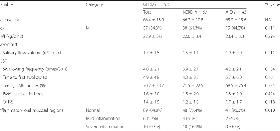
The numbers of NERD and reflux oesophagitis (grade A–D) instances were 62 and 43, respectively. Oral mu-cosal region inflammation showed a negative correlation; however, there were no relationships between the GERD grading and the other evaluated items (Table 4).
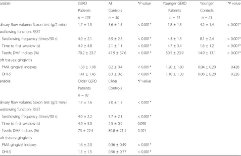
Comparison between 1) GERD patients and all controls; 2) younger GERD patients and younger controls; 3) older GERD patients and older controls
Regarding the comparison between GERD patients and all controls, both of salivary flow volume and swallowing function in GERD patients were signifi-cantly lower than in all controls (P < 0.001). DMF in-dices, PMA gingival indexes and OHI-S in GERD patients were significantly higher than in all controls (P < 0.001). Regarding the comparison between youn-ger GERD patients and younyoun-ger controls, both of sal-ivary flow volume and swallowing function in GERD patients were significantly lower than in all controls (P < 0.001). DMF indices in GERD patients were signifi-cantly higher than in younger controls (P < 0.001). How-ever, no significant differences were seen in PMA gingival indexes and OHI-S. Regarding the comparison between older GERD patients and older controls, salivary flow vol-ume and swallowing frequency were significantly lower a Salivary flow volume (g/2min)
f OHI-S
e PMA gingival indexes
d DMF indices
c Time to first swallow (s)
b Swallowing frequency (times/ 30 s)
P< 0.001
P< 0.001
P= 0.001
P< 0.001
P= 0.038 P< 0.001
P= 0.033 P< 0.001
P< 0.001 P< 0.001
P< 0.001
P= 0.007
than in all controls (P< 0.001). Further, no significant dif-ference was seen in time to first swallow and DMF indices. On the other hand, PMA gingival indexes and OHI-S in older GERD patients were significantly higher than in older controls (P< 0.001) (Table 5).
Discussion
The medical interview results showed that oral dryness was the most common complaint among the GERD pa-tients (59/105; 56.2%), supporting the findings that GERD patients had a significant risk of suffering from xerostomia [29]. Oral dryness is induced by reduced sal-ivary flow volume [29]. In this study, the salsal-ivary flow volume in the GERD patients was significantly reduced, as was observed in our preliminary study [1], which sug-gested that this was a cause for the oral dryness. All GERD patients included in this study manifested the
typical reflux symptoms. Therefore, this result may indi-cate some correlations between acid reflux as typical GERD symptoms and oral dryness as the atypical symp-toms. Swallowing function in the GERD patients was also significantly reduced in this study, as was also ob-served in our previous study [1].
Dental caries is one of the major oral disorders similar to the periodontal disease [9, 10]. Therefore, the risks of dental caries were also evaluated. As a result, the DMF indices in the GERD patients were significantly higher than in the younger controls, but lower than in the older controls, supporting that idea that dental caries increases with age [9]. Our study revealed no significant relation-ship between risks of dental caries and GERD.
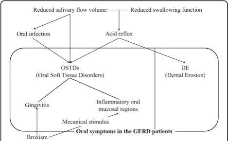
The PMA gingival indexes levels, as determined by gingival inflammation evaluation items, in the GERD pa-tients were significantly higher than in both control groups. The OHI-S levels, as determined by oral hygiene evaluation items, in the GERD patients were also signifi-cantly higher than in both control groups. Saliva pro-tects gingiva by cleaning the tissue and functioning as an antimicrobial agent [14]. Therefore, gingivitis in GERD patients may be induced by reduced salivary flow volume (Fig. 2). Relationships between periodontal disease including gingivitis and GERD have been con-troversial [14, 15]. The most reasonable explanation for these relationships was reduced salivary flow vol-ume [14].
One of the exacerbation factors of periodontal disease is bruxism, which can increase the rate of progression of
Table 4Associations between GERD grading, which were divided into NERD and reflux oesophagitis (grade A-D) subgroups, and the evaluated items
Variable Category GERDn = 105 *Pvalue
Total NERDn = 62 A-Dn = 43
Age (years) 66.4 ± 13.0 66.7 ± 10.8 65.9 ± 15.6 NA
Sex M 57 (54.3%) 38 (61.3%) 19 (44.2%) 0.111
BMI (kg/cm2) 22.9 ± 3.6 22.6 ± 3.4 23.4 ± 3.8 0.294
Saxon test
Salivary flow volume (g/2 min.) 1.7 ± 1.5 1.5 ± 1.1 1.9 ± 2.0 0.211
RSST
Swallowing frequency (times/30 s) 4.0 ± 2.1 3.9 ± 2.1 4.2 ± 2.1 0.584
Time to first swallow (s) 4.9 ± 4.8 4.3 ± 3.7 5.7 ± 6.0 0.161
Teeth; DMF indices (%) 70.2 ± 23.7 71.5 ± 22.5 68.5 ± 25.4 0.535
PMA gingival indexes 1.6 ± 2.0 1.5 ± 2.0 1.8 ± 2.0 0.424
OHI-S 1.4 ± 1.5 1.2 ± 1.3 1.7 ± 1.7 0.118
Inflammatory oral mucosal regions Normal 89 (84.8%) 48 (77.4%) 41 (95.3%) 0.010
Mild inflammation 6 (5.7%) 4 (6.5%) 2 (4.7%)
Severe inflammation 10 (9.5%) 10 (16.1%) 0 (0.0%)
GERDGastroesophageal reflux disease,NERDNon-erosive reflux disease,MMale,NANot applied,BMIBody mass index,RSSTRepetitive saliva swallowing test,
DMFindices: Decayed, missing, and filled indices,PMA gingival indexesPapillary, marginal, and attached gingival indexes,OHI-SSimplified oral hygiene indices *Pvalues of Wilcoxon rank-sum test or Fisher’s exact test were determined as significant byP< 0.05
Table 3Oral mucosal region inflammation (16 GERD patients)
Oral mucosal region Total Mild inflammation Severe inflammation
Tongue 8 2 6
Right buccal mucosa 4 0 4
Left buccal mucosa 6 2 4
Hard palate 1 1 0
Soft palate 2 1 1
Upper vestibular 1 1 0
Lower vestibular 2 2 0
pre-existing periodontal disease by mechanically destroy-ing periodontal tissues [30, 31]. Mengatto [32] reported that when a GERD group (n= 19) was compared with a non-GERD group (n= 26), a statistically higher prevalence of sleep bruxism in the GERD group (14/19; 73.7%) was observed compared with the non-GERD group (6/26; 23.1%,P= 0.017). In our present study, the GERD patients (18/105: 17%) showed a significantly higher frequency of bruxism than that of the younger controls (2/25: 8%) and older controls (none). Therefore, gingivitis in some GERD patients may be exacerbated by bruxism (Fig. 2).
It has been reported that sleep bruxism results from acid reflux, occurring via arousal, often together with swallowing [24, 32, 33]. In this study, the bruxism diag-noses were based on participant self-reports, and more detailed objective evaluations, frequency and onset times of the bruxism were then required to demonstrate the concurrence with GERD.
Although the etiology regarding periodontal disease and GERD were not absolutely revealed, the reduced sal-ivary flow volume suggests an association, as it is com-monly recognized as one of the exacerbation factors of gingivitis [13, 14]. Furthermore, gingivitis seen in GERD
patients can be caused by gastric acid reflux, similar to DE [1] (Fig. 2).
In this study, 10 GERD patients were completely eden-tulous. In the edentulous patients, we were unable to evaluate the gingiva. However, their oral mucosal re-gions, except for the gingiva, were evaluated. Inflamma-tory oral mucosal regions were found only in the GERD patients (16/105; 15.2%). The functions of saliva are anti-microbial action, cleansing and maintaining mucosal integrity [13]. Therefore, inflammatory oral mucosal re-gions including oral infection, for example, Candida in GERD patients may be caused by reduced salivary flow volume (Fig. 2).
Inflammatory oral mucosal regions in the GERD pa-tients were only reported in the palatal regions, as de-tected by morphometry [16, 17]. On the other hand, in our present study, inflammation was observed at all sites, including the tongue, buccal mucosa, and palatal regions. Inflammatory oral mucosal regions in the GERD patients may be damaged by gastric acid reflux that spreads into the oral cavity. In particular, inflammation of palatal mucosa can be affected by direct damage from the acid (Fig. 2). Mechanical stimulus by bruxism should Table 5Comparison between 1) GERD patients and all controls; 2) younger GERD patients and younger controls; 3) older GERD patients and older controls
Variable GERD All *Pvalue Younger GERD Younger *Pvalue
Patients Controls Patients Controls
n = 105 n = 50 n = 13 n = 25
Salivary flow volume; Saxon test (g/2 min.) 1.7 ± 1.5 3.6 ± 1.5 < 0.001* 1.8 ± 1.3 4.2 ± 1.4 < 0.001* Swallowing function; RSST
Swallowing frequency (times/30 s) 4.0 ± 2.1 6.9 ± 2.5 < 0.001* 4.3 ± 1.3 8.1 ± 2.4 < 0.001*
Time to first swallow (s) 4.9 ± 4.8 2.1 ± 1.1 < 0.001* 4.7 ± 3.4 1.6 ± 1.2 < 0.001*
Teeth; DMF indices (%) 70.2 ± 23.7 47.9 ± 37.6 < 0.001* 50.5 ± 23.9 14.9 ± 13.1 < 0.001*
Soft tissues; gingivitis
PMA gingival indexes 1.58 ± 1.98 0.2 ± 0.4 < 0.001* 1.20 ± 1.80 0.04 ± 0.20 0.428
OHI-S 1.41 ± 1.45 0.3 ± 0.6 < 0.001* 1.10 ± 1.30 0.08 ± 0.28 0.226
Variable Older GERD Older *Pvalue
Patients Controls
n = 92
Salivary flow volume; Saxon test (g/2 min.) 1.7 ± 1.6 3.0 ± 1.3 < 0.001* Swallowing function; RSST
Swallowing frequency (times/30 s) 4.0 ± 2.2 5.7 ± 2.1 < 0.001*
Time to first swallow (s) 4.9 ± 5.0 2.5 ± 0.9 0.090
Teeth; DMF indices (%) 73 ± 22.4 80.8 ± 21.1 0.101
Soft tissues; gingivitis
PMA gingival indexes 1.6 ± 2.0 0.36 ± 0.49 < 0.001*
OHI-S 1.5 ± 1.5 0.56 ± 0.77 < 0.001*
GERDGastroesophageal reflux disease,RSSTRepetitive saliva swallowing test,DMF indicesDecayed, missing, and filled indices,PMAgingival indexes Papillary, marginal, and attached gingival indexes,OHI-SSimplified oral hygiene indices
be considered. The bruxism is possible to be also co-cause of check-biting [30, 31] (Fig. 2). Especially, the tongue and bilateral buccal mucosa that the most com-mon regions of inflammation were seen in this study, may be affected by the mechanical stimulus, for exam-ples, sharp of teeth edges anatomically. On the other hand, the oral floor may be prevented from the refluxing acid by the tongue and the major salivary glands; sublin-gal gland, therefore no inflammation was seen [1, 16, 17, 34]. Inflammation was evaluated only optical findings in this study. Therefore it is desirable to be evaluated by scientific methods in the future.
No significant difference was found in any of the evaluated items by GERD grading, which was similar to our prelimin-ary study [1]. Notably, inflammation severity in the oral mu-cosa and esophagus showed negative correlations. Herbert failed to demonstrate statistical differences between GERD and NERD patients for the prevalence of acidic lesions in the oral cavity and their localization [22]. Though oral and esophagus mucosa is covered by squamous membrane his-tologically, the degree of keratinization of oral mucosa is completely different from that of esophagus [34]. Especially, the epithelium of the oral mucous membrane is of the strati-fied squamous variety, which may be keratinized, parakerati-nized or nonkeratiparakerati-nized, depending on the location [34–36]. Therefore, the soft tissue damages by acid reflux evaluated in this study, such as redness, erosion and ulcer, may be different between the oral and esophagus region. Other causes of inflammatory oral mucosal regions seen in GERD patients except for the acid reflux, including reduced
salivary flow volume and the bruxism may also affect these results.
In this study, the number of cases was enlarged from 40 to 105 in GERD patients, from 15 to 25 in younger and older controls each, in a 5 years sampling interval [1]. Not only the numbers of subjects were enlarged, but also BMI and inflammatory oral mucosal regions were evaluated in addition to our preliminary study [1]. Fur-thermore, GERD patients were divided into younger and older groups and compared with both controls respect-ively. According to this result, it could be reconfirmed that salivary flow volume and swallowing function were reduced by not only aging but also GERD. In fact, the older the GERD patients were, the more severe gingivitis might be affected by GERD as a new finding in this study. Therefore, we suggest that OSTDs may be added to DE as the extraesophageal syndromes of GERD.
We recognize some limitations in this study. Firstly, no significant associations in BMI, alcohol and tobacco use were observed. These may be explained by sampling, study design (cross-sectional study), small sample size and less statistical power. Secondly, we did not perform an adjusted analysis with the participant’s background information for comparisons between the GERD patient group and the control groups. Thirdly, we did not con-sider disease duration and GERD treatment. Thus, in the future, larger prospective studies and detailed ana-lyses are needed to validate our findings. Finally, GERD diagnosis was made on the presence of typical reflux symptoms and GIF without any information regarding pH of saliva. One of the past reports demonstrated sig-nificant salivary pH reduction in patients with chronic laryngopharyngitis by GERD before and after treatment [37]. In the future, the correlations with acid exposure, a number of acid reflux events, and potentially proximal extent of reflux should be evaluated objectively.
Our analyses have not addressed the problem for mul-tiple comparisons, and the adjusted a P-value was not calculated. However, because the aim of our study is an exploratory study to generate the hypothesis of relation-ship between GERD and oral hygiene, we believe that we do not need to use the adjusted procedure for multiple comparisons used in confirmatory trials.
Conclusions
OSTDs were associated with GERD, similar to what was observed for DE.
Abbreviations
BMI:Body mass index; DE: Dental erosion; DMF indices: Decayed, missing, and filled indices; GERD: Gastroesophageal reflux disease; GIF: Gastrointestinal fiberscope; M: Male; NA: Not applied; NERD: Non-erosive reflux disease; OHI-S: Simplified oral hygiene indices; OSTDs: Oral soft tissue disorders; PMA gingival indexes: Papillary, marginal, and attached gingival indexes; RSST: Repetitive saliva swallowing test; SD: Standard deviation DE
(Dental Erosion) OSTDs
(Oral Soft Tissue Disorders)
Gingivitis Inflammatory oral mucosal regions
Bruxism
Reduced swallowing function
Acid reflux Reduced salivary flow volume
Oral symptoms in the GERD patients Mecanical stimulus
Oral infection
Acknowledgements
We greatly thank Professor Masanori Fukushima (Translational Research Informatics Centre, Foundation for Biomedical Research and Innovation) for his valuable comments and criticism.
Funding
This work was financially supported by grant from Department of Internal Medicine, Oral and Maxillofacial Surgery in Shimane University Hospital, and Translational Research Informatics Center, Foundation for Biomedical Research and Innovation.
Availability of data and materials
The dataset supporting the conclusions of this article can be made available upon request.
Authors’contributions
JS conceived and designed the experiments. MW, HY and YK collected clinical materials. MW and HY performed the experiments. EN performed statistical analysis. MW, EN, HY, TK, YN, AY, MV, YK and JS interpreted the data. MW, EN and JS wrote the draft of the manuscript. EN, HY, TK, YN, AY, MV, YK and JS contributed to critical revision of the manuscript. All authors have read and approved the final version for publication.
Ethics approval and consent to participate
The ethics committee of the Shimane University Hospital approved the study (protocol No. 398, No. 1082, No. 1217, No. 1750 and No. 1764). All participants signed informed consent.
Consent for publication Not applicable.
Competing interests
The authors declare that they have no competing interests.
Publisher’s Note
Springer Nature remains neutral with regard to jurisdictional claims in published maps and institutional affiliations.
Author details
1Department of Oral and Maxillofacial Surgery, Shimane University Faculty of Medicine, 89-1 Enya-cho, Izumo, Shimane 693-8501, Japan.2Translational Research Informatics Center, Foundation for Biomedical Research and Innovation, 1-5-4, Minatojima minamimachi, Kobe, Hyogo 650-0047, Japan. 3Department of Oral and Maxillofacial Surgery, Matsue City Hospital, 32-1 Nosira-cho, Matsue, Shimane 690-8509, Japan.4Pathologisches Institut Klinikum Bayreuth GmbH, Preuschwitzer Str. 101, 95445 Bayreuth, Germany. 5
Department of Internal Medicine, Shimane University Faculty of Medicine, 89-1 Enya-cho, Izumo, Shimane 693-8501, Japan.
Received: 10 June 2017 Accepted: 31 July 2017
References
1. Yoshikawa H, Furuta K, Ueno M, Egawa M, Yoshino A, Kondo S, Nariai Y, Ishibashi H, Kinoshita Y, Sekine J. Oral symptoms including dental erosion in gastroesophageal reflux disease are associated with decreased salivary flow volume and swallowing function. J Gastroenterol. 2012;47:412–20. 2. Helm JF, Dodds WJ, Pelc LR, Palmer DW, Hogan WJ, Teeter BC. Effect of
esophageal emptying and saliva on clearance of acid from the esophagus. New Engl J Med. 1984;310:284–8.
3. Alfaro EV, Aps JKM, Martens LC. Oral implications in children with gastroesophageal reflux disease. Curr Opin Pediatr. 2008;20:576–83. 4. Vakil N, Van Zanten SV, Kahrilas P, Dent J. Jones R. Global Consensus Group
The Montreal definition and classification of gastroesophageal reflux disease: a global evidence-based consensus Am J Gastroenterol. 2006;101: 1900–20.
5. Pauwels A. Dental erosions and other extra-oesophageal symptoms of gastro-oesophageal reflux disease: Evidence, treatment response and areas of uncertainty. United European Gastroenterol J. 2015;3:166–70.
6. Geng-Ru W, Hui Z, Zhong-Gao W, Guang-Shui J, Cheng-Hao G. Relationship between dental erosion and respiratory symptoms in patients with gastro-oesophageal reflux disease. J Dent. 2010;38:892–8.
7. Nagler RM, Hershkovich O. Relationships between age, drugs, oral sensorial complaints and salivary profile. Arch Oral Biol. 2005;50:7–16.
8. Yokoyama M, Mitomi N, Tetsuka K, Tayama N, Niimi S. Role of Laryngeal Movement and Effect of Aging on Swallowing Pressure in the Pharynx and Upper Esophageal Sphincter. Laryngoscope. 2000;110:434–9.
9. Eslamipour F, Borzabadi-Farahani A, Asgari I. The relationship between aging and oral health inequalities assessed by the DMFT index. Eur J Paediatr Dent. 2010;11:193–9.
10. Wei SH, Lang KP. Periodontal epidemiological indices for children and adolescents: I. gingival and periodontal health assessments. Pediatr Dent. 1981;3:353–60.
11. Motohashi M, Nakajima I, Aboshi H, Honda K, Yanagisawa M, Miyata T, Maeno M, Kuwata F, Sidaphone B, Ngonephady S, Sitthiphanh A, Kingsada SO, Otsuka K. The oral health of children in a rural area of the Lao People’s Democratic Republic. J Oral Sci. 2009;51:131–5.
12. Silva MA, Damante JH, Stipp AC, Tolentino MM, Carlotto PR, Fleury RN. Gastroesophageal reflux disease: new oral findings. Oral Surg Oral Med Oral Pathol. 2001;91:301–10.
13. Llena-Puy C. The rôle of saliva in maintaining oral health and as an aid to diagnosis. Med Oral Patol Oral Cir Bucal. 2006;11:449–55.
14. Jun YS, Hyung HK, Eun JC, Tae YK. The Relationship between
Gastroesophageal Reflux Disease and Chronic Periodontitis. Gut Liver. 2014; 8:35–40.
15. Muñoz JV, Herreros B, Sanchiz V, Amoros C, Hernandez V, Pascual I, Mora F, Minguez M, Bagan JV, Benages A. Dental and periodontal lesions in patients with gastro-oesophageal reflux disease. Dig Liver Dis. 2003;35:461–7. 16. Ranjitkar S, Smales RJ, Kaidonis JA. Oral manifestations of gastroesophageal
reflux disease. J Gastroenterol Hepatol 2012;27:21-27.
17. Silva MA, Damante JH, Stipp AC, Tolentino MM, Carlotto PR, Fleury RN. Gastroesophageal reflux disease: New oral findings. Oral Surg Oral Med Oral Pathol Oral Radiol Endod. 2001;91:301–10.
18. Fujiwara M, Eguchi Y, Fukumori N, Eguchi H, Tomonaga M, Yoshioka T, Hyakutake M, Sakanishi Y, Kyoraku I, Sugioka T, Fujimoto K, Kusano M, Yamashita S. The Symptoms of Gastroesophageal Reflux Disease Correlate with High Body Mass Index, the Aspartate Aminotransferase/Alanine Aminotransferase Ratio and Insulin Resistance in Japanese Patients with Non-alcoholic Fatty Liver Disease. Intern Med. 2015;54:3099–104. 19. Aro P, Ronkainen J, Talley NJ, Storskrubb T, Bolling-Sternevald E, Agréus L.
Body mass index and chronic unexplained gastrointestinal symptoms: an adult endoscopic population based study. Gut. 2005;54:1377–83. 20. Chattopadhyay DK, Greaney MG, Irvin TT. Effect of cigarette smoking on the
lower oesophageal sphincter. Gut. 1977;18:833–5.
21. Shao-hua C, Jie-wei W, You-ming L. Is alcohol consumption associated with gastroesophageal reflux disease? J Zhejiang Univ Sci B. 2010;11:423–8. 22. Herbert D, Thomas M, Stefan W, Marco K, Anna R, Monther B, Anton S.
Erosive esophageal reflux vs. non erosive esophageal reflux: oral findings in 71 patients. BMC Oral Health. 2015;15:84.
23. Sugiura T, Iwakiri K, Kotoyori M, Kobayashi M. Relationship between severity of reflux esophagitis according to the Los Angeles classification and esophageal motility. J Gastroenterol. 2001;36:226–30.
24. Miyawaki S, Tanimoto Y, Araki Y, Katayama A, Fujii A. Takano- Yamamoto T. Association Between Nocturnal Bruxism and Gastroesophageal Reflux. Sleep. 2003;7:888–92.
25. Shizukuishi K, Nagaoka S, Kinno Y, Saito M, Takahashi N, Kawamoto M, Abe A, Jin L, Inoue T. Scoring analysis of salivary gland scintigraphy in patients with Sjögren's syndrome. Ann Nucl Med. 2003;17:627–31.
26. Tamura F, Mizukami M, Ayano R, Mukai Y. Analysis of feeding function and jaw stability in bedridden elderly. Dysphagia. 2002;17:235–41.
27. Greene JC. Oral hygiene and periodontal disease. Am J Public Health Nations Health. 1963;53:913–22.
28. Greene JC, Vermillion JR. The simplified oral hygiene index. J Am Dent Assoc. 1964;68:7–13.
29. Campisi G, Lo Russo L, Di Liberto C, Di Nicola F, Butera D, Vigneri S, Compilato D, Lo Muzio L, Di Fede O. Saliva variations in gastro-oesophageal reflux disease. J Dent. 2008;36:268–71.
31. Davies SJ, Gray RJ, Linden GJ, James JA. Occlusal considerations in periodontics. Br Dent J. 2001;191:596–604.
32. Mengatto CM, Dalberto CS, Scheeren B, Barros SG. Associaion betwen sleep bruxism and gastroesophageal reflux disease. J Prosthet Dent. 2013;5:349–55. 33. Ohmure H, Oikawa K, Kanematsu K, Saito Y, Yamamoto T, Nagahama H,
Tsubouchi H, Miyawaki S. Influence of experimental esophageal acidification on sleep bruxism: a randomized trial. J Dent Res. 2011;90:665–71. 34. Pace F, Pallotta S, Tonini M, Vakil N, Bianchi PG. Systematic review:
gastro-oesophageal reflux disease and dental lesions. Aliment Pharmacol Ther. 2008;27:1179–86.
35. Nevill BW, Damm DD, Allen CM, Bouquot JE. Oral&Maxillofacial Pathology. 2nd ed. Philadelphia: W. B. Saunders; 2001.
36. Sekine J. Fokussierte zytologische Diagnose und Behandlung von Erkrankungen der Mundhöhle. Verdauungskrankheiten. 2017;35:92–108. 37. Eckley CA, Costa HO. Comparative study of salivary pH and volume in adults
with chronic laryngopharyngitis by gastroesophageal reflux disease before and after treatment. Braz J Otorhinolaryngol. 2006;72:55–60.
• We accept pre-submission inquiries
• Our selector tool helps you to find the most relevant journal
• We provide round the clock customer support
• Convenient online submission
• Thorough peer review
• Inclusion in PubMed and all major indexing services • Maximum visibility for your research
Submit your manuscript at www.biomedcentral.com/submit