AbstrAct
Background: the prevalence of synchronous or
metastatic tumours in patients with head and neck squamous cell carcinoma (HNscc) ranges from 6-20 % and has implications for prognosis and management of the primary disease. there is no consensus about the role of ct chest prior to definitive treatment patients with HNscc.
Methods: A systematic review of all ct chest studies
in relation to HNscc was performed, together with a review of our local database.
Results: 24 studies were identified in addition to our
local data. Prevalence of positive ct chest was 7.93 %. Patients were significantly more likely to have a positive ct chest with N2 or N3 neck disease (P=0.0062), stage III or IV disease (P=0.0001) and significantly less likely with tumours of the oral cavity (P=0.0007).
Conclusions: We advocate ct chest as part of the
initial investigations for patients with HNscc.
INtroductIoN
Head and neck squamous cell carcinoma (HNscc) is the 6th commonest cancer worldwide, and the commonest
in the Indian subcontinent. between 6-20% of patients with newly diagnosed HNscc will have synchronous or metastatic tumours at initial presentation. these most commonly arise within the head and neck itself, but may also present in the lungs or oesophagus. Furthermore, the presence of distant metastases has implications for the disease free survival of these patients, and may influence their management, particularly when major surgery and reconstruction is planned. the incidence of pulmonary metastases in these patients has traditionally been reported to be 1-2%, but the utilisation of modern screening tool, particularly spiral computer tomography (ct) of the chest has suggested that the prevalence of lung metastases is higher.1
It is important to identify patients with distant metastases as part of the staging process for newly diagnosed HNscc. traditionally the chest was imaged by a plain radiograph but this has low sensitivity, although specificity is high. ct chest
The Role of Chest Computed Tomography in
Staging of Oropharyngeal Cancer:
A Systematic Review
*N.M.H. McLeod FDSRCSEd, *A. Jess MFDS, *R. Anand FRCS (OMF),
‡E. Tilley, ◊B. Higgins, *P.A. Brennan M.D.
*Department of Oral & Maxillofacial Surgery, ‡Department of Radiology, Queen Alexandra Hospital Cosham Portsmouth PO6 3LY.
is currently the gold standard in imaging patients with suspected thoracic malignancy. However, it involves a considerable dose of radiation (on average 8msv- the equivalent of 400 chest X-rays). some centres routinely undertake ct chest in all HNscc patients,2-10 while others selectively screen those with
clinical stage III or IV disease, or using other clinical criteria such as presence of nodal disease.1,11-24 In
patients with HNscc, most chest malignancy occurs in the lung apices or mediastinal lymph nodes. As a result, it has been our local policy to perform a limited ct chest from the thoracic inlet to level of pulmonary veins in patients presenting with t1 and t2 tumours (this can be done without moving the patient or giving further contrast). A full ct chest is used in high risk patients, such as those with t3 and t4 disease and in those patients with multiple or large neck node metastases.
the prevalence of positive ct chest in patients with HNscc in the literature ranges from 1.92 % to 37.5 %. 14,24
We performed a systematic review of all publications to date, together with a review of our own patient database to identify the prevalence of positive ct chest findings in HNscc and to provide some recommendations about the appropriateness of ct chest in these patients. We examined subgroups based on factors such as tumour site, tNM classification and disease stage, to determine if any specific risk factors could be identified to select patients who would benefit from ct chest more than others.
MetHods
All publications relating to the use of ct chest in staging HNscc were identified by searching Medline and cinahl databases using the terms; oral, pharynx, larynx, squamous cell carcinoma, ct chest. references were hand searched for further relevant publications. recent conference abstracts were also searched.
All studies were included which contained data on ct chest either alone or in comparison with other imaging modalities (for example plain X-ray or Positron emission tomography (Pet). All references were searched for data pertaining to prevalence of synchronous bronchogenic primary malignancy or metastatic HNscc within the chest, sensitivity and specificity of ct chest for malignancy, and tumour data (t classification, N classification, disease stage,
primary tumour site and differentiation). data obtained from abstracts of oral or poster presentations at recent conferences and our local data was included in the analysis to ensure the highest study population was considered.
All studies were graded as level II or level III evidence.
Patient selection criteria were also noted (all patients or selected patients, new or recurrent disease).
Patient data was extracted where possible and grouped by t classification, N classification, disease stage, primary tumour site and differentiation, as per the study description .
data was analysed with statsdirect software (© statsdirect Ltd, cheshire, uK). Group proportions were assessed using chi-squared, Fisher’s exact and Mantel-Heanszel calculations as appropriate. study heterogencicity was estimated with cochrane Q calculation and inter-group bias with egger’s calculation. differences in data reporting meant that many of the identified publications had to be selectively excluded from subset analysis.
our own head and neck cancer database was searched for all patients with head and neck (not cutaneous) scc who had undergone full ct chest during the period 2002-2007. ct chests were reviewed for the presence of malignancy at the time of diagnosis of HNscc.
resuLts
review of local database:
between July 2002 and March 2007, we identified 195 patients with newly diagnosed head and neck scc who had undergone partial (n=118 ) or full (n=77) ct chest as part of their staging investigations.
of those patients who underwent partial ct chest, 5 (4%) had positive findings for chest malignancy, while 10 (13%) had a positive ct chest following a full ct chest. Patients who underwent partial ct chest were significantly less likely to have a positive ct chest than those who underwent a full ct chest (or 0.2982, 95% cI 0.0893 to 0.9093, P=0.0307). Interestingly, in 8 out of the 10 patients who had positive findings on full chest scans, the abnormality was within the upper thorax, which would have been identified by partial ct scanning.
some patients underwent the incorrect scanning protocol and when full and partial scan were compared taking in to account tumour size (t1 or t2 compared to t3 or t4), the type of scan did not significantly affect the likelihood of a positive result (or 1.3518 95% cI 0.3956 to 4.6191).
systematic review:
twenty-two published studies were identified, together with two abstracts from conferences in addition to the data from our own database. there were 14 prospective and 11 (including our own) retrospective studies.
ct chest was performed in all patients presenting with head and neck squamous cell carcinoma in 16 papers, while 9 groups used ct chest selectively for high risk groups identified according to various criteria, including tumour size, neck node status, disease stage, and whether it was primary or recurrent disease.
overall the data represented some 4062 patients with 322 true positive ct chest. Analysing studies which reported outcomes of all scans including false negative and false positive staging ct chest scans reveals the sensitivity of ct chest
for identifying synchronous tumours or metastasis to be 84.62% and the specificity to be 93.50%.
the pooled point prevalence of positive ct chest in patients with HNscc is 7.93 % (95% cI 7.10 to 8.76). there was significant heterogenicity of study results (cochran Q 213.04 (P<0.0001)) and therefore any assumptions based on pooled data must be interpreted with caution.
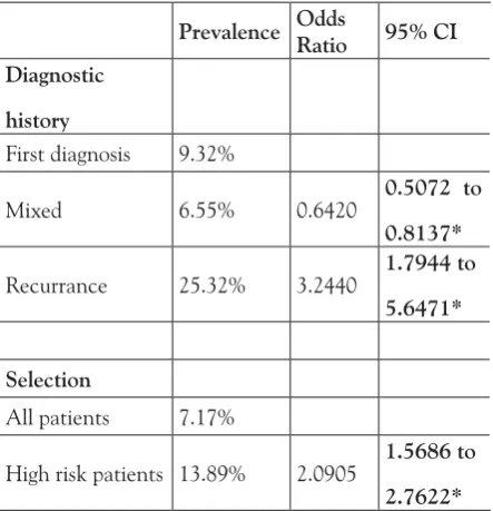
Figure 1 demonstrates the published prevalence of positive ct chest findings with studies grouped according to the author’s use of patients with new,
recurrent or mixed HNscc.
there was a significantly higher point prevalence of positive ct chest in studies examining patients diagnosed with a recurrent HNscc compared to those with a first diagnosis of HNscc or mixed groups of patients (chi square = 43.1118, P < 0.0001) (table 1). In studies which included identifiable data for new and recurrent diagnosis patient groups there was no evidence of study heterogenicity (cochran Q 1.6726, P = 0.64) or group bias (egger 1.6370, P = 0.0918)
there was a significantly higher point prevalence of positive ct chest in studies which undertook ct chest in selected patients and not all patients presenting with new HNscc (13.89% compared to 7.17%). (table 1)
the style and detail of the reported data varied greatly
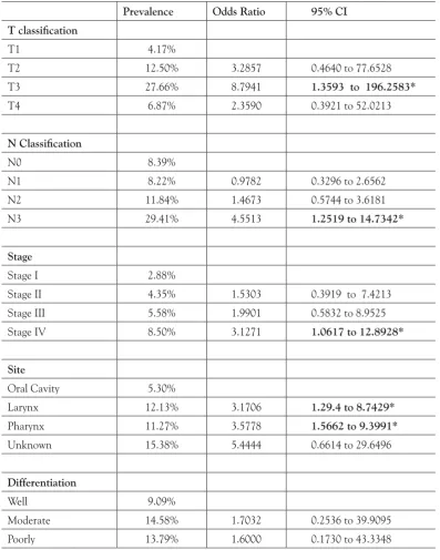
the proportion of patients with positive ct chest with different t classifications (chi square 15.98, P=0.0011), principally due to an increased prevalence in patients with t3 tumours (table 2). there was no evidence of group bias (egger -1.0069, P=0.37).
the prevalence of positive ct chest in patients with t1 and t2 tumours grouped together was not significantly lower than that for patients with t3 and t4 tumours grouped. (prevalence 9.52% compared to 12.50%, or = 1.6096, 95% cI = 0.7351 to 3.3247 ).
NecK LyMPH Nodes
(N-cLAssIFIcAtIoN)
there were 7 studies (440 patients) that included data on the disease N-classification. A total of 52 (11.82%) patients had positive ct chest findings. there was a significant difference in the proportion of patients with positive ct chest with different N classifications (chi-square = 8.1000, P = 0.044), the prevalence increasing with increasing N classification (table 2). there was no evidence of group bias (egger 2.2364, P=0.07).
Patients with N0 or N1 neck node classification were significantly less likely than those with N2 or N3 necks to have a positive ct chest (prevalence 7.42% compared to 16.54%, or = 0.4431, 95% cI = 0.2237 to 0.8775).
dIseAse stAGe
there were 13 studies (1359 patients) that included data on disease stage. A total of 110 (8.09%) patients had positive ct chest. there was no significant difference in the proportion of patients with positive ct chest based on the individual disease stage (chi-square = 3.8305, P = 0.28) (table 2). there was no evidence of group bias (egger -1.0812, P=0.36).
When Patients with stage I or II disease were grouped, the prevalence of positive ct chest was significantly lower than those with disease stage III or IV grouped (Prevalence 3.47% compared to 10.47%, or = 0.4053, 95% cI = 0.12264 to 0.7254).
sIte oF PrIMAry dIseAse
eleven studies (1149 patients) included data on the site of primary disease. A total of 110 (9.57%) patients had Table 1 subgroup analysis based on patient selection
criteria (* = p<0.05)
Prevalence Odds Ratio 95% CI Diagnostic
history
First diagnosis 9.32%
Mixed 6.55% 0.6420 0.5072 to
0.8137*
recurrance 25.32% 3.2440 1.7944 to 5.6471*
Selection
All patients 7.17%
High risk patients 13.89% 2.0905 1.5686 to 2.7622*
and therefore only selected data could be included in further sub-group analysis. results of the different subgroups analyses are shown in table 2.
PrIMAry tuMour sIze (t-stAGe)
positive ct chest findings. significant differences in the prevalence of positive ct chest findings existed according to site (chi-square = 10.8367, P = 0.0126) (table 2).
A primary tumour in the oral cavity was significantly less
likely to be associated with a positive ct chest than all other sites grouped (prevalence 5.30% compared to 11.67%, or 0.4233, 95% cI 0.2416 to 0.7104, P = 0.0007).
Table 2. subgroup analysis of grouped data. (* = p<0.05)
Prevalence Odds Ratio 95% CI
T classification
t1 4.17%
t2 12.50% 3.2857 0.4640 to 77.6528
t3 27.66% 8.7941 1.3593 to 196.2583*
t4 6.87% 2.3590 0.3921 to 52.0213
N Classification
N0 8.39%
N1 8.22% 0.9782 0.3296 to 2.6562
N2 11.84% 1.4673 0.5744 to 3.6181
N3 29.41% 4.5513 1.2519 to 14.7342*
Stage
stage I 2.88%
stage II 4.35% 1.5303 0.3919 to 7.4213
stage III 5.58% 1.9901 0.5832 to 8.9525
stage IV 8.50% 3.1271 1.0617 to 12.8928*
Site
oral cavity 5.30%
Larynx 12.13% 3.1706 1.29.4 to 8.7429*
Pharynx 11.27% 3.5778 1.5662 to 9.3991*
unknown 15.38% 5.4444 0.6614 to 29.6496
Differentiation
Well 9.09%
Moderate 14.58% 1.7032 0.2536 to 39.9095
tuMour ceLL dIFFereNtIAtIoN
three studies (238 patients) included data on the degree of primary tumour differentiation. A total of 30 (12.6%) patients had positive ct chest findings. there was no significant difference between degree of tumour differentiation and existence of positive ct chest findings (chi-square = 0.2487, P = 0.8831) (table 2)
dIscussIoN
the presence of a synchronous bronchogenic tumour or chest metastases when a patient presents with HNscc has implications for the prognosis of the patient, and may have a major impact on their management.
Whilst in some cases the chest lesion may be amenable to resection or chemo-radiotherapy and therefore the treatment of the primary HNscc unaffected, in other cases the patient will be deemed to have incurable disease and extensive resection or aggressive chemo-radiotherapy of the primary HNscc may be inappropriate. Whilst there are questions as to the survival benefit of population chest malignancy screening with either cXr or ct chest the impact of a positive investigation on the management of HNscc makes screening for chest malignancy in this population group important. Mazer et al,25 Finlay et al26 and young
et al27 have all demonstrated a clear survival benefit
in patients with HNscc with lung metastases who underwent surgical resection of their lung metastases. screening of the chest for synchronous or metastatic HNscc is therefore clearly indicated.
the scottish Intercollegiate Guidelines Network (sIGN) have advocated that all patients with HNscc should undergo ct chest as part of their staging investigations, despite their being little evidence to support the benefit to this move.28 the National
Institute of clinical excellence (NIce) document ‘Improving outcomes in Head and Neck cancer’ states that “all patients with upper aerodigestive tract (uAt) cancers should have chest X-rays. other forms of imaging are necessary to assess the stage and spread of the tumour, and specialist ultrasound, computed tomography (ct) and magnetic resonance imaging (MrI) should be available. Positron emission tomography (Pet) imaging should be used, if available, when it is important to differentiate between benign and malignant lung nodules. It is anticipated that the role of Pet will increase over the course of the next decade”.29
Plain chest radiography, usually in the form of a plain postero-anterior chest film has traditionally been used for both population screening for primary chest malignancy and screening for chest malignancy in the patients with other primary tumours.
studies where cXr results were compared to those of ct chest demonstrate a pooled sensitivity of cXr of 42.68% with a specificity of 98.47%.2,4,5,12-16,18 cXr is
therefore only half as sensitive as ct chest, although its specificity is comparable.
the sensitivity and specificity of chest ct currently make it the gold standard in screening for chest malignancy although its use for population screening for
chest malignancy is limited by the lack of evidence of a survival benefit in unselected population groups.30
the sensitivity of Pet –ct is high (96-100%), making it much more sensitive than cXr and slightly more so than ct chest in identifying chest malignancy, but its specificity (77.8%) is poor compared to either modality.8,18,31,32 When combined with its high cost
and limited availability, particularly in europe and Asia, these findings do not support its use for routine staging for chest malignancy in patients with HNscc at present.
In addition to the advantages of detecting malignancy not evident on a plain cXr, which may change both patient management and prognosis, there are disadvantages of ct chest which must be considered.1
these include additional radiation and the risk of false positive results, necessitating other investigations whilst increasing patient anxiety still further.16 Ideally these
risks could be minimized and the benefit of ct chest maximized by identifying those patients who are most at risk of malignant lung pathology, but as yet there is no consensus on the best approach to identifying higher risk patients.32
t stage alone is not a reliable indicator of likelihood of identifying chest malignancy. Whilst there is an increasing prevalence of chest malignancy with increasing t classification from t1 to t3 tumours, those with t4 tumours have a lower prevalence. t classification does not simply reflect tumour size. t4 tumours are defined as invading local structures, for example the mandible or maxilla in oral cancer, irrespective of size. the lower prevalence in t4 tumours may indicate over-staging of tumours due to their proximity to other structures or the different biological behaviour of different tumours. the ability of small tumours to metastasize, whilst others will become locally very advanced before metastasizing is recognised.
Patient who have N2 or N3 neck disease are more likely to have synchronous or metastatic chest malignancy as are those with stage III or stage IV disease, and these indices could be used to select high risk patients. However it is not clear in the published studies whether the authors were using initial clinical classifications of neck lymph node disease or pathological staging, which would result in greater disease staging, after investigations are completed.
unfortunately there was insufficient data in the studies analysed to include pathological parameters such as extra-capsular spread (ecs), and position of the metastatic nodes. However, it is generally agreed that these variables as well as the number of nodes involved are associated with increased risk, and most authorities would cite three or more nodes as having particularly poor prognosis.33-36
Whichever selection criteria were used, when study data was pooled the point prevalence of positive ct chest was at least 2.88% for all sub-groups (stage I tumours) (table 1, 2). this is higher than the prevalence of chest malignancy in studies on the use of ct chest in screening high risk patients for chest malignancy (0.3 to 2.3%) and for breast malignancy in screened patients (0.60%).30,37 And whilst the current cost:benefit or
survival benefit arguments have not favoured the development of a population based chest malignancy screening program, one does exist in most developed countries for breast malignancy.
coNcLusIoNs
Improvements in treatment of HNscc have resulted in better loco-regional control, however the mortality
rates have barely improved over the last 30 years, and most would attribute this to the development of distant metastases.33-36,38 Furthermore, HNscc cells can be
found in the bone marrow in these patients, although the significance of this finding is unclear.39 the early
detection of distant metastases therefore is important because of its possible effects on the treatment planning of the patient. Major ablative and reconstructive surgery should only be considered where there is a reasonable prospect of effecting a cure and whilst the presence of distant metastases would not in itself contra-indicate surgery, treatment of the distant site would need to be considered as well.12,13 this may take to form of thoracic
surgery or chemo-radiotherapy.
ct chest is currently the gold standard investigation for primary and metastatic chest malignancy.
because of the difficulties in accurately identifying patients at higher risk of having synchronous or metastatic chest malignancy and the survival and cost benefits in identifying the presence of synchronous or metastatic chest malignancy, we would advocate the use of ct chest in all patients presenting with HNscc.
reFereNces
1. Houghton dJ, Mcgarry G, stewart I, Wilson JA, Mackenzie K. chest computerised tomongraphy scanning in patients presenting with head and neck cancer. clin otolaryngol 1998:23;348-50. 2. de bree r, deurloo ee, snow Gb, Leemans cr. screening
for distant metastases in patients with head and neck cancer. Laryngoscope 2000:110:397-401.
3. Nilssen eLK, Murthy P, Mcclymont L, denholm s. radiological staging of the chest and abdomen in head and neck squamous cell carcinoma - are computed tomography and ultrasound necessary. J Laryngol otology 1999:113:152-154.
4. tan LKs, Greener cc, seikaly H, rasseki H, calhoun KH. role of screenig chest computed tomography in patients with advanced head and neck cancer. otolaryngol Head Neck surg 1999:120:689-92.
5. Warner Gc, cox GJ. evaluation of chest radiography ersus chest computer tomography in screening for pulmonary malignancy in advanced head and neck cancer. J otoloaryngol. 2003:32(3): 107-109.
6. brouwer J, de bree r, Hoekstra os,et al. screening for distant metastases in patients with head and neck cancer: is chest computed tomography sufficient. Larygoscope 2005: 115: 1813-1817. 7. Keski-santi Ht, Markkola Ato, Makitie AA, back LJJ, Atula ts.
ct of the chest and abdomen in patienst with newly diagosed head and neck squamous cell carcinoma. Head Neck 2005:27; 909-15 8. teknos tN, rosenthal eL, Lee d, taylor r, Marn cs. Positron
emission tomography in the evaluation of stage III and IV head and neck cancer. Head Neck 2001:23:1056-60.
pulmonary metastases from squamous carcinoma of the head and neck. Am J surg 1988;156:238-42.
26. Finley rK, Verazin Gt, driscoll dL et al. results of surgical resection of pulmonary metastases of squamous cell carcinoma of the head and neck. Am J surg 1992; 164: 594-8.
27. younes ry, gross JL, silva JF, Fernandez JAP, Kowalski LP. surgical treatment of lung metastases of head and neck tumours. Am J surg 1997:174; 499-502.
28. scottish Intercollegiate Guidelines Network. diagnosis and management of head and neck cancer. A national clinical guideline. www.sign.ac.uk./pdf/sign90.pdf
29. National Institute for clinical excellence. Guidance on cancer services. Improving outcomes in head and neck cancer. www. guidance.nice.org.uk/csghn
30. ellis Jr, Gleeson FV. New concepts in lung cancer screening.curr opin Pulm Med 2002;8:270-4.
31. schmid dt, stoeckli sJ, bandhauer F, Huguenin P, schmid s, von schulthess GK, Goerres GW. Impact of positron emission tomography on the initial staging and therapy in locoregional advanced squamous cell carcinoma of the head and neck. Laryngoscope 2003;113:888-91.
32. brouwer J, de bree r, Hoekstra os, Langenijk JA, castelijns JA, Leemans cr. screening for distant metastases in patients with head and neck cancer: what is current clinical practice? clin otoloaryngol 2005; 30:438-443.
33. Leon X, Quer M, orus c, del Prado Venegas M, Lopez M distant metastases in head and neck cancer patients who achieved loco-regional control. Head Neck 2000;22:680-6.
34. Alvi A, Johnson Jt development of distant metastasis after treatment of advanced-stage head and neck cancer. Head Neck 1997;19:500-5.
35. Leemans cr, tiwari r, Nauta JJ, van der Waal I, snow Gb regional lymph node involvement and its significance in the development of distant metastases in head and neck carcinoma. cancer 1993;71:452-6.
36. shingaki s, suzuki I, Kobayashi t, Nakajima t. Predicting factors for distant metastases in head and neck carcinomas: an analysis of 103 patients with locoregional control.J oral Maxillofac surg 1996;54:853-7.
37. richardson A, Graham P, brown t, smale P, cox b. breast cancer detection rates and standardised detection rates in the New zealand breast cancer screening program. J Med screen 2004:11(2); 65-9. 38. spector JG, sessions dG, Haughey bH, chao Ks, simpson J, el Mofty s, Perez cA. delayed regional metastases, distant metastases, and second primary malignancies in squamous cell carcinomas of the larynx and hypopharynx. Laryngoscope 2001;111:1079-87. 39. Partridge M, brakenhoff r, Phillips e, Ali K, Francis r, Hooper r,
Lavery K,brown A, Langdon J detection of rare disseminated tumor cells identifies head and neck cancer patients at risk of treatment failure. clin cancer res 2003;9:5287-94.
10. tesche s. Habermann cr, sagowski c, Wenzel s, Metternich Fu. the value of chest ct-scanning for staging of Progressed or recurrent Head and Neck squamous cell carcinomas (HNscc). Laryngo-rhino-otol 2006: 85;93-98.
11. Mercader VP, Gatenby rA, Mohr rM, Fisher Ms, caroline dF. ct surveillace of the thorax in patients with squamous cell carcinoma of the head and neck: a preliminary experience. J comp Assist tomography 1997:21(3): 412-417.
12. Houghton dJ, Hughes ML, Garvey c, et al. role of chest ct scanning in the management of patients presenting with head and neck cancer. Head Neck 1998:20:614-8
13. reiner b, siegel e, sawyer r. the impact of routine ct of the chest on the diagnosis and management of newly diagnosed squamous cell carcinoma of the head and neck. AJr 1997: 169:667-671. 14. Halpern J. the value of chest ct scan in the work-up of head and
neck cancers. J Med 1997:28 (3): 191-198.
15. ong tK, Kerawal cJ, Martin Ic, stafford FW. the role of thorax imaging in staging head and neck squamous cell carcinoma .J craniomaxilofac surg 1999:27:339-44.
16. Keith dJW, ongtK, Martin Ic. the role of thoracic computed tomography in staging newly-diagnosed oral squamous cell carcinoma. br J oral Maxilofac surg 2006:44:198-202.
17. Arunachalam Ps, Putnam G, Jennings P, Messersmith r, robson AK. role of computerized tomography (ct) scan of the chest in patients with newly diagnosed head and neck cancers. clin otolaryngol 2002:27:409-411.
18. Wax MK, Myers L, Gabalski ec, Husain s, Gona JM, Nadi H. Positron emission tomography in the evaluation of synchronous lung lesions in patients with untreated head and neck cancer. Arch otolaryngol Head Neck surg 2002: 128: 703-7.
19. Loh Ks, brown dH, baker Jt et al. A rational approach to pulmonary screening in newly diagnosed head and neck cancer. Head Neck 2005; 27; 990-4.
20. Jäckel Mc, reischl A, Huppert P. efficacy of radiologic screening for distant metastases and second primaries in newly diagnosed patients with head and neck cancer. Laryngoscope. 2007;117(2):242-7. 21. Glynn F, brennan s, o’Leary G. ct staging and surveillance of the
thorax in patients with newly diagnosed and recurrent squamous cell carcinoma of the head and neck: is it necessary? eur Arch otorhinolaryngol. 2006: 263(10):943-5.
22. Ghosh s, Kumar A, roland N et al. detection of pulmonary tumours in Head and Neck cancer Patients. Poster presented at british Association of Head &Neck oncologists Annual Meeting, 2007 April; London, uK.
23. Morrison J, Markose G, carton At, Hislop Ws. thoracic computed tomography in newly diagnosed oral carcinomas. br J oral Maxillofac surg 2007; 45(7): e32.
24. bisase b, Kerawala c, Lee J. the role of computed tomography of the chest in the staging of early squamous cell carcinoma of the tongue. br J oral Maxillofac surg 2007: in press.