R E S E A R C H
Open Access
Plantar pressure changes in hindfoot relief
devices of different designs
F. Mazur
1, B. Swoboda
1, H. D. Carl
2, C. Lutter
3, M. Engelhardt
4, M. W. Hoppe
4,5, T. Hotfiel
1*†and C. Grim
4†Abstract
Background:It is frequently observed that overloading the foot can impair bone and soft tissue healing and can lead to harmful sequelae (i.e. ulcers, stress reactions) in context of pre-existing tissue disabilities. In terms of offloading, hindfoot relief devices are commonly applied as a non-operative treatment as well as after various surgical procedures for hindfoot disorders. Despite their common use, there is a paucity of data comparing different orthotic devices with respect to changes in plantar pressure distributions. The aim of this study was to investigate plantar loadings in hindfoot relief devices of different designs.
Methods:Twenty-five healthy participants (13 women, 12 men; (mean ± SD) age 37 ± 14 years; BMI 23 ± 4 kg/m2) were recruited. Plantar pressure distributions were collected using i.) a neutral shoe, ii.) a hindfoot relief shoe (HRS) and iii.) a hindfoot relief orthosis (HRO). Peak pressure values were measured via dynamic pedobarography during walking and were analysed from four different plantar regions: the hindfoot, midfoot, metatarsal I-V and forefoot. As a reference standard, the normal walk using neutral shoes served as the condition for full weight-bearing.
Results:Concerning the hindfoot, using the HRS as well as the HRO resulted in significant decreases in plantar pressures compared to baseline values that were obtained with the neutral shoe (−52% for the HRS and−52% for the HRO,p< 0.001). Significant increases in peak pressures were found in the midfoot region for both devices (HRS: 32%,
p= 0.002; HRO: 47%,p< 0.001). For the metatarsal region, peak pressures were found to decrease significantly (HRS:− 52%,p< 0.001; HRO: -17%,p= 0.034). With respect to the forefoot, a significant reduction in peak pressures using the HRS (−41%,p< 0.001) was detected, whereas the HRO did not lead to significant changes (−4%,p= 0.691).
Conclusions:Both the HRO and HRS significantly reduced plantar hindfoot pressure, corresponding to a relative decrease of nearly 50% of the baseline. Nevertheless, the adjacent midfoot zone displayed a significant increase in plantar pressure values for both devices. Supported by these findings, physicians should cautiously consider a substantial increase in midfoot loading, especially in patients affected by additional midfoot injuries or accompanying impairments of tissue healing.
Level of evidence:IV, Case series.
Keywords:Plantar pressure, Hindfoot relief shoes, Plantar ulcers, Kinetics, Biomechanics, Pedobarography, Foot, Stress fractures
* Correspondence:thilo.hotfiel@fau.de
†T. Hotfiel and C. Grim contributed equally to this work.
1Division of Orthopaedic Rheumatology, Department of Orthopaedic
Surgery, Friedrich-Alexander-University Erlangen-Nuremberg, Rathsberger Str. 57, D-91054 Erlangen, Germany
Full list of author information is available at the end of the article
delays in fracture healing (Claes & Heigele, 1999; Reike et al.,1997; Genc et al.,2016). Hindfoot relief devices have commonly been used in the post-surgical rehabilitation process, following various procedures such as the repair of calcaneal fractures, ligament reconstructions, corrective osteotomies, and trauma surgery of the hindfoot (Carl et al.,2006; Hodge et al.,1999; Schepers et al.,2008; Bohl et al.,2017; Groot et al.,2013; Cavanagh & Bus,2011; Kraus et al.,2014; Bus et al.,2009). In cases of tarsal bone marrow oedema, stress reactions or stress fractures, hindfoot relief devices allow a mobilization under limited weight-bearing conditions that are encouraged to promote healing without overloading the tissue (Pauser et al., 2011). Additionally, hindfoot relief devices are used to improve the healing process for plantar ulcers and wound healing disorders due to trauma, peripheral arterial disease, neuropathic disabil-ities and rheumatoid arthritis (Pauser et al., 2011; Götz et al.,2016; da Conceição et al.,2015). Offloading the hind-foot is mostly carried out by hindhind-foot relief shoes (HRSs) and hindfoot relief orthoses (HROs) (Hunt et al., 1987; Hahn et al., 2014). Nevertheless, commonly available devices display fundamentally different designs and concepts. Despite the common use of pressure relief devices, there is a paucity of data comparing their offload-ing effects related to biomechanical aspects.
Dynamic pedobarography is a modality that has been widely validated as a method to evaluate plantar pressure under dynamic conditions (Skopljak et al.,2014). Owing the ability to record consecutive steps in one measure-ment, insole-based pedobarography has become an important tool for the evaluation of foot loads during the application of insoles, orthoses or other types of footwear (Skopljak et al., 2014; Westphal et al., 2016; Kluger et al., 2014; Lorkowski et al., 2015). By this approach, the offloading effects of forefoot relief shoes in surgical or non-surgical terms have been extensively investigated (Carl et al., 2006; Kraus et al., 2014; Bus et al., 2009). In contrastthere has been a paucity of data comparing plantar pressure patterns in HRSs of various designs (Hahn et al.,2014). To our knowledge, there has been no study assessing foot load pattern in HRSs in comparison to HROs. Knowledge regarding the resulting loads during the rehabilitation and healing processes are nevertheless of high clinical interest. We focused on mean peak pressure pattern via dynamic pedobarography in an
plaints. Exclusion criteria were any history of lower limb surgery, significant leg length discrepancy, lower limb malalignment or history of acute or overuse injuries of the lower limb.
Every participant was examined according to full range of ankle motion and ankle stability. Two volunteers were excluded from the analysis, as they did not fulfil the clusion criteria (one participant had a lateral ankle in-stability; one presented midfoot pain).
Data acquisition
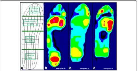
Pedobarographic data were obtained using the pedar-X system (novel GmbH, Munich, Germany), consisting of insoles holding 99 separate pressure sensors that operate at a frequency of 50 Hz. Peak pressure values (kPa, high-est values during each step and region) were obtained from 12 steps per foot during walking, following previ-ously published protocols (Arts & Bus, 2011). The sizes of the measurement insoles were adjusted individually based on each participant’s foot size. The plantar foot was subdivided into four anatomical regions (Westphal et al., 2016), representing the hindfoot (0–30% length, 0–100% width), midfoot (31–60% length, 0–100% width), metatarsal I-V (61–80% length, 0–100% width) and forefoot (81–100% length, 0–100% width) (Fig.1). A total of three trials, each with different devices, were performed. Measurements were taken indoors on a level surface, while walking speeds were kept constant at 3.5 km/h using a photo-barrier (Baur et al., 2018; Burnfield et al.,2004).
In advance of every trial, the volunteers performed a 10–15 min walk to become accustomed to each device. During the first trial the participants were asked to walk with a neutral shoe (Fuss und Schuh Breidbach® Inc., Fulda, Germany) (Fig. 2a) to define baseline values and equalize conditions of full weight-bearing. The shoe was established as a reference shoe for dynamic pedobaro-graphy (Kluger et al., 2014). It is composed of 4 mm polyethylene-vinyl acetate and has a heel pitch of 0 mm; elastic velcro buckles allow adjustment and fixation around the foot.
shoe consists of a wedge-designed sole with a 5° slope and measures approximately 5 cm at the highest point. The sole is made of polyethylene-vinyl acetate. The symmet-rical shoe can be used for both the left and right sides.
The third trial utilized an HRO (“Dr. Settner/Münch”®, Otto Bock Health Care Germany GmbH (OBHCD)) (Fig.2c). This orthosis is based on a modular system that allows a customized individual adaption. The relief zone is approximately 25–30% of the entire foot length. The HRO incorporates an outsole thickness of 1 cm height. It is based on a modular system and includes a further inner insert of approximately 4 cm peak height (peak heights are exemplary given for size “L”). For both de-vices, the size was adjusted individually based on the manufacturers’instructions.
During the second and the third trial a conventional available running shoe (The Faas 500, Puma Inc., Herzo-genaurach, Germany), categorised as a “neutral running shoe”, was applied at the contralateral side (Kluger et al.,
2014). According to the manufacturer this shoe has no pronation or supination support. For each participant only one foot was determined for data analyses (Vette et al.,2019; Gray et al.,2014).
Statistical analysis
For each participant kinetic data were computed as the mean peak pressure value for each specific region (mean value of each trial) using the novel multiprojects-ip soft-ware package (Novel GmbH, Munich, Germany). Within the defined specific region, the sensor with the highest
Fig. 1Demonstrating the subdivision of the plantar surface into four anatomical regions (a). Exemplary graphical illustration of mean peak pressure values assessed on the different devices (b-d);b: neutral shoe;c: hindfoot relief shoe;d: hindfoot relief orthosis
regarded as statistically significant.
Ethics approval and consent to participate
The local Ethics Committee approved the study with no requirements (Ref. No. 57_17 B; University of Erlangen-Nuremberg). All patients were informed re-garding the purpose, benefits and risks of the inves-tigation prior to signing an institutionally approved informed consent form to participate in the study.
Results
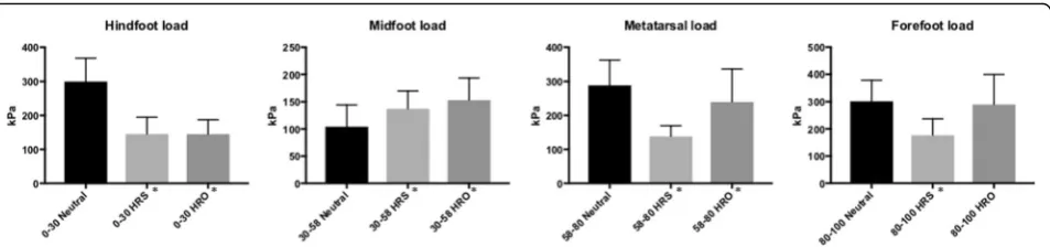
Descriptive results are listed in Table 1 and graphically illustrated in Figs.1and3.
Neutral shoe
Peak pressure values (mean ± SD) obtained in the neutral shoe were 300 ± 68 kPa under the hindfoot, 104 ± 40 kPa under the midfoot, 288 ± 74 kPa under the metatarsal zone and 302 ± 77 kPa under the forefoot. Concerning the entire foot, a pressure value of 346 ± 66 kPa was measured.
HRS and HRO Hindfoot
The HRS revealed 145 ± 50 kPa, indicating a statistically significant reduction in hindfoot peak pressure of 52% in comparison to the baseline value obtained in the neutral shoe (p< 0.001). The HRO showed a reduction in hind-foot peak pressure of 52% (145 ± 43 kPa) which was also significantly different from the baseline (p< 0.001). The HRO and HRS peak pressures were not significantly different (p= 0.960).
Metatarsal zone peak pressures were significantly lower with the HRS as well with the HRO (138 ± 32 kPa; p< 0.001 and 240 ± 96 kPa; p= 0.034, respectively). The comparison between the HRO and the HRS revealed a significant reduction for the HRS compared with the HRO (p< 0.001).
Forefoot
Regarding the forefoot, the HRS had a significant reduc-tion in peak pressure to 59% of the baseline value (177 ± 60 kPa;p< 0.001). The HRO showed 96% of the base-line value (290 ± 110 kPa), which was not significantly different from the baseline;p= 0.692); HRO values were significantly different from those of HRS (p< 0.001).
Discussion
Despite the wide use of HRS and HRO in clinical practice, there is a paucity of data representing biomech-anical changes of plantar pressure distribution using commonly applied offloading devices, and outcomes are even less often investigated. It is hypothesized that the clinical effects of hindfoot relief orthoses are based on offloading effects to the plantar tissue (Hahn et al.,
2014). Nevertheless, to date, no study has compared such biomechanical tissue responses between HRO and HRS. Offloading effects of forefoot relief devices are already benchmarked and well-studied and have helped to transfer biomechanical principles to clinical implica-tions (Bus et al.,2016; Cavanagh & Bus,2011; Bus et al.,
2009). To our knowledge, the present study is the first to assess plantar pressure distributions via dynamic ped-obarography in hindfoot relief devices of various designs comparing data to conditions of full weight-bearing. Moreover, for the first time we demonstrated a hindfoot peak pressure reduction with an HRO.
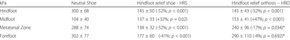
Table 1Absolute peak pressure values in kPa (mean ± SD) for the described anatomical regions and percentage changes compared to baseline
kPa Neutral Shoe Hindfoot relief shoe - HRS Hindfoot relief orthosis–HRO
Hindfoot 300 ± 68 145 ± 50 (-52%;p< 0.001) 145 ± 43 (-52%;p< 0.001)
Midfoot 104 ± 40 137 ± 33 (+32%;p= 0.02) 153 ± 41 (+47%;p< 0.001)
Metatarsal Zone 288 ± 74 138 ± 32 (-52%;p< 0.001) 240 ± 96 (-17%;p= 0.034)*
Forefoot 302 ± 77 177 ± 60 (-41%;p< 0.001) 290 ± 110 (-4%;p= 0.692)*
Our study revealed several main findings. First, we demonstrated significant offloading effects for the hind-foot area, and second, we observed significantly elevated peak pressures for the adjacent midfoot region both for the HRO and HRS.
Surprisingly, our study demonstrated that there were no significant differences among hindfoot relief devices of different designs. The decrease in the plantar pressure to the hindfoot that was observed for the HRS may be explained by the midsole concept that represents a 5° sloped, wedge-designed hindfoot relief zone. Offloading effects of the HRO may be achieved by the lever-type shaft construction. Thus, both devices display funda-mentally different offloading concepts. Nevertheless, their offloading effects were nearly similar (p> 0.05), and we cannot recommend one or the other of these devices based on the offloading effects. However, there were significant differences corresponding to the metatarsal and forefoot region. Our results showed significantly higher peak pressure reductions in the metatarsal and forefoot region for the HRS (−52% and−41%) com-pared to the HRO (−17% and−4%). Nevertheless, no device displayed elevated values in comparison to baseline. Hindfoot peak pressure reductions of nearly 50% of the baseline obtained in this study were com-parable to those of a previous investigation by Hahn et al., who evaluated different types of HRSs (Hahn et al., 2014). The authors reported a decrease of hind-foot load of 90% (0–15% of sole) and 18% (15–30% of sole) for the devices used in the study (Hahn et al., 2014). A weakness of that study was that HROs were not included. With respect to the offloading effects of forefoot relief shoes (FRS), peak pressure re-ductions of 38 to 58% have been reported (Bus et al.,
2009). Previous studies and reviews by Bus et al. have already provided evidence of forefoot offloading con-cepts concerning ulcer prevention (Bus & Valk, 2008; Bus, 2016). Based on our results we confirmed the significant peak pressure reduction using an HRS ob-served by Hahn et al. with comparable values (Hahn et al., 2014).
Clinical implications
Although limited weight-bearing is often required by sur-geons’specifications, there have been no evidence-based rehabilitation guidelines that determine exact values of weight-bearing graduations in accordance with operative or non-operative interventions (Wild et al., 2016). In rehabilitation after lower limb surgery there is a lack of unified, evidence-based rehabilitation concepts (Pfeifer et al.,2015). However, if total offloading to the hindfoot is required, our results indicate that neither HROs nor HRSs are able to alleviate plantar pressure at all, as 50% of the baseline must be considered. Furthermore, increased mid-foot load must be cautiously considered. Our data indi-cated significant peak pressure increases at the midfoot region while using hindfoot relief devices. Similar pressure shifts have been described for forefoot relief devices (Mueller et al.,2016; Cousins et al.,2013; Birtane & Tuna,
2004). Regarding the HRS (München shoe), Hahn et al. reported only a 5% increase, whereas our study demon-strated a significant 32% increase for the midfoot region. An increased midfoot vulnerability using orthotic devices was already reported for an ankle-foot orthosis (Vacoped®) by Pauser et al. (Pauser et al.,2012). Based on the existing investigations identifying increasing plantar pressure in the midfoot, as in our findings, the midfoot area appears to be a sensitive area for adapting increasing foot loads (Hotfiel et al., 2017). Regarding the localization of stress fractures, to which peak pressure is a commonly accepted risk factor, the midfoot area displayed the highest inci-dence in contrast to the tarsal bone, sesamoid or toe phal-anx (Hotfiel et al., 2017). In particular, patients with accompanying midfoot injuries or neuropathic or diabetic diseases should undergo a regular clinical examination to avoid further damage.
Interestingly our results offered differences in peak pressure patterns between the HRS and HRO in regard to the metatarsal and forefoot region. Hence clinical implications should be considered here as well. The significant lower peak pressure using the HRS could be relevant for patients with simultaneous complaints to the metatarsal or forefoot regions.
modifications are useful to compensate for elevated mid-foot loads particularly in hindmid-foot relief devices.
Pedobarography
Dynamic pedobarography was chosen for the assessment of foot loading because it has been established as a use-ful adjunct to clinical research for the recognition of plantar pressure conditions (Baur et al.,2018; Westphal et al., 2016; Hahni et al., 2016; Mehlhorn et al., 2017). Hindfoot weight-bearing was defined as a limitation of loads on the plantar surface, assessed by dynamic pedo-barography. Although this definition is widely accepted, there is no clear-cut evidence that foot load actually is a sufficient surrogate parameter for weight-bearing condi-tions in regard to intraosseous or intraarticular loading (Wild et al.,2016; Schaefer et al.,2015). We decided to assess peak pressure in accordance with the vast major-ity of previous investigations evaluating plantar loading under various conditions (Hotfiel et al.,2017).
Study limitations
This study has few limitations. First our results do not allow statements regarding estimation of gait stability or comfort while using the orthotic devices. Pain or dis-comfort could be a trigger for unintentional overload of the contralateral foot. In this context, we did not assess kinematic data of the hip, knee and ankle to observe in which position of the gait cycle peak pressures develop. Altered biomechanics of the limb may play a role in the change of foot loading. Second, our study was comprised of healthy participants and not patients. When designing the study, we could not rule out the possibility that some settings exceeded a certain limitation of weight bearing, and patients might have been jeopardized. How-ever, future investigations including selected patients (in-juries as well as pre-existing disabilities), are needed to confirm findings which were obtained in this study. In these studies further functional kinetic parameters (i.e. normal impulse-based measures (Vette et al., 2019)), that may provide differential information on loading should be implemented too.
Conclusions
Taken together, our results suggest that hindfoot relief shoes and orthoses significantly decrease plantar peak
The present work was performed in fulfilment of the requirements for obtaining the degree of Doctor of Medicine.
Funding Not applicable.
Availability of data and materials
The datasets used and/or analysed during the current study are available from the corresponding author on reasonable request.
Authors’contributions
TH, HDC and BS designed the study. FM and TH performed the data acquisition. FM, TH, CG, CL and MH interpreted the data. FM, TH, MH, CG, ME and CL have made major contributions in drafting and writing the manuscript. All authors read and approved the final manuscript.”
Ethics approval and consent to participate
The local Ethics Committee approved the study with no requirements (Ref. No. 57_17 B; University of Erlangen-Nuremberg). All patients were informed regarding the purpose, benefits and risks of the investigation prior to signing an institutionally approved informed consent form to participate in the study.
Consent for publication Not applicable
Competing interests
The authors declare that they have no competing interests.
Publisher’s Note
Springer Nature remains neutral with regard to jurisdictional claims in published maps and institutional affiliations.
Author details 1
Division of Orthopaedic Rheumatology, Department of Orthopaedic Surgery, Friedrich-Alexander-University Erlangen-Nuremberg, Rathsberger Str. 57, D-91054 Erlangen, Germany.2Department of Orthopaedic and Trauma
Surgery, Martha-Maria Hospital, Nuremberg, Germany.3Department of
Orthopaedic and Trauma Surgery, Sports Orthopaedics and Sports Medicine, Klinikum Bamberg, Bamberg, Germany.4Department of Orthopaedics,
Trauma and Hand Surgery, Klinikum Osnabrück, Osnabrück, Germany.
5Department of Movement and Training Science, University of Wuppertal,
Wuppertal, Germany.
Received: 18 October 2018 Accepted: 17 January 2019
References
Arts MLJ, Bus SA. Twelve steps per foot are recommended for valid and reliable in-shoe plantar pressure data in neuropathic diabetic patients wearing custom made footwear. Clinical Biomechanics (Bristol, Avon). [online] Department of Rehabilitation, Academic Medical Center, University of Amsterdam, the Netherlands.M.L.Arts@amc.uva.nl: Elsevier science; 2011; 26(8): 880–884. Available from: doi:https://doi.org/10.1016/j.clinbiomech.2011. 05.001
Birtane M, Tuna H (2004) The evaluation of plantar pressure distribution in obese and non-obese adults. Clin Biomech (Bristol, Avon). [Online] Great Britain: Elsevier Science B.V., Amsterdam.; (10):1055 Available from:https://www. clinbiomech.com/article/S0268-0033(04)00162-7/fulltext.https://doi.org/10. 1016/j.clinbiomech.2004.07.008
Bohl DD, Ondeck NT, Samuel AM, Diaz-Collado PJ, Nelson SJ, Basques BA, et al. Demographics, Mechanisms of Injury, and Concurrent Injuries Associated With Calcaneus Fractures: A Study of 14 516 Patients in the American College of Surgeons National Trauma Data Bank. Foot & Ankle Specialist. [Online] Department of Orthopaedic Surgery, Rush University Medical Center, Chicago, Illinois (DDB, BAB).; Department of Orthopaedics and Rehabilitation, Yale School of Medicine, New Haven, Connecticut (NTO, AMS, PJDC, SJN, MPL, JNG).: Sage Publications; 2017;10(5): 402–410. Available from: doi:https:// doi.org/10.1177/1938640016679703
Burnfield JM, Few CD, Mohamed OS, Perry J. The influence of walking speed and footwear on plantar pressures in older adults. Clinical Biomechanics. 2004;19: 78–84. Available from:https://doi.org/10.1016/j.clinbiomech.2003.09.007 Bus SA. The Role of Pressure Offloading on Diabetic Foot Ulcer Healing and Prevention of Recurrence. Plastic and reconstructive surgery. 2016;138(3 Suppl): 179S–187S. Available from: doi:https://doi.org/10.1097/PRS. 0000000000002686
Bus SA, Valk GD (2008) The effectiveness of footwear and offloading
interventions to prevent and heal foot ulcers and reduce plantar pressure in diabetes : a systematic review. Available from 24(October 2007):162–180. https://doi.org/10.1002/dmrr
Bus SA, van Deursen RW, Armstrong DG, Lewis JEA, Caravaggi CF, Cavanagh PR. Footwear and offloading interventions to prevent and heal foot ulcers and reduce plantar pressure in patients with diabetes: a systematic review. Diabetes Metab Res Rev. [online] Department of Rehabilitation Medicine, academic medical Centre, University of Amsterdam, Amsterdam, the Netherlands.: Wiley-Blackwell; 2016;32 Suppl 1: 99–118. Available from: doi: https://doi.org/10.1002/dmrr.2702
Bus SA, van Deursen RWM, Kanade RV, Wissink M, Manning EA, van Baal JG et al (2009) Plantar pressure relief in the diabetic foot using forefoot offloading shoes. Gait & Posture 29:618–622. Available from.https://doi.org/10.1016/j. gaitpost.2009.01.003
Carl H-D, Pfander D, Swoboda B. Assessment of plantar pressure in forefoot relief shoes of different designs. Foot & Ankle International. [Online] Division of Orthopaedic Rheumatology, Department of Orthopaedic Surgery, University of Erlangen-Nuremberg, Rathsbergerstrasse 57, D-91054 Erlangen, Germany. Hans-Dieter.Carl@ortho-rheuma.med.uni-erlangen.de: Sage Publications; 2006; 27(2): 117–120. Available from:https://journals.sagepub.com/doi/abs/10.1177/ 107110070602700208?journalCode=faib.https://doi.org/10.1177/
107110070602700208
Cavanagh PR, Bus SA (2011) Off-Loading the Diabetic Foot for Ulcer Prevention and Healing. Plastic & Reconstructive Surgery 127:248S Available from:https://www.jvascsurg.org/article/S0741-5214(10)01328-5/ fulltext.https://doi.org/10.1016/j.jvs.2010.06.007
Claes LE, Heigele CA. Magnitudes of local stress and strain along bony surfaces predict the course and type of fracture healing. Journal Of Biomechanics. [Online] Department Unfallchirurgische Forschung und Biomechanik, University of Ulm, Germany. claes@sirius.medizin.uni-ulm.de: Elsevier Science; 1999;32(3): 255–266. Available from:https://www.sciencedirect.com/science/ article/pii/S0021929098001535.https://doi.org/10.1016/S0021-9290(98)00153-5 Cousins SD, Morrison SC, Drechsler WI. Foot loading patterns in normal
weight, overweight and obese children aged 7 to 11 years. Journal Of Foot And Ankle Research. [online] School of Health, sport and bioscience, University of East London, Stratford, London, England.S.C. Morrison@uel.ac.uk.: BioMed central; 2013;6(1): 36. Available from: doi: https://doi.org/10.1186/1757-1146-6-36
da Conceição CS, Gomes Neto M, Mendes SMD, Sá KN, Baptista AF (2015) Systematic review and meta-analysis of effects of foot orthoses on pain and disability in rheumatoid arthritis patients. Disability and Rehabilitation. 37(14): 1209–1213. Available from.https://doi.org/10.3109/09638288.2014.961654 Genc Y, Gultekin A, Duymus TM, Mutlu S, Mutlu H, Komur B (2016) Original
research: Pedobarography in the assessment of postoperative calcaneal fracture pressure with gait. The Journal of Foot and Ankle Surgery 55:99–105. Available from.https://doi.org/10.1053/j.jfas.2015.07.018
Götz J, Grifka J, Baier C. [Hindfoot deformities in adults. Conservative and surgical treatment]. Der Orthopade. [Online] Orthopädische Universitätsklinik Regensburg im Asklepios-Klinikum Bad Abbach, Kaiser-Karl-V. Allee 3, 93077,
Bad Abbach, Deutschland.juergen-goetz@gmx.de.: Springer-Verlag; 2016; 45(1): 97–108. Available from: doi:https://doi.org/10.1007/s00132-015-3203-z Gray K, Gibbons P, Little D, Burns J (2014) Bilateral clubfeet are highly correlated:
a cautionary tale for researchers. Clinical orthopaedics and related research 472(11):3517–3522. Available from.https://doi.org/10.1007/s11999-014-3776-6 Groot R, De FAJ, Schepers T, Roerdink WH (2013) Complications following the
extended lateral approach for calcaneal fractures do not influence mid- to long-term outcome. Injury 44(11):1596–1600. Available from.https://doi.org/ 10.1016/j.injury.2013.06.014
Hahn T, Carl H-D, Jendrissek A, Brem M, Swoboda B, Rummel P, et al. Assessment of plantar pressure in Hindfoot relief shoes of different designs. J. Am. Podiatr. Med. Assoc.. [online] American podiatric medical association, Inc.; 2014;104(1): 19–23. Available from: doi:https://doi.org/10. 7547/0003-0538-104.1.19
Hahni M, Hirschmuller A, Baur H (2016) The effect of foot orthoses with forefoot cushioning or metatarsal pad on forefoot peak plantar pressure in running. Journal of foot and ankle research 9:44. Available from.https://doi.org/10. 1186/s13047-016-0176-z
Hodge MC, Bach TM, Carter GM (1999) Novel award first prize paper: orthotic management of plantar pressure and pain in rheumatoid arthritis. Clinical Biomechanics 14:567–575. Available from. https://doi.org/10.1016/S0268-0033(99)00034-0
Hotfiel T, Carl HD, Wendler F, Jendrissek A, Heiss R, Swoboda B. Plantar pressures increase with raising body weight: a standardised approach with paired sample using neutral shoes. J. Back Musculoskelet. Rehabil.. 2017;30(3): 583– 589. Available from: doi:https://doi.org/10.3233/BMR-150442
Hunt G, C, Fromherz WA, Gerber LH, Hurwitz SR (1987) Hindfoot pain treated by a leg-Hindfoot orthosis: a case report. Physical Therapy 67(9). Available from). https://doi.org/10.1093/ptj/67.9.1384
Kluger AK, Carl H-D, Jendrissek A, Swoboda B, Hotfiel T. Introduction of a neutral shoe to assess reference values for dynamic pedobarography.
Biomedizinische Technik / Biomedical Engineering VO - 59. [online] Walter de Gruyter GmbH & co. KG; 2014;(3): 213. Available from: doi:https://doi.org/ 10.1515/bmt-2013-0078
Kraus TM, Graf F, Mitternacht J, Döbele S, Stöckle U, Siebenlist S (2014) Vacuum shoe system vs.forefoot offloading shoe for the management of metatarsal fractures. A prospective, randomized trial. MMW Fortschritte Der Medizin 156(Suppl : 1):1–17 Available from:https://link.springer.com/article/10. 1007%2Fs15006-014-2877-1
Lorkowski J, Grzegorowska O, Kotela I. [The use of Pedobarographic examination to biomechanical evaluation of foot and ankle joint in adult - own experience]. Ortopedia, Traumatologia, Rehabilitacja. [online] Klinika Ortopedii i Traumatologii, Centralny Szpital Kliniczny MSW w Warszawie, Polska.: Medsport press; 2015;17(2): 207–213. Available from: doi:https://doi.org/10.5604/15093492.1157136 Mehlhorn AT, Walther M, Yilmaz T, Gunst L, Hirschmuller A, Sudkamp NP et al
(2017) Dynamic plantar pressure distribution, strength capacity and postural control after Lisfranc fracture-dislocation. Gait & posture 52:332–337. Available from.https://doi.org/10.1016/j.gaitpost.2016.11.043
Mueller S, Carlsohn A, Mueller J, Baur H, Mayer F. Influence of obesity on foot loading characteristics in gait for children aged 1 to 12 years. Plos One. [online] university outpatient clinic, Sports Medicine & Sports Orthopaedics, University of Potsdam, Potsdam, Germany.: public library of science; 2016;11(2): e0149924–e0149924. Available from: doi:https://doi. org/10.1371/journal.pone.0149924
Pauser J, Carl H-D, Swoboda B, Jendrissek KA. [Insufficiency fractures of the feet and lower limbs in rheumatoid arthritis]. Zeitschrift Fur
Rheumatologie. [Online] Abteilung für Orthopädische Rheumatologie, Friedrich-Alexander-Universität Erlangen-Nürnberg, Im Waldkrankenhaus St. Marien, Rathsberger Str. 57, 91054, Erlangen, Deutschland. johannespauser@web.de: Springer; 2011;70(10): 866–873. Available from: doi:https://doi.org/10.1007/s00393-011-0889-0
Pauser J, Jendrissek A, Brem M, Gelse K, Swoboda B, Carl H (2012) Foot loading with an ankle-foot orthosis : the accuracy of an integrated physical strain trainer:1411–1415. Available from.https://doi.org/10.1007/s00264-012-1501-1 Pfeifer CG, Grechenig S, Frankewycz B, Ernstberger A, Nerlich M, Krutsch W (2015)
Analysis of 213 currently used rehabilitation protocols in foot and ankle fractures. Injury 46(Suppl 4):S51–S57. Available from.https://doi.org/10.1016/ S0020-1383(15)30018-8
Sarajevo, Sarajevo, Bosnia and Herzegovina.: Academy of Medical Sciences of Bosnia and Herzegovina; 2014;22(6): 374–378. Available from: doi:https://doi.org/10.5455/aim.2014.22.374-378
Vette AH, Funabashi M, Lewicke J, Watkins B, Prowse M, Harding G et al (2019) Functional, impulse-based quantification of plantar pressure patterns in typical adult gait. Gait & posture 67:122–127. Available from.https://doi.org/ 10.1016/j.gaitpost.2018.09.029
Westphal E, Carl H-D, Krinner S, Grim C, Swoboda B, Hotfiel T. Plantar force deviations in dynamic pedobarography - the role of insole and platform based systems as influencing factors. Sports Orthopaedics and Traumatology VO - 32. 2016;(4): 380. Available from: doi:https://doi.org/ 10.1016/j.orthtr.2016.10.007