O R I G I N A L A R T I C L E
Open Access
Forensic evaluation of sex estimation via
measurements of adult index and ring
finger lengths using postmortem
computed tomography
Tomoya Ikeda
1,2*, Kazunori Miyamoto
2, Naoto Tani
1,2, Shigeki Oritani
1, Tomomi Michiue
1,2, Fumiya Morioka
1and Takaki Ishikawa
1,2Abstract
Background:Sex estimation from fragmented or isolated human bones found during excavations is imperative not
only in the field of forensics but also paleoanthropology. This study investigated the possibility of sex estimation from computed tomography (CT) data for the lengths of the index and ring fingers. The scans were obtained using a multislice ECLOS-16 CT scanner (Hitachi Medical Co., Tokyo, Japan), and the images were analyzed using a SYNAPSE VINCENT volume analyzer (Fujifilm Medical Co., Tokyo, Japan). For 205 cases, the authors measured the total length of the distal, middle, and proximal phalanges (P) and of the metacarpal bones for the index and ring fingers of both hands. Right heart serum testosterone and estradiol levels in 92 cases were also measured by an electrochemiluminescence immunoassay and a chemiluminescence immunoassay, respectively.
Result:The difference in P between the index and ring fingers was significantly greater in men than in women for both hands; receiver operating characteristic analysis showed an optimal cutoff of 6.0 mm (sensitivity, 0.64 and specificity, 0.64). Blood testosterone levels were moderately correlated with this difference.
Conclusion:A value of 6.0 mm for the ring finger–index finger difference for the left hand distinguished men from women, and these results are affected by testosterone levels. The findings from this report in the field of forensics and paleoanthropology indicated that the CT data-assessed morphometry of the phalanges could be used as an objective index for sex estimation.
Keywords:Forensic science, Forensic anthropology, Sex estimation, Computed tomography, Index finger length,
Ring finger length, Japanese population
Background
Forensic anthropology, along with DNA analyses of sex and genetic polymorphisms, is essential for the estimation of sex, age, and stature in the identification of human remains and single bones (Aldegheri and Agostini1993; Duyar and Pelin 2003; Ozaslan et al. 2003; Petrovecki et al. 2007; Zeybek et al. 2008). Moreover, paleoanthropology enables the estimation of sex from a bone fragment and/or isolated human bones from excavations (Alarcon et al. 2016;
Slizewski et al. 2013). However, one of the greatest limita-tions to the application of the anatomical sex estimation method is its inability to evaluate some of the skeletal elements required for the calculation. Indeed, a major chal-lenge in analyzing skeletal remains, particularly archaeo-logical samples, is the high incidence of missing and/or non-measurable elements (Auerbach 2011). Therefore, there are a number of factors that contribute to the vari-ability in the preservation and use of archaeological human remains for research. In this process, radiology is used to detect anatomical characteristics, specific bone pathologies, and foreign bodies (including surgical materials), as well as identifying sex-related differences and age-dependent * Correspondence:[email protected]
1Department of Legal Medicine, Osaka City University Medical School, Osaka 545-8585, Japan
2Forensic autopsy section, Medico-legal Consultation and Postmortem Investigation Support Center (MLCPI-SC), Osaka, Japan
changes, and providing measurements for stature estima-tion (Hasegawa et al. 2009; Rainio et al. 2001). However, until recently, it has been difficult to make such assess-ments from a single small bone from a cadaver, especially given that the bone may have been moved away from the remainder of the cadaver, such as by an animal or insects (Campobasso and Introna2001).There is therefore a lack of scientific data about small bones in the field of forensic medicine.
In addition to conventional radiology, computed tomo-graphy (CT) may be useful for the documentation and reconstruction of skeletal data in autopsy routines. Post-mortem imaging has been reported as useful for identifying individuals, and investigations are ongoing about the practi-cality of imaging various bones for the estimation of sex, height, and weight (Giurazza et al. 2012; Hishmat et al.
2014; Torimitsu et al. 2014a; Torimitsu et al. 2014b; Torimitsu et al.2015c). Several studies have demonstrated the successful application of CT to virtual bone measure-ment for estimating sex and stature (Djorojevic et al.2014; Hishmat et al. 2015; Inamori-Kawamoto et al. 2016; Macaluso and Lucena 2014; Rodriguez et al. 2014; Torimitsu et al.2015b; Torimitsu et al.2014c; Torimitsu et al.2015a; Verhoff et al.2008).
Previous studies in the forensic science field have re-ported osteometric data about index and ring finger lengths for sex estimation in different modern populations (Agnihotri et al.2015; Kanchan et al.2010; Kanchan and Pradeep Kumar 2010; Kanchan et al.2010; Krishan et al.
2013), but, as yet, such studies have not involved postmor-tem CT data. Furthermore, to the best of our knowledge, there have been no published studies in the forensic science field that have investigated the influence of sex hormones (such as androgen and estrogen) on the lengths of the index and ring fingers. In the present study, there-fore, the authors investigated the possibility of sex estima-tion from CT data about index and ring finger lengths, as well as the influence of sex hormones on finger length.
Materials and methods
Postmortem CT data.
Postmortem CT scans are performed at our institution immediately before autopsy as part of the scope of rou-tine casework such as personal identification, injury tact, and pathophysiological analysis. In this study, we used autopsy cases of post-adolescent Japanese subjects (age ≥20 years) of known sex, age, height, weight, and stat-ure, for which complete finger bone CT data were avail-able. Cases with advanced decomposition, evident fracture, destruction, or advanced osteoarthrosis were excluded. Cases were excluded if the details regarding sex and height could not be definitively verified due to postmortem destruction, and no case included in this study qualified for exclusion. Samples for 205 cases
(102 men and 103 women; age range, 20–95 years; me-dian age, 60 years) were collected during a 5-year period from July 2011 to September 2015 (Table1).
Postmortem interval, defined as the time elapsed from estimated time of death to autopsy ranged from 10 to 370 h (median, 33 h) and was calculated based on autopsy findings, case history, and circumstantial evidence; thus, possible error in postmortem interval ranged up to hours depending on elapsed time in cases of unwitnessed deaths. Necessary data were extracted from autopsy documents, pathological findings, and circumstantial evidence.
Whole-body PMCTs were routinely performed immedi-ately before forensic autopsy using a 16-row multidetector CT scanner (ECLOS, Hitachi Medical Co., Tokyo, Japan) as part of routine casework. Spiral CT was performed using the following conditions: 120 kV; 250 mA; 1.25-pitch factor, 2.5 × 4 mm collimation, and 1.0 × 1.25 mm thickness; field of view, 500 mm. A CT data ana-lysis system, volume analyzer Synapse Vincent (Fujifilm Medical Co., Ltd., Tokyo, Japan), was used to reconstruct three-dimensional (3D) images of the finger bone in situ for virtual length measurement which was extracted by manual cursoring. These analyses were performed by two forensic pathologists and one radiographer. The reprodu-cibility of virtual reconstruction and measurement was checked by two independent observers. CT attenuation [Hounsfield units (HUs)] was performed using the corre-lation of HU with bone-specific length.
Measurements.
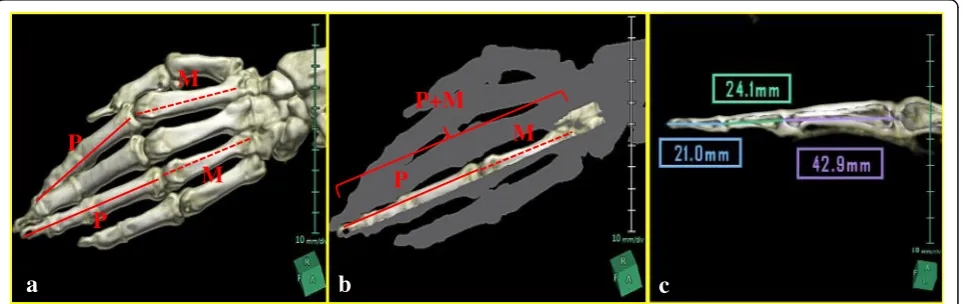
For each finger (left and right index and ring fingers), the authors measured the following: P, the length from the bottom prominence to the tip of the distal phalanx (i.e., the total length of the distal, middle, and proximal phalanges);M, the length of the metacarpal bones in the longitudinal direction; and P+M, the total of lengths
P and M (Fig. 1a-c). Moreover, the differences in P, M and P+M between the finger (R) and index finger (I) were calculated for each hand (R–I). The analysis tested differences between the sexes for all these lengths and their correlations with the levels of sex hormones testos-terone and estradiol.
Biochemical analyses.
(median 275 pg/ml) for the women (except for any that had been pregnant at the time of death.
Toxicological analyses.
Blood carboxyhemoglobin concentrations for the fire fa-talities were determined using a CO-oximeter system (Maeda et al. 1997; Maeda et al. 1996). Drugs, such as an amphetamine and antipsychotropics, were detected by gas chromatography/mass spectrometry.
Statistical analyses.
The statistical analyses were performed using Microsoft Excel and SPSS (version 17.0; SPSS, Inc., Chicago, IL, USA). The Kruskal–Wallis test, Bland–Altman plots, Pearson’s correlation analysis, and Mann–Whitney U tests were used, as appropriate, for nonparametric com-parisons of two datasets and multiple group analyses of all examination items. Linear regression analysis was used to estimate stature based on the individual bone
parameters. In these analyses, p–values < 0.05 were con-sidered statistically significant. Receiver operating char-acteristic analysis was used to establish cutoff points for each individual bone parameter for the optimal estima-tion of sex (Vexler et al. 2008); cases above and below the cutoff values were deemed male and female, respect-ively. The accuracy of sex estimation using these cutoff values was examined by dividing the number of cases identified by the total number of cases.
Results
Relationships between bone lengths and general characteristics
There was no significant correlation between the lengths of the index or ring fingers and postmortem time (p> 0.05). In detail, the authors compared the postmortem period with under more than two days (≤ 48 h: n= 150, > 48 h: n= 55), there was no statis-tical difference (left index finger: p= 0.3098, left ring Table 1Case profiles
Cause of death n (Male / Female)
Age (years) Height (cm) Weight (kg) Postmortem time (h)
Median Min Max Median Min Max Median Min Max Median Min Max
Blunt injury 59 (35 / 24) 62 20 93 161 138 180 54.5 30.2 105 34 14 145
Sharp instrument injury 13 (7 / 6) 55 38 86 160 150 169 61.5 31.9 86.2 28 10 41
Drowning 11 (6 / 5) 51 33 85 160 144 172 56.1 34.4 67.5 36 17 206
Fire fatalities 2 (1 / 1) 55 65 158 168 52.5 75.7 17 24
Asphyxia 22 (10 / 12) 49 21 86 164 143 174 54.6 34.6 73.5 33 14 301
Hypothermia 5 (1 / 5) 83 47 87 149 140 159 39.9 30.3 55.7 39 26 110
Hyperthermia 21 (8 / 13) 71 28 95 157 136 170 46.4 23.6 72.4 33 15 109
Sudden cardiac death 18 (10 / 8) 69 29 84 161 145 181 54.3 43.2 82.3 35 16 181
Other natural death 54 (24 / 30) 51 24 93 160 140 179 53.6 27.5 99.8 32 10 370
Total 205 (102 / 103) 59 20 95 160 136 181 54.2 23.6 105 33 10 370
P
M
P+M
M
P
M
P
a
b
c
finger: p= 0.5318, right index finger: p= 0.3638, right ring finger p= 0.3186). Furthermore, about the same person (84 years, male, height 168 cm, body weight 59.6 Kg), as a result of having performed the measurement (24 h later) twice, there was not the difference that was slight to a peripheral bone (first time: 16.70 mm, second time 16.70 mm), middle phalanx (first time: 21.30 mm, second time 21.30 mm), proximal phalanx (first time: 40.10 mm, second time 40.1 mm). On the other hand, after having left the same person unattended posthu-mously for several months, it is not a permissible thing ethically to measure the length of the finger again. There-fore, the authors were not able to examine a post mortem change in detail.
P (the total length of the three phalanges) was moder-ately to strongly correlated with height (r= 0.439–0.545,
p< 0.0001) for the left and right index and ring fingers of both sexes. However,Pshowed no significant correlation with body mass index, bodyweight, or age (p> 0.05).
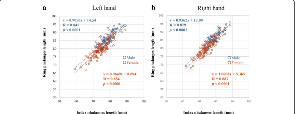
There were strong correlations between P for the ring and index fingers in both men (left: r= 0.847,p< 0.0001 and right:r= 0.879,p< 0.0001) and women (left:r= 0.854,
p< 0.0001 and right:r= 0.887,p< 0.0001; Fig.2).
M (the length of the metacarpal bones) was slightly to moderately correlated with height (r= 0.534–0.702,
p< 0.0001) and weight (r= 0.217–0.330, p< 0.05) for the left and right index and ring fingers of both sexes. In men, but not women, there was a slight negative correlation be-tweenM and age (left index,r= 0.260,p <0.01; left ring,
r= 0.310,p< 0.01 and right index,r= 0.289,p< 0.01; right ring,r= 0.246,p <0.05).
Relationships between bone lengths and sex
Values for P, M, and P+M for both sexes are given in Table2. Values ofPfor the left and right index and ring
fingers were significantly greater for the male than the female (left index, p< 0.0001; right index, p< 0.001; left ring, p< 0.0001; right ring, p< 0.0001). Similarly, values ofMfor the left and right index and ring fingers were sig-nificantly greater for the male than the female (left index,
p< 0.0001; right index, p< 0.0001; left ring, p< 0.0001; right ring, p< 0.0001). Moreover, P + M values for the male were significantly longer than for the female (left index, p< 0.0001 and right index, p< 0.0001; left ring,
p< 0.0001 and right ring: male, p< 0.0001).
Relationships between R−I differences and sex
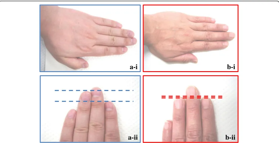
The differences inP between the ring and index fingers, R − I, are shown in Table 3. These differences were significantly greater for the male than for the female (left, p< 0.0001; right, p< 0.001). The presence of a sex difference in the left hand was clearer when we com-paredPbetween the right and left hands (Fig.3).
However, there were no significant differences between the sexes for the R–I difference inM(p> 0.05) orP + M (p> 0.05; Tables2and3).
Cutoff values to distinguish men from women
Figure 4 shows differences in phalangeal length (ring finger–index finger) in male and female. In the receiver operating characteristic analysis, the optimal cutoff value for distinguishing men from women was a value of 6.0 mm for the R–I difference in P for the left hand. This showed a sensitivity of 0.64 and a specificity of 0.64 (Fig. 4). There was statistical significant difference in right and left index finger of the male (p< 0.05), without other fingers.
The area under the curve (AUC) in this study was 0.673. Discriminant function equations is −0.0004 ×2– 50.795× + 317.91 = 0, The x-axis was a difference of the
b
a
length of the left finger, and the y-axis was a difference of the length of the right finger, and the plotted the whole data. Afterwards, the authors measured the differ-enced the male and female boundary line by the cluster analysis. As the results, it was D = 2579.62 (D > 0).
Relationships between finger lengths and levels of sex hormones
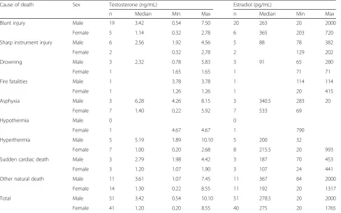
Measurements of testosterone levels in the right heart serum (postmortem period < 2 days) of 51 male and 41 female found significantly higher levels in the male than in the female (Table4). There was not the correlation of testosterone and finger length in the elderly male and fe-male both finger, respectively (> 70 years,p> 0.05).
There was a slight positive association between P of the left ring finger and serum testosterone in men (n= 51,
r= 0.34, p< 0.05; y = 0.1472×−8.8372, where x is length in mm and y is testosterone level in ng/ml). No significant associations with testosterone were found for M for the right index and ring fingers in men, forP + Min men, or for any finger lengths in women.
In the men, there was a slight correlation between serum testosterone and the R–I difference for P in the left hand (r= 0.29, p< 0.05) although there were differ-ences in P in the right hand. In the women, there were no correlations between testosterone levels and R–I dif-ferences inP(Fig.5).
Higher testosterone levels were detected in cases with a positive R–I difference in P + M(i.e., the total length of the ring finger was longer than that for the index finger).
Measurements of right heart serum estradiol levels in 51 male and 40 female showed no significant correlation with R–I differences inPfor either hand.
Discussion
The smaller index to ring finger (2D:4D) ratio has been considered as a ‘male finger pattern’ and is associated with blood testosterone levels (Aycinena et al. 2014, Robertson et al. 2008, Warrington et al.2018). However, there is no report that examined the sex estimation using this method in a forensic medicine field.
A lot of testosterone participating are reported in the length of the finger by sex differences so far in the field of embryology and/or life sciences. However, in the field of forensic medicine, there is no report considered about sex hormone and the length of the finger. This examin-ation is the first report that proved that the length of the finger with the sex differences depends on testosterone in the field of forensic medicine. In this study, one of the important points includes that we do not use the long bones such as a thigh bone or the humerus. Osteoar-throsis may develop in the long bone with aging accor-ding to the indication a reviewer. However, the transformation of the bone of the finger is not caused other than the special diseases such as rheumatism, amyotrophic lateral sclerosis (ALS) or the hyperpara-thyroidism (Kalichman et al.2018; Zhang W et al.2008, Parkin Kullmann and Pamphlett, 2017, Rhara et al. 2015). In this study, effective points with the using short bone such as the finger include that there is little osteoarthrosis.
This study found moderate-to-strong correlations be-tween height and the total lengths of the phalanges(P), the total lengths of the metacarpal bones (M), andP+M in the index fingers of both hands. TheP,M, andP+M lengths in the index and ring fingers of both hands were longer in male than in female, and the R–I differences in
Pin both hands were also greater in the male. However, Table 2Bone lengths
Finger Sex Left Hand Right hand
Phalanx (mm) Metacarpal (mm) Total length (mm) (Phalanx+Metacarpal)
Phalanx (mm) Metacarpal (mm) Total length (mm) (Phalanx+Metacarpal)
Med. Min Max Med. Min Max Med. Min Max Med. Min Max Med. Min Max Med. Min Max
Index finger Male 78.9 59.2 89.3 62.9 50.5 72.4 141.8 109.7 161.7 78.7 62.0 88.6 63.7 50.2 71.2 142.1 112.2 159.2
Female 74.5 62.6 83.1 60.4 51.0 69.2 134.4 116.0 148.2 74.4 65.1 84.1 60.3 50.5 67.3 134.6 115.6 149.3
Ring finger Male 85.6 67.5 95.4 55.4 42.7 62.9 140.7 110.2 157.4 85.5 69.2 95.8 54.7 42.9 61.9 140.0 112.1 157.7
Female 79.6 67.1 93.8 52.0 42.8 58.9 131.9 111.9 149.1 80.2 67.9 92.2 51.8 44.7 61.2 132.5 113.3 152.7
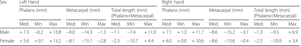
Table 3Difference in lengths (ring finger–index finger)
Sex Left Hand Right hand
Phalanx (mm) Metacarpal (mm) Total length (mm) (Phalanx+Metacarpal)
Phalanx (mm) Metacarpal (mm) Total length (mm) (Phalanx+Metacarpal)
Med. Min Max Med. Min Max Med. Min Max Med. Min Max Med. Min Max Med. Min Max
Male + 7.3 −0.2 + 13.8 −8.0 −14.3 −1.3 −1.1 −7.4 + 11.0 + 7.1 + 1.3 + 11.7 −8.6 −15.2 −3.1 −1.3 −9.5 + 6.9
results indicated that this difference did not differ in females. This result held even for cases with a long post-mortem time. Thus, the authors concluded that calcula-ting the R–I difference for P in the ring finger is potentially useful for sex prediction in complicated fo-rensic work in a variety of situations. In this study, the optimal cutoff value for distinguishing men from women based on the R–I difference for P was estimated to be 6.0 mm. However, our studies showed a sensitivity 0.64, specificity 0.64, and AUC 0.673. Thus, it was not able to
make refer to that the expected values of this study were high.
Generally, image analysis inherently low accuracy levels in contrast to DNA analysis that provides high accuracy for sex estimation. In a previous study, an accuracy of 61.7%- 87.1% was obtained using the calcaneus and talus as materials for sex estimation (Inamori-Kawamoto et al. 2016). Furthermore, sensi-tivity and specificity of 0.85 and 0.92, respectively, was achieved when the authors used the human
a-i
a-ii
b-i
b-ii
Fig. 3Relationship between the difference in ring and index finger lengths and the sex of the individual.a-ianda-ii: The figures show the length of the finger of the male.b-iandb-ii: The figures show the length of the finger of the female
patella for sex estimation (Michiue et al. 2017). How-ever, image analysis does offer some advantages for human sex estimation. These include its utility for analyzing highly decomposed bodies (Kranioti et al.
2009) and bone fragments and/or isolated bones (Croker et al. 2016), and it can easily be updated new
data on evolution and/or degeneration (Memarian et al. 2017) .
Previous studies have suggested that testosterone in-fluenced differences in ring finger and index finger lengths, and that this would be expected to apply to a wide range of circumstances (Bijleveld and Baalbergen Table 4Serum concentrations of testosterone and estradiol in the right heart
Cause of death Sex Testosterone (ng/mL) Estradiol (pg/mL)
n Median Min Max n Median Min Max
Blunt injury Male 19 3.42 0.54 7.50 20 263 20 2000
Female 5 1.14 0.32 2.78 6 365 203 720
Sharp instrument injury Male 6 2.56 1.92 4.56 5 88 78 382
Female 2 0.32 2.78 2 129 202
Drowning Male 3 2.32 0.78 5.83 3 91 65 280
Female 1 1.65 1.65 1 71 71
Fire fatalities Male 1 3.78 3.78 1 114 114
Female 1 1.26 1.26 1 20 415
Asphyxia Male 3 6.28 4.26 8.15 3 340.5 283 20
Female 7 1.40 0.22 5.92 7 533 69
Hypothermia Male 0 0
Female 1 4.67 4.67 1 790
Hyperthermia Male 5 5.19 1.89 10.10 5 200 32
Female 7 1.00 0.20 2.68 8 215.5 20 993
Sudden cardiac death Male 3 2.79 1.98 4.42 3 187 70 453
Female 3 1.20 1.07 1.90 3 107 24 441
Other natural death Male 11 3.61 1.07 7.45 11 367 84 2000
Female 14 1.30 0.22 8.55 11 192 20 1317
Total Male 51 3.42 0.54 10.10 51 278.5 20 2000
Female 41 1.20 0.20 8.55 40 275 20 1765
b
a
2017; Parkin Kullmann and Pamphlett, 2017). In the present study, measurements of right heart serum levels of testosterone levels in 92 cases of men and women showed higher levels in the men who had longer ring than index finger phalanx lengths.
There was no correlation between finger bone lengths and age. However, except for elderly persons, there were associated with increase in testosterone concentration and the finger lengths. Moreover, a relationship between serum testosterone and P for the left hand was also found although this did not apply to the right hand. The reason for this difference between the hands is unclear, but it may reflect the degree of work performed by the fingers due to handedness, or the possibility that a de-crease in age-related testosterone influences is supposed. There were no significant correlations between finger lengths and blood estradiol levels. The correlation be-tween finger length and testosterone levels decreases as individuals become elderly. An explanation for the lack of correlation between finger length and testosterone levels in women is that testosterone may be masked by estrogen in premenopausal women (Matsui et al. 2013) although this masking decreases after the menopause, when testosterone levels have been reported to increase (Matsui et al.2013) (Burger et al.2002).
Conclusion
A value of 6.0 mm for the ring finger–index finger dif-ference for the left hand distinguished men from women, and these results are affected by testosterone levels. The findings from this report in the field of foren-sics and paleoanthropology indicated that the CT data-assessed morphometry of the phalanges could be used as an objective index for sex estimation.
Abbreviations
CT:Computed tomography;I: The index finger;M: The length of the metacarpal bones in the longitudinal direction;P+M: The total of lengthsP andM;P: The length from the bottom prominence to the tip of the distal phalanx;R: The ring finger
Acknowledgements
Not applicable.
Ethical Approval and Consent to participate.
There are no ethical approval and consent to participate. The need for approval was waived.
Competing interests.
There are no financial competing interests in our study.
Authors’Contribution.
T. Ikeda, T. Michiue, and T. Ishikawa analyzed the data and drafted the manuscript. K. Miyamoto, S. Oritani, and T. Ishikawa designed the study, and directed implementation and data collection. N. Tani, and K. Miyamoto collected the data, and K. Miyamoto provided necessary logistical support. Endnotes.
We are pleased to submit the above manuscript for consideration for publication as an original research article in Egyptian Journal of Forensic Sciences.
We believe that the results of this study should provide valuable information for your readers.
The manuscript has not been published elsewhere and is not being considered for publication in another journal. All authors have read and approved submission of the manuscript.
We look forward to learning the results of peer review in due course.
Funding
There are no funding in our study.
Availability of data and materials.
Our data can be found in the main paper.
Consent for publication.
Not applicable.
Publisher’s Note
Springer Nature remains neutral with regard to jurisdictional claims in published maps and institutional affiliations.
Received: 11 January 2018 Accepted: 10 July 2018
References
Agnihotri AK, Jowaheer AA, Soodeen-Lalloo AK (2015) Sexual dimorphism in inger length ratios and sex determination - a study in indo-Mauritian population. J Forensic Legal Med 35:45–50
Alarcon JA, Bastir M, Rosas A (2016) Variation of mandibular sexual dimorphism across human facial patterns. Homo 67:188–202
Aldegheri R, Agostini S (1993) A chart of anthropometric values. J Bone Joint Surg Br 75:86–88
Auerbach BM (2011) Methods for estimating missing human skeletal element osteometric dimensions employed in the revised fully technique for estimating stature. Am J Phys Anthropol 145:67–80
Aycinena D, Baltaduonis R, Rentschler L (2014) Risk preferences and prenatal exposure to sex hormones for ladinos. PLoS One 9:e103332
Bijleveld E, Baalbergen J (2017) Prenatal exposure to testosterone (2D:4D) and social hierarchy together predict voice behavior in bankers. PLoS One 12: e0180008
Burger HG, Dudley EC, Robertson DM, Dennerstein L (2002) Hormonal changes in the menopause transition. Recent Prog Horm Res 57:257–275
Campobasso CP, Introna F (2001) The forensic entomologist in the context of the forensic pathologist's role. Forensic Sci Int 120:132–139
Croker SL, Reed W, Donlon D (2016) Comparative cortical bone thickness between the long bones of humans and five common non-human mammal taxa. Forensic Sci Int 260:104 e101–104 e117
Dettling A, Skopp G, Graw M, Haffner HT (2008) The influence of sex hormones on the elimination kinetics of ethanol. Forensic Sci Int 177:85–89 Djorojevic M, Roldan C, Garcia-Parra P, Aleman I, Botella M (2014) Morphometric
sex estimation from 3D computed tomography os coxae model and its validation in skeletal remains. Int J Legal Med 128:879–888
Duyar I, Pelin C (2003) Body height estimation based on tibia length in different stature groups. Am J Phys Anthropol 122:23–27
Ezaki K, Nakagawa M, Taniguchi Y, Nagano Y, Teshima Y, Yufu K, Takahashi N, Nomura T, Satoh F, Mimata H (2010) Gender differences in the ST segment: effect of androgen-deprivation therapy and possible role of testosterone. Circ J 74:2448–2454
Giurazza F, Del Vescovo R, Schena E, Battisti S, Cazzato RL, Grasso FR, Silvestri S, Denaro V, Zobel BB (2012) Determination of stature from skeletal and skull measurements by CT scan evaluation. Forensic Sci Int 222(398):e391–e399 Hasegawa I, Uenishi K, Fukunaga T, Kimura R, Osawa M (2009) Stature estimation
formulae from radiographically determined limb bone length in a modern Japanese population. Leg Med (Tokyo) 11:260–266
Hishmat AM, Michiue T, Sogawa N, Oritani S, Ishikawa T, Fawzy IA, Hashem MA, Maeda H (2015) Virtual CT morphometry of lower limb long bones for estimation of the sex and stature using postmortem Japanese adult data in forensic identification. Int J Legal Med 129:1173–1182
Inamori-Kawamoto O, Ishikawa T, Michiue T, Mustafa A.M, Sogawa N, Kanou T, Oritani S, Maeda H (2016) Possible application of CT morphometry of the calcaneus and talus in forensic anthropological identification. Int J Legal Med 130: 575–585
Kalichman L, Batsevich V, Kobyliansky E (2018) 2D:4D finger length ratio and radiographic hand osteoarthritis. Rheumatol Int 38:865–870
Kanchan T, Kumar GP, Menezes RG, Rastogi P, Rao PP, Menon A, Shetty BS, Babu YP, Monteiro FN, Bhagavath P et al (2010) Sexual dimorphism of the index to ring finger ratio in south Indian adolescents. J Forensic Legal Med 17:243–246 Kanchan T, Pradeep Kumar G (2010) Index and ring finger ratio--a morphologic
sex determinant in south-Indian children. Forensic Sci Med Pathol 6:255–260 Kranioti EF, Vorniotakis N, Galiatsou C, Iscan MY, Michalodimitrakis M (2009) Sex
identification and software development using digital femoral head radiographs. Forensic Sci Int 189(113):e111–e117
Krishan K, Kanchan T, Asha N, Kaur S, Chatterjee PM, Singh B (2013) Estimation of sex from index and ring finger in a north Indian population. J Forensic Legal Med 20:471–479
Macaluso P.J Jr., Lucena J (2014) Stature estimation from radiographic sternum length in a contemporary Spanish population. Int J Legal Med 128: 845–851 Maeda H, Fukita K, Oritani S, Ishida K, Zhu BL (1997) Evaluation of post-mortem
oxymetry with reference to the causes of death. Forensic Sci Int 87:201–210 Maeda H, Fukita K, Oritani S, Nagai K, Zhu BL (1996) Evaluation of post-mortem
oxymetry in fire victims. Forensic Sci Int 81:201–209
Matsui S, Yasui T, Tani A, Kunimi K, Uemura H, Yamamoto S, Kuwahara A, Matsuzaki T, Irahara M (2013) Associations of estrogen and testosterone with insulin resistance in pre- and postmenopausal women with and without hormone therapy. Int J Endocrinol Metab 11:65–70
Memarian A, Aghakhani K, Mehrpisheh S, Fares F (2017) Gender determination from diagnostic factors on anteroposterior pelvic radiographs. J Chin Med Assoc 80(3):161–168
Michiue T, Hishmat AM, Oritani S, Miyamoto K, Amin MF, Ishikawa T, Maeda H (2017) Virtual computed tomography morphometry of the patella for estimation of sex using postmortem Japanese adult data in forensic identification. Forensic Sci Int.https://doi.org/10.1016/j.forsciint.2017.11.029
Ozaslan A, Iscan MY, Ozaslan I, Tugcu H, Koc S (2003) Estimation of stature from body parts. Forensic Sci Int 132:40–45
Parkin Kullmann JA, Pamphlett R (2017) Does the index-to-ring finger length ratio (2D:4D) differ in amyotrophic lateral sclerosis (ALS)? Results from an international online case-control study. BMJ Open 7:e016924
Petrovecki V, Mayer D, Slaus M, Strinovic D, Skavic J (2007) Prediction of stature based on radiographic measurements of cadaver long bones: a study of the Croatian population. J Forensic Sci 52:547–552
Rainio J, Lalu K, Ranta H, Penttila A (2001) Radiology in forensic expert team operations. Leg Med (Tokyo) 3:34–43
Rodriguez S, Gonzalez A, Simon A, Rodriguez-Calvo MS, Febrero-Bande M, Cordeiro C, Munoz-Barus JI (2014) The use of computerized tomography in determining stature and sex from metatarsal bones. Leg Med (Tokyo) 16:252–257
Slizewski A, Schonau E, Shaw C, Harvati K (2013) Muscle area estimation from cortical bone. Anat Rec (Hoboken) 296:1695–1707
Torimitsu S, Makino Y, Saitoh H, Ishii N, Hayakawa M, Yajima D, Inokuchi G, Motomura A, Chiba F, Iwase H (2014a) Stature estimation in Japanese cadavers using the sacral and coccygeal length measured with multidetector computed tomography. Leg Med (Tokyo) 16:14–19
Torimitsu S, Makino Y, Saitoh H, Sakuma A, Ishii N, Hayakawa M, Inokuchi G, Motomura A, Chiba F, Hoshioka Y (2015a) Stature estimation in Japanese cadavers based on scapular measurements using multidetector computed tomography. Int J Legal Med 129:211–218
Torimitsu S, Makino Y, Saitoh H, Sakuma A, Ishii N, Hayakawa M, Inokuchi G, Motomura A, Chiba F, Hoshioka Y et al (2015b) Stature estimation in Japanese cadavers based on the second cervical vertebra measured using multidetector computed tomography. Leg Med (Tokyo) 17:145–149 Torimitsu S, Makino Y, Saitoh H, Sakuma A, Ishii N, Hayakawa M, Yajima D,
Inokuchi G, Motomura A, Chiba F (2014b) Stature estimation based on measurements of the sternal medullary cavity using multidetector computed tomography images of Japanese cadavers. Forensic Sci Int 242:299 e291–299 e295
Torimitsu S, Makino Y, Saitoh H, Sakuma A, Ishii N, Hayakawa M, Yajima D, Inokuchi G, Motomura A, Chiba F et al (2014c) Stature estimation based on radial and ulnar lengths using three-dimensional images from
multidetector computed tomography in a Japanese population. Leg Med (Tokyo) 16:181–186
Torimitsu S, Makino Y, Saitoh H, Sakuma A, Ishii N, Hayakawa M, Yajima D, Inokuchi G, Motomura A, Chiba F et al (2015c) Stature estimation in Japanese cadavers based on pelvic measurements in three-dimensional multidetector computed tomographic images. Int J Legal Med 129:633–639 Verhoff MA, Ramsthaler F, Krahahn J, Deml U, Gille RJ, Grabherr S, Thali MJ, Kreutz
K (2008) Digital forensic osteology--possibilities in cooperation with the Virtopsy project. Forensic Sci Int 174:152–156
Vexler A, Schisterman EF, Liu A (2008) Estimation of ROC curves based on stably distributed biomarkers subject to measurement error and pooling mixtures. Stat Med 27:280–296
Warrington NM, Shevroja E, Hemani G, Hysi PG, Jiang Y, Auton A, Boer CG, Mangino M, Wang CA, Kemp JP et al (2018) Genome-wide association study identifies nine novel loci for 2D:4D finger ratio, a putative retrospective biomarker of testosterone exposure in utero. Human Mol Genet 12 (in press) Zeybek G, Ergur I, Demiroglu Z (2008) Stature and gender estimation using foot
measurements. Forensic Sci Int 181(54):e51–e55