RESEARCH
Viral antigens detectable in CSF exosomes
from patients with retrovirus associated
neurologic disease: functional role of exosomes
Monique R. Anderson
1,2, Michelle L. Pleet
3, Yoshimi Enose‑Akahata
2, James Erickson
3, Maria Chiara Monaco
4,
Yao Akpamagbo
3, Ashley Velluci
2, Yuetsu Tanaka
5, Shila Azodi
2, Ben Lepene
6, Jennifer Jones
7, Fatah Kashanchi
3and Steven Jacobson
2*Abstract
Background: HTLV‑1 infects over 20 million people worldwide and causes a progressive neuroinflammatory disor‑ der in a subset of infected individuals called HTLV‑1 associated myelopathy/tropical spastic paraparesis (HAM/TSP). The detection of HTLV‑1 specific T cells in the cerebrospinal fluid (CSF) suggests this disease is immunopathologically mediated and that it may be driven by viral antigens. Exosomes are microvesicles originating from the endosomal compartment that are shed into the extracellular space by various cell types. It is now understood that several viruses take advantage of this mode of intercellular communication for packaging of viral components as well. We sought to understand if this is the case in HTLV‑1 infection, and specifically if HTLV‑1 proteins can be found in the CSF of HAM/ TSP patients where we know free virus is absent, and furthermore, if exosomes containing HTLV‑1 Tax have functional consequences.
Results: Exosomes that were positive for HTLV‑1 Tax by Western blot were isolated from HAM/TSP patient PBMCs (25/36) in ex vivo cultures by trapping exosomes from culture supernatants. HTLV‑1 seronegative PBMCs did not have exosomes with Tax (0/12), (Fisher exact test, p = 0.0001). We were able to observe HAM/TSP patient CSF (12/20) con‑ taining Tax+ exosomes but not in HTLV‑1 seronegative MS donors (0/5), despite the absence of viral detection in the
CSF supernatant (Fisher exact test p = 0.0391). Furthermore, exosomes cultivated from HAM/TSP PBMCs were capable of sensitizing target cells for HTLV‑1 specific CTL lysis.
Conclusion: Cumulatively, these results show that there are HTLV‑1 proteins present in exosomes found in virus‑free CSF. HAM/TSP PBMCs, particularly CD4+CD25+ T cells, can excrete these exosomes containing HTLV‑1 Tax and may
be a source of the exosomes found in patient CSF. Importantly, these exosomes are capable of sensitizing an HTLV‑1 specific immune response, suggesting that they may play a role in the immunopathology observed in HAM/TSP. Given the infiltration of HTLV‑1 Tax‑specific CTLs into the CNS of HAM/TSP patients, it is likely that exosomes may also contribute to the continuous activation and inflammation observed in HAM/TSP, and may suggest future targeted therapies in this disorder.
Keywords: Exosomes, Nanotraps, Spontaneous proliferation, Specific lysis, HTLV‑1 Tax, Cell‑free virus
© The Author(s) 2018. This article is distributed under the terms of the Creative Commons Attribution 4.0 International License (http://creat iveco mmons .org/licen ses/by/4.0/), which permits unrestricted use, distribution, and reproduction in any medium, provided you give appropriate credit to the original author(s) and the source, provide a link to the Creative Commons license, and indicate if changes were made.
Open Access
*Correspondence: jacobsons@ninds.nih.gov
2 Viral Immunology Section, Neuroimmunology Branch, National Institute
for Neurological Disease and Stroke, National Institutes of Health, 10 Center Drive Rm 5C103, Bethesda, MD 20892, USA
Background
Human T cell lymphotropic virus type 1 (HTLV-1) is a human delta retrovirus that targets the immune system,
primarily infecting CD4+ T cells [1, 2]. Infection for
most individuals results in little to no clinical sequelae, although ~ 1–5% of patients will develop a chronic, pro-gressive myelopathy termed HTLV-1 associated
myelop-athy/tropical spastic paraparesis (HAM/TSP) [3]. HAM/
TSP patients typically present with bladder/bowel dys-function, lower limb spasticity, ataxia and paresthesias
after decades of infection [4]. Patient immune cells are
characterized by continuous proliferation, activation, and
lack of immune suppression [5, 6]. While immunologic
studies have demonstrated a chronic infiltration of
acti-vated CD4+ and CD8+ T cells into the central nervous
system (CNS) with disease progression [7, 8], HTLV-1
has not been shown to actively infect neurons,
oligoden-drocytes, or microglia in vivo [9, 10].
Similar to other viruses that establish persistent infec-tions, HTLV-1 is adept at replicating while evading the immune system; it tightly controls the expression of viral genes in cells to escape potential immune recognition
[11, 12]. However, there are multiple reports indicating
detection of viral proteins extracellularly [13–15]. Indeed,
while neither the provirus nor the viral transcript has been demonstrated in non-cell associated material such
as serum and cerebrospinal fluid (CSF) [16], HTLV-1
specific T cells have been found within HAM/TSP CSF,
parenchyma and spinal cord [17, 18]. Intact extracellular
viral antigens suggest that viral proteins may be exported by viral co-opting of the exosomal pathway.
Exosomes are unilamellar extracellular vesicles that originate from the endosomal compartment in all cells
[19]. Exosomes begin as intraluminal vesicles (ILVs)
which aggregate to form multivesicular bodies (MVBs) as they progress through the endosomal sorting
com-plex required for transport (ESCRT) pathway [20, 21].
Ultimately, once the MVBs fuse with the plasma mem-brane in an alternate pathway to lysosomal degradation, ILVs are released to the extracellular space where they
are subsequently termed exosomes [22, 23]. In
appear-ance, exosomes are cup-shaped vesicles that range in size
from 30 to 100 nm [24, 25], although the definition has
recently expanded to include all vesicles less than 150 nm
[26], as they all originate from the ESCRT/endosomal
compartment [27, 28]. These vesicles have been shown
to shuttle intracellular proteins, mRNAs, and miRNAs to neighboring and distal sites in a recently discovered form
of intracellular communication [29]. Within the immune
system, exosomes have been shown to play a role in T cell activation via dendritic cell (DC) presentation of
repro-cessed internalized exosomal antigens [30, 31] or capture
and presentation of exosomes at the cell surface [21, 32].
Alternatively, T cells produce exosomes that can educate
DCs [33]. In particular, regulatory T cells (Tregs) which
are a type of T cell important in immune homeostasis and suppression of activated immune responses, are a prolific source of exosomes in the T cell compartment
[34]. Tregs produce exosomes containing miRNAs which
have been shown to aid in suppression of activated T cells
as well as reduce the ability of DCs to activate T cells [34,
35]. CD4+CD25+ T cells, including Tregs, also are the
primary reservoir of HTLV-1 in infected individuals [6,
36]. As Treg dysfunction has been implicated in HAM/
TSP [5, 37, 38], assessment of exosomes from the
regula-tory T cells subset becomes important.
In addition to CD4+CD25+ T cells, HTLV-1 is capable
of infecting several other cell types of the immune
sys-tem, including dendritic cells, CD8+ T cells, and
mono-cytes [1, 39–41]. As exosomes have been demonstrated
to play a vital role in the intercellular communication
that occurs in the immune system [21, 42], an
under-standing of how virus-infected immune cells change exosomal function is important. Several human viruses have been shown to incorporate viral components into exosomes. Herpesviruses, flaviviruses, bunyaviruses, and lentiviruses have all demonstrated incorporation of viral proteins and miRNAs into exosomes or alteration of
host contents of immune cell derived exosomes [43–46].
HTLV-1 also appears to employ this mechanism in vitro
[47], however it is unclear what, if any, role incorporation
of viral components into exosomes might play ex vivo in the pathogenesis of HTLV-I associated diseases. In addi-tion to their role in the immune system, exosomes have also been shown to cross the blood–brain barrier (BBB)
into the CNS and into other immune privileged sites [48,
49]. In patients, exosomes from the periphery have been
implicated as progenitors of neuroinflammation, there-fore it is essential to elucidate if exosomes can be found in the CNS of HAM/TSP patients and if these exosomes
are similarly neuroinflammatory [50].
Here we demonstrated that Nanotrap® particle-isolated
of exosomes containing viral antigen may be a biomarker of viral exposure particularly in sites (i.e., CSF) that are absent of cell-free virus.
Results
Nanotrapping exosomes from tissue culture supernatants
Previously, the utility of Nanotrap® (NT) particles
(NT80) has been shown in capturing viral protein-con-taining exosomes from HTLV-1 infected cell line super-natants, while other particles were shown to be more
specific for viral capture (NT86) [47, 51]. However, more
recent applications of NTs have successfully used the
combination of two particles (NT80 + 82) for the
con-centration of exosomes from extracellular milieu [51,
52]. Therefore, to test the efficacy of this combination
with HTLV-1 exosomes, we initially isolated exosomes
from tissue culture supernatants from HUT102 (HTLV-1 infected) and Jurkat (HTLV-1 uninfected) cell lines using
Nanotrap® technology. NT80 + 82 particles pulled down
exosomes from both cell lines, regardless of HTLV-1 infection status, as demonstrated by the presence of Alix,
CD63, and Actin (Fig. 1a, lanes 3 and 4). Control NT
par-ticles, NT86 (known to isolate viruses but not exosomes
[53]), was unable to isolate exosomes from tissue culture
supernatant, as shown by a lack of Alix, CD63, or Actin
(Fig. 1a, lanes 5 and 6).
Next, exosomes nanotrapped from several T cell lines were assessed for CD81, another exosomal marker. As
shown in Fig. 1b, using an electrochemical based ELISA,
CD81 signals for exosomes isolated by NT particles from 1 mL of tissue culture media from Jurkat, C8166, and HUT102 cells was equal to or greater than the CD81
a
d
b
c
0 0.1 0.2 0.3 0.4 0.5 0.6
Jurkat C81 MT-2 HUT102
AchE
(U/µL)
Supernatant NT Concentrated
Acn CD63 Alix
1 2 3 4 5 6
MT-2 MW Jurk
at
HUT102 Jurk
at
HUT102
NT80+82 Ctrl NT 86
NTs
100nm 100nm
Noise Noise
Pre-NT + NT Post NT
Noise
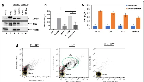
Fig. 1 Nanotrapping exosomes from tissue culture supernatants. a CD63, Alix, and Actin levels from exosomes nanotrapped by either NT80 + 82 particles (lanes 3 and 4) or by NT86 particles (Ctrl NT 86; lanes 5 and 6) from Jurkat (HTLV‑1 uninfected) and HUT102 (HTLV‑1 infected) tissue culture supernatants were analyzed by Western blot. MT‑2 whole cell extracts and molecular weight (MW) ladder are shown (lanes 1 and 2). b
signal detected from ultracentrifuged Jurkat exosomes
from 10 mL of starting material (Fig. 1b). These data
sig-nify that Nanotrap® particles can be used to concentrate
exosomes to equal or greater levels than those isolated from 10 times the same starting material by conventional ultracentrifugation.
To further characterize the efficiency of Nanotrap®
particles in exosomal capture, samples were assessed for acetylcholinesterase (AchE) activity before and after
NT80 + 82 concentration [54, 55]. Prior to NT isolation,
exosomes were present at low levels in tissue culture supernatant of Jurkat, C81 (C8166), MT-2, and HUT102 cells, as demonstrated by low levels of AchE activity
(Fig. 1c, blue bars). However, after nanotrapping with
NT80 + 82, there was a fourfold increase in AchE
activ-ity consistent with an increase in exosome concentration
(Fig. 1c, orange bars).
Samples were additionally analyzed by nanoFACS, a technique which quantifies vesicles by forward and side
scatter (FSC, SSC) [56]. As seen in Fig. 1d, there was a
population of vesicles approximately 100 nm in size in tissue culture supernatants before nanotrapping (left panel). After the addition of NT particles to the super-natant, these NT particles could be visualized by a shift
in FSC vs. SSC, indicating an increase in size (Fig. 1d,
middle panel). Moreover, the decrease in the 100 nm population was visualized after the addition of the NT particles, signifying extraction of the exosomes by the
NTs (Fig. 1d, middle panel). After trapping, the 100 nm
vesicles (exosomes) were removed from the tissue cul-ture supernatant, leaving the post-NT sample absent of
this population (Fig. 1d, right panel). In addition,
exo-some visualization was performed by laser capture of the Brownian motion of extracellular vesicles with Nanosight
(Additional file 1: Figure S1). Pre-nanotrapped samples
showed a population of 2 × 10⁹ 109 nm vesicles along
with 5 × 10⁸ 311, 477 and 583 nm vesicles (left panel).
NTs alone showed a population of particles sized 215– 600 nm in size, as expected (middle panel). However, the resultant nanotrapped exosomes and NTs after pulldown were denoted by peaks from 215 to 600 nm (right panel).
Additionally, a population of ~ 3.5 × 10⁹ 145 nm vesicles
had been pulled from the initial tissue culture super-natant. Analysis of the vesicles from pre- and post-NT supernatants showed that small (< 150 nm) vesicles were reduced by nanotrapping, while medium (200–300 nm) and large (> 300 nm) vesicles increased after nanotrapped vesicles were removed from the media. This may indicate the selective nanotrapping of small vesicles (exosomes). Collectively, these results support nanotrapping with
NT80 + 82 particles as an efficient method for exosome
isolation.
HTLV‑1 infected cell lines have exosomes containing HTLV‑I Tax protein
As shown in Fig. 1, all cell lines tested produced exosomes
in culture, regardless of infection status. However, it has also been previously shown that viral infection can alter exosome cargo. Indeed, HTLV-1 infected cell lines were shown to produce exosomes that contained HTLV-1 Tax protein, which could be isolated by ultracentrifugation
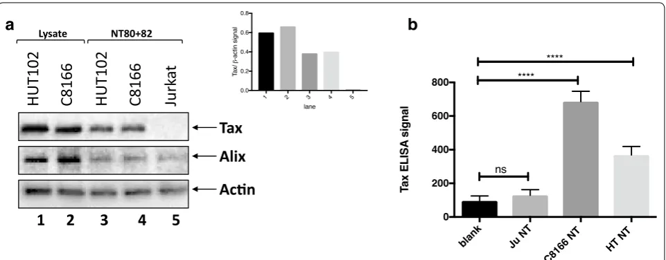
and NT80 particles [47]. Here, we used 1 mL of cell-free,
filtered (0.22 μm) tissue culture supernatant to demon-strate that of the nanotrapped exosomes isolated from Jurkat, C8166, and HUT102 cells, HTLV-1 Tax protein was only detectable by Western blot from the exosomes isolated from the HTLV-1 infected cell lines C8166 and
HUT102 (Fig. 2a, lanes 3 and 4). Levels of Tax were
nor-malized to the levels of Actin to further emphasize that relatively high levels of Tax in comparison to Actin were present in infected cell exosomes, while there were
unde-tectable levels present from uninfected cells (Fig. 2a,
inset). In addition, exosomes containing HTLV-1 Tax were also confirmed using ELISA. Exosomes were freeze-thawed after nanotrapping to open the vesicles, after which only exosomes isolated from the HTLV-1 infected cell lines C8166 and HUT102 showed ELISA reactivity
signal for HTLV-1 Tax above background (Fig. 2b). These
data indicate that use of NT80 + 82 particles to
concen-trate exosomes from HTLV-1 infected cells can allow one to capture exosome-associated Tax with reliable specificity.
Exosome production from cultured PBMCs
We next investigated peripheral blood mononuclear cells (PBMCs) as a potential source of exosomes in HAM/
TSP, since HTLV-1 primarily infects immune cells [57].
Exosomes were isolated from normal donor (ND) and HAM/TSP PBMCs after a short term 5-day culture. Through a process of spontaneous lymphoproliferation, it is well known that HAM/TSP PBMCs can proliferate in culture without the addition of exogenous antigens or
cytokines [58, 59]. However, ND PBMCs do not
prolifer-ate without the addition of a stimulating agent, such as IL-2. To analyze the growth of our HAM/TSP PBMCs in comparison to unstimulated ND cells over time, we
per-formed a 3H-thymidine uptake assay. Maximum
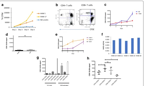
prolifer-ation was seen after 5 days in culture, as demonstrated by
peak 3H-thymidine uptake by representative HAM/TSP
patients compared to an ND (Fig. 3a). Moreover, both
CD4+ and CD8+ T cells from HAM/TSP patients
pro-liferated as seen by CFSE diminution on flow cytometry
(Fig. 3b). Exosome production from proliferating PBMCs
stimulated ND PBMCs (Fig. 3c). No significant differ-ence between CD81 signals from IL-2 stimulated ND and
HAM/TSP PBMCs was detected (Fig. 3d). To directly
compare exosomes produced from HAM/TSP PBMCs from 2 different patients, CD81 levels were measured by ELISA over 5 days. Maximum exosome detection was once again found to be at 5 days for both patient PBMCs
(Fig. 3e). Vesicles isolated from several HAM/TSP PBMC
5 days cultures by NT pulldown were further confirmed as exosomes by detection of AchE activity from samples
(Fig. 3f). Water served as a negative control for which
no AchE activity could be detected (data not shown). All samples showed roughly similar levels of AchE activity. From these results, 5 days was therefore chosen as the optimal time point for analysis of exosomes isolated from PBMC short-term cultures.
HAM/TSP patients are associated with an activated immune response, and here we showed that PBMCs from HAM/TSP patients produce exosomes during
active proliferation (Fig. 3e, f). Given prior reports that
demonstrated that exosome production increases after
activation of mouse and human PBMCs [60], it was of
interest to determine which T cell subset’s exosome pro-duction was most influenced by activation. Therefore, the effect of CD3 activation on T cell exosome
produc-tion was first evaluated in NDs (Fig. 3g). As it has
previ-ously been shown that Treg cells (CD4+CD25+) are both
prolific producers of exosomes and a primary target for
HTLV-1 infection [6, 34, 36], we first focused our analysis
on this subset of T cell. ND PBMCs were either activated by CD3 treatment prior to sorting into T cell subsets, or sorted first into T cell subpopulations and then
main-tained with IL-2 in culture. As shown in Fig. 3g, relative
to IL-2 treated T cell cultures, CD3 activation increased
exosome production from predominantly CD4+CD25+
and CD8+CD25+ T cell subsets, implicating Treg cells as
a predominant producer of exosomes in activated PBMC T cells.
As HTLV-1 in HAM/TSP patients is known to
pre-dominantly infect and activate CD4+ T cells [10], we
next analyzed the production of exosomes in HAM/
TSP T cell subsets. Similar to exogenously CD3−
acti-vated ND PBMCs, endogenously stimulated HAM/TSP
CD4+CD25+ produce more exosomes than CD4+CD25−
cells (Fig. 3h). However, in contrast to activated ND
PBMCs, CD8+CD25− T cell exosomes from HAM/
TSP patients produced a higher CD81 signal while
CD8+CD25+ T cell exosomes produced minimal CD81
signal. This may be indicative of a different CD8+ T cell
subset that could potentially be responsible for greater
release of exosomes in comparison to the CD8+CD25+
T cells in HAM/TSP patients. Together, these results
indicate that CD4+CD25+ T cells (Tregs) are likely one
of the main culprits in the generation of exosomes dur-ing both T cell activation and HTLV-1 infection in HAM/ TSP patients.
a
b
Alix
Tax
Acn
1 2
3
4
5
HUT102
C816
6
HUT102
C816
6
Jurk
at
NT80+82 Lysate
Cultured HAM/TSP PBMCs produce exosomes containing HTLV‑1 Tax
Previously, it has been shown that HTLV-1 infected cell lines produce exosomes that contain Tax protein
[47]. Likewise, we similarly showed that Tax-containing
exosomes could be captured from HTLV-1 infected cell
lines using NT80 + 82 particles (Fig. 2). However, to our
knowledge, Tax has not been shown to be present in exosomes from primary HTLV-1 infected cells. Along these lines, we cultured HAM/TSP PBMCs from two patients for 1–5 days to determine if HTLV-1 Tax pro-tein could similarly be detected by ELISA from the
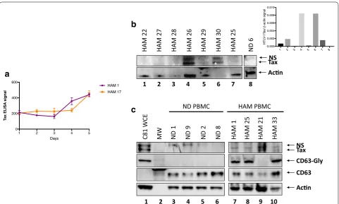
sub-sequent nanotrapped exosomes. As shown in Fig. 4a,
cultured PBMCs from the 2 HAM/TSP patients pro-duced exosomes containing HTLV-1 Tax that increased over this 5-day period. This trend corresponds nicely to the total exosome production (shown by CD81 lev-els) previously seen from the same patients’ cultured
PBMCs (Fig. 3e). Tax was additionally observed in the
majority of HAM patient exosomes at varying levels by
Western blot (Fig. 4b, c). As expected, exosomes isolated
from ND PBMCs did not contain Tax protein. Tax sig-nal above Actin was observed in every lane except 3 and 8, representing 1 HAM/TSP patient and a ND
respec-tively (Fig. 4b, inset). Data in Fig. 4c shows that, similar
to Fig. 4b, the majority of HAM/TSP patient PBMCs
con-tained varying levels of Tax. Interestingly, levels of gly-cosylated CD63 (CD63-Gly) differed strikingly between ND and HTLV-1 infected samples. Only samples infected with HTLV-1, including the C81 whole cell lysates, pos-sessed CD63-Gly, whereas exosomes from all samples had associated unmodified CD63. Cross-sectional anal-ysis of the HAM/TSP cohort by Western Blot demon-strated that 69.4% (25/36) were positive for HTLV-1 Tax in exosomes isolated from cultured PBMCs, while no Tax
positive (Tax+) exosomes were produced from cultured
PBMC of HTLV-1 seronegative controls (0/12) (Fisher
exact test: p-value = 0.0001).
c
3H CP
M
a
b
0 102 103 104 105 <FITC-A>: CFSE 0
102 103 104 105
<A
PC-A>
: CD
4
3.48 67.6
24.8 4.12
CD4+ T cells
0 102 103 104 105 <FITC-A>: CFSE 0
102 103 104 105
<P
er
C
P
-C
y5-5-A
>:
C
D8
4.24 19.5
72.9 3.36
CD8+ T cells
CFSE
0 0.001 0.002 0.003 0.004 0.005 0.006
HAM 19 HAM 21 HAM 4 HAM 25 HAM 30
AchE
uU/m
L
d
e
f
g
h
0 50000 100000 150000 200000
Day 2 Day 3 Day 4 Day 5
HAM 1 HAM 17 ND unsm
Fig. 3 Exosome production from cultured PBMCs. a Normal donor (ND) or HAM/TSP (HAM) PBMCs were cultured in exosome free media and measured over 2–5 days for 3H‑thymidine uptake in counts per minute (CPM). NDs were left unstimulated by IL‑2 or CD3 (ND unstim). b
Carboxyfluorescein succinimidyl ester (CFSE) signal by HAM/TSP PBMCs was measured after 5 days in culture. One representative graph is pictured.
c ELISAs were performed over 5 days for CD81 from nanotrapped (NT80 + 82) exosomes from ND and HAM PBMCs. ND PBMCs were maintained with IL‑2 to induce proliferation. d Exosomes were nanotrapped (NT80 + 82) after 5 days culture of HAM or ND PBMCs (n = 5), followed by ELISA for CD81. ND PBMCs were maintained with IL‑2 to induce proliferation. e A CD81 ELISA was performed from nanotrapped (NT80 + 82) exosomes isolated from HAM 1 and HAM 17 patient samples over 5 days in culture. f Vesicles isolated from HAM PBMC (HAM 4, 19, 21, 25, and 30) short‑term cultures by NT pulldown were measured for AchE activity. g ND PBMCs were sorted immediately after thawing (IL‑2 only) or after activation for 1 day with anti‑CD3 100 ng/mL and 100 IU/mL IL‑2 (activated). h HAM/TSP PBMCs were cultured for 24 h prior to FACS sorting into CD4+CD25−,
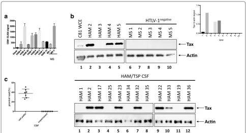
Detection of exosomes containing HTLV‑1 Tax in CSF of HAM/TSP patients
HAM/TSP is a neuropathologically mediated disorder and therefore the detection of exosomes in the CSF is of significant interest. Cell-free CSF was obtained from 4 HAM/TSP patients and 5 HTLV-1 seronegative multiple sclerosis (MS) control patients and directly trapped by
NT80 + 82 to be analyzed by Western blot for the
pres-ence of HTLV-1 Tax. Measurable, but variable exosome levels were detected from both HAM/TSP and MS CSF
samples as quantified by CD81 ELISA reactivity (Fig. 5a).
Moreover, 3 of 4 initial HAM/TSP CSF samples had exosomes with detectable Tax compared to none of the
control MS CSF samples (Fig. 5b). This was verified by
assessing Tax above Actin signal for the upper Western blot panel, where signal was only detected from lanes
2, 4, and 5 (Fig. 5b, inset). An additional 12 HAM/TSP
CSF samples are shown in Fig. 5c in which 7 are
posi-tive for Tax containing exosomes. It is remarkable to note the significant levels of Tax observed in some of
the HAM/TSP CSF samples by Western blot. While Tax was detected at low levels from HTLV-1 infected cell line
extracts (Fig. 5b, lane 1), Tax from HAM/TSP patient
CSF exosomes was far more abundant, when present. However, not all HAM/TSP CSF exosomes tested were
Tax+ as detectable by Western blot (12/20 tested, 60%).
Interestingly, the detection of exosomes containing Tax from HAM/TSP CSF did not correlate with the total numbers of exosomes isolated (data not shown). As it is well established that HTLV-1 is a cell-associated virus
and restricted to cells in the CSF [61], we tested the
pro-viral load of the cell pellet vs. the supernatant of several HAM/TSP CSF samples. We confirmed that while the cell pellet showed expected levels of HTLV-1 provirus,
the CSF supernatant was HTLV-1 virus-free (Fig. 5d).
These data therefore show the presence of exosomes con-taining HTLV-1 Tax in the CSF of HAM/TSP patients in the supernatant compartment from which no HTLV-1 virus can be detected.
a
b
HAM 27
HAM 22 HAM 28 HAM 26 HAM 29 HAM 30 HAM 25 ND
6
1 2 3 4 5 6 7 8
1 2 3 4 5 6 7 8 9 10
C81
WC
E ND PBMC
MW ND
1
ND
9
ND
2
ND
8
HAM
1
HAM 25 HAM 21 HAM 33
Tax NS CD63-Gly CD63 Acn
HAM PBMC
Tax Acn
c
NS
Since subsets of HAM/TSP patient PBMCs (Fig. 4b)
and CSF samples (Fig. 5b) were shown to contain
HTLV-1 Tax+ exosomes, we asked if there were
correla-tions between these two compartments. All tested HAM/
TSP patients (12/12) with Tax+ exosomes in the CSF
were also positive for Tax in exosomes from cultured ex vivo PBMCs (data not shown). For CSF samples from HAM/TSP patients that were negative for Tax in the CSF, 3/8 were also negative in the exosomes isolated from ex vivo PBMCs (data not shown). These results suggest
that Tax+ exosomes in the CSF could reflect levels of
sys-temic infection.
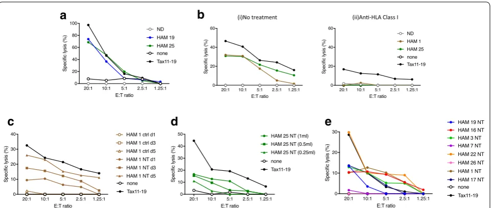
HAM/TSP exosomes can sensitize targets to antigen‑specific responses
As exosomes containing HTLV-1 Tax in CSF were
detect-able in the majority of HAM/TSP patients (Fig. 5b, c),
it was of interest to determine if Tax+ exosomes were
immunologically functional. Therefore, we investi-gated the potential of exosomes produced by HAM/TSP PBMCs to sensitize targets for HTLV-1 Tax-specific CTL lysis. HTLV-1 Tax is highly immunogenic and HTLV-1 Tax-specific CTLs have been detected in brain and spinal
cord from HAM/TSP biopsy and autopsy cases [18, 62–
64]. To determine if exosomes containing Tax could
sensi-tize target cells for lysis by Tax-specific CTLs, autologous lymphoblasts from a HAM/TSP patient were pulsed with exosomes generated from HAM/TSP patients or NDs. A
representative experiment is shown in Fig. 6a. Exosomes
containing Tax from 2 HAM/TSP patient PBMCs signifi-cantly sensitized target cells for CTL lysis at levels
com-parable to control Tax peptide [65, 66] pulsed targets,
whereas Tax− exosomes from representative NDs (n = 4)
did not sensitize targets for CTL-mediated lysis (Fig. 6a).
Donor antigen presenting cells (APC) pulsed with Tax11-19 peptide (black circle) yielded 100% specific lysis at 20:1 E:T ratio and decreased at lower E:T ratios. HAM/TSP PBMC-derived exosomes (blue and green circle) yielded close to 80% specific lysis of donor APCs at 20:1 E:T ratio and decreased at lower E:T ratios. ND (gray open circle) did not sensitize target donor APCs for lysis at any E:T
ratio. CD8+ antigen specific T cell lysis is known to be
HLA class I restricted. As shown in Fig. 6b(i, ii), anti-HLA
class I antibodies blocked lysis of control Tax11-19 pep-tide (black circle) and HAM/TSP exosome-pulsed targets (brown and green circles).
Tax
a
b
c
1 2 3 4 5 6 7 8 9 10
C81
WC
E
HAM
2
HAM
3
HAM
4
HAM
5
MS 1 MS 2 MS 3 MS 4 MS 5
HTLV-1nega ve
HAM
1
HAM
2
HAM 17 HAM 25 HAM 23 HAM 34 HAM 32 HAM 35 HAM 22 HAM 33 HAM 19 HAM 36
HAM/TSP CSF
1 2 3 4 5 6 7 8 9 10 11 12
Tax
Acn Acn
To demonstrate that these observations were spe-cific for exosomes containing Tax from HAM/TSP patients, control nanotraps (ctrl NT86) were used and failed to isolate exosomes that could sensitize targets for
CTL lysis (Fig. 6c, brown open symbols) compared to
exosomes isolated with NT80 + 82 (Fig. 6b, brown closed
symbols). As shown previously, exosome production increased daily (1–5 days) with ex vivo culture of HAM/ TSP PMBCs with a corresponding increase in Tax
detec-tion from isolated exosomes (Figs. 3e, 4a). This increase
in Tax+ exosomes additionally correlated with increased
CTL lysis (Fig. 6c, NT day 1, day 3, day 5). Specifically,
samples that were nanotrapped with NT80 + 82 particles
(NT) at day 1 (brown closed circle) yielded 10%, while NT day 3 yielded 20% specific lysis, and NT day 5 yielded 30% specific lysis of targets at 20:1 and decreased at lower E:T ratios. Samples that were nanotrapped with control NT86 particles do not capture exosomes (ctrl) at day 1, day 3, and day 5 and were therefore unable to sensitize targets for lysis at any E:T ratio after pulsing of targets
(Fig. 6c, brown open circle). Further characterization of
the Tax+ exosomal sensitization of targets for CTL lysis
is shown in Fig. 6d, which demonstrated a
dose-depend-ent response of sensitization with increasing concdose-depend-entra-
concentra-tions (250 μL to 1 mL) of Tax+ exosomes. Nanotrapping
from the standard 1 mL (green closed circle) of tissue culture supernatant yielded ~ 18% specific lysis, while nanotrapping from 500 μL (green closed square) yielded
15% specific lysis of targets and exosomes isolated from 250 μL (green closed triangle) of starting tissue culture supernatant yielded 10% specific lysis of targets at an E:T of 20:1. Each sample concentration showed decreased specific lysis as lower E:T ratios. While most HAM/TSP PBMC derived exosomes were able to sensitize targets for CTL response, the magnitude of this response
var-ied among patients (Fig. 6e). CTL assay using HAM/TSP
patient PBMC-derived exosomes showed that exosomes from 2/11 HAM/TSP patients did not sensitize targets
for specific lysis at any E:T ratio tested (Fig. 6e,
represent-ative of 8 HAM/TSP patients). Collectively, these results demonstrate that exosomes containing HTLV-1 Tax can contribute to a functional immune response by sensitiz-ing target cells to specific lysis by antigen-specific CTLs.
Discussion
There is significant interest in the role of exosomes in viral diseases and as a potential biomarker in these dis-orders. Many viruses enter cells through the endosomal compartment and coopt proteins involved in endosome/ exosome formation and shuttling, using them for viral
particle egress [23]. Indeed, herpesviruses, flaviviruses,
bunyaviruses, and lentiviruses incorporate viral messages
into exosome cargo [23]. It has been suggested that these
released exosomes may dysregulate the immune response
[67, 68]. Since the observation that HTLV-1 Tax could
be found in exosomes produced by HTLV-1 infected cell
a
c
d
(i)No treatment (ii)An-HLA Class I
b
e
lines in vitro [47], it became important to characterize exosomes ex vivo from patients with HTLV-1 associated neurologic disease. HAM/TSP, a neurologic disorder associated with higher levels of HTLV-1 proviral loads
[69], is characterized by a widespread inflammatory
immune response and decreased immune suppression [5,
70].
Here we show that PBMCs from HAM/TSP patients produced exosomes containing HTLV-1 Tax
pro-tein (Fig. 3) in ex vivo cultures, consistent with reports
demonstrating Tax in exosomes produced by HTLV-1
infected cell lines [47]. HTLV-1 Tax is a pleiotropic
transactivating protein known to be critical in immune
activation detected in HAM/TSP [71]. It is also highly
immunogenic, which results in a strong Tax-specific
immune response [72–74]. HTLV-I Tax-specific CTLs
have been shown to be elevated in peripheral blood and even higher in CSF of HAM/TSP patients, in part driven by high levels of HTLV-I proviral loads demonstrated
in these compartments [18, 62, 75]. These observations
have supported the hypothesis that these antigen-specific responses are associated with the neuropathogenesis of HAM/TSP, and therefore may be reasonable targets for immune interventions that either directly target the effector CTL or the antigen that stimulates this response. Activated T cells are a hallmark of HAM/TSP, but within the CNS there is a paucity of HTLV-1 infected cells, and
even if present, have been postulated to be
nonproduc-tively infected [9].
While HTLV-1 proviral DNA levels are significantly increased in T cells in the CSF of HAM/TSP patients
compared to peripheral blood [63, 76], no cell-free virus
can be demonstrated in CSF supernatant [61, 77]. This
is not unexpected since HTLV-1 is known to be highly
cell-associated [78]. However, we could demonstrate
exosomes containing HTLV-1 Tax in HAM/TSP CSF
supernatant (Fig. 5). The presence of Tax in exosomes in
CSF supernatants suggests that exosomes either cross the blood–brain barrier (BBB) after production by infected cells in the periphery, or by infiltrating infected T cells
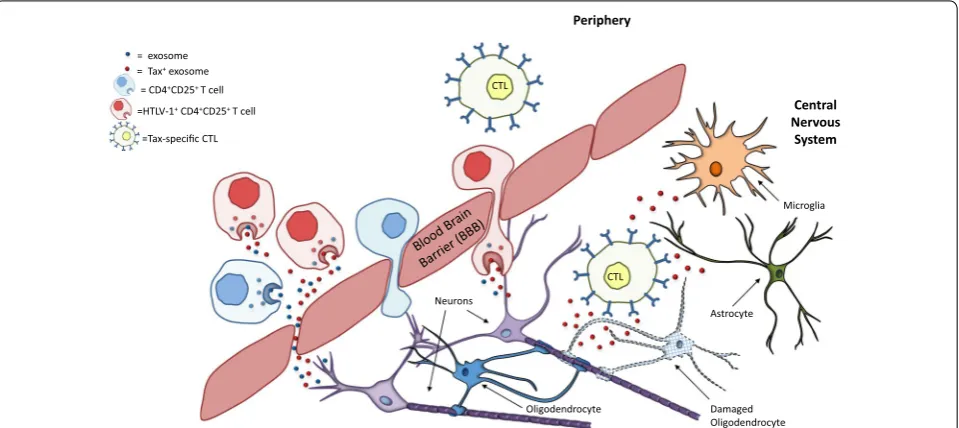
within the CSF (schematic representation Fig. 7). While
monocytes and macrophages comprise a significant
por-tion of exosome producpor-tion in the periphery [79, 80], the
vast majority of immune cells infected in HAM/TSP are
activated T cells [5], making them a likely source of the
Tax+ exosomes found in our samples. Indeed, HAM/
TSP CD4+CD25+ subset produces significantly more
exosomes than HAM/TSP CD4+CD25− T cells (Fig. 3h)
and CD8+CD25+ T cells. The CD4+CD25+ subset also
includes regulatory T cells (Tregs), a subset known to be
dysfunctional in HAM/TSP [5], and has been reported to
be a predominant source of exosomes in mice [34].
While Tax+ exosome detection was apparent in HAM/
TSP CSF (12/20), we could not find evidence of Tax+
= exosome = Tax+ exosome
= CD4+CD25+ T cell
=HTLV-1+ CD4+CD25+ T cell
=Tax-specific CTL
Oligodendrocyte Neurons
Damaged Oligodendrocyte
Central Nervous
System Periphery
CTL
CTL
Astrocyte
Microglia
Fig. 7 Hypothesized contribution of exosomes to HAM/TSP immunopathogenesis. HTLV‑1 infected PBMCs, especially HTLV‑1 infected CD4+CD25+
T cells, produce exosomes containing HTLV‑1 Tax. These cells are present in the periphery and produce Tax+ exosomes in the periphery that can
potentially cross the blood brain barrier (BBB). However, these infected T cells are also capable of crossing the BBB and producing exosomes in the CNS. Both scenarios may explain the presence of Tax+ exosomes in HAM/TSP patient CSF. These Tax+ exosomes can then be taken up by CNS
exosomes in a small group of asymptomatic carrier (AC) CSF samples (data not shown) (and, as expected, there were none detected from HTLV-1 negative patients
(0/5, Fig. 5b, c). Of interest, we were able to detect
Tax+ exosomes in the CSF sample of an individual
ini-tially classified as an AC who began developing clinical signs and symptoms of HAM/TSP on the date the CSF was obtained (unpublished observation). Collectively, these observations suggest that the detection of Tax in exosomes isolated from the CSF could serve as an addi-tional biomarker of progression to HAM/TSP. The lack of Tax protein present in the exosomes from all HAM/TSP CSF samples probably reflects the diversity in the HAM/ TSP patient population and the biology of the disease.
In HAM/TSP, HTLV-1 Tax in exosomes may also serve as a source of antigen in the disease. As shown in this present study, HAM/TSP PBMCs produced exosomes
containing Tax in culture (Fig. 4), and these ex vivo
Tax-containing exosomes were able to sensitize target cells
for Tax-specific CTL lysis (Fig. 6). Exosomes
contain-ing viral proteins may therefore be an underappreciated source of antigen that drives continued immune activa-tion in HAM/TSP. Histologically, antigen-specific CTLs have been demonstrated in the CNS and leptomeninges in HAM/TSP patients, suggesting that this disease is
immunopathologically mediated [17, 64]. Our
observa-tion that Tax+ exosomes can be detected in the CSF of
HAM/TSP patients suggests exosomal transference of HTLV-1 antigens to antigen presenting cells (APCs) may also occur in the CNS, and could play a role in perpetu-ating neuroinflammation in HAM/TSP through damage to various CNS resident cells such as astrocytes,
micro-glia or oligodendrocytes (Fig. 7). Sensitization of CTLs
by Tax+ exosomes suggests a potential role in HAM/TSP
disease initiation and/or progression. Future experiments targeting diminution of Tax content in exosomes through knockdown or silencing methods will be of value since recently it has been shown that Tax expression is
essen-tial for HAM/TSP PBMC survival [81].
The observations in this study on the detection of HTLV-1 viral antigens in exosomes found in virus-free CSF supernatant has implications for other virally asso-ciated neuroinflammatory diseases. For example, Ebola virus has been shown to be associated with long-term neurologic sequelae even though the virus is thought to
have been cleared [81, 82], and while Ebola viral proteins
have been detected within exosomes in vitro [83], this
has not yet been shown to be the case within patient CSF material. As we have demonstrated that HAM/TSP CSF supernatant, which is HTLV-1 negative, contains HTLV-1
Tax+ exosomes, exosomes containing viral antigens
could be a potential biomarker for neurologic disorders associated with viral infections in which the virus may be
absent in CSF. Indeed, recent data has demonstrated that exosomes containing HIV antigens are still present in the serum of patients under highly active antiretroviral
ther-apy (HAART) for whom there is no detectable virus [53],
indicating that tracking of exosomal cargo could monitor viral reservoirs. Furthermore, exosomes could be a tool for pathogen discovery in a wide variety of undiagnosed aseptic meningitides and encephalitides for which a virus
is suspected but undetectable [84, 85]. The etiology for
over 50% of meningitis and encephalitis cases is
cur-rently unknown [86, 87]. Collectively, the observations
presented in this report on exosomes containing viral antigens in the CSF of patients with HTLV-1 associated neurologic disease will be of relevance to many neuroin-flammatory diseases in which viruses are thought to play a role.
Conclusions
The understanding of exosomes and their contribution to human disease is a quickly expanding field. Evidence suggests that exosomes play a large role in immunity and infection. Some viruses have been shown to alter exo-some cargo with viral components in another mechanism of immune evasion. Here we show that HTLV-1 virus incorporates HTLV-1 Tax, an important viral regulatory protein, into exosomes found in the CSF and produced by HTLV-1 infected PBMCs. These exosomes could sen-sitize cells for lysis by HTLV-1 specific CTLs which are indicated in HAM/TSP neuropathology. This suggests that exosomes may have a functional role in CNS damage in HAM/TSP patients.
Methods
Cell lines
HTLV-1 infected cell lines HUT102, MT-2 and C8166 and the uninfected Jurkat cell line were maintained in exosome-free (Exo-free) complete RPMI. Specifically,
FBS was spun at > 90,000×g for 2 h and filtered through
a 0.22 μm MCE filter (EMD Millipore, Darmstadt, Ger-many), after which it was considered exosome-free. Exo-free CRPMI consisted of RPMI 1640 supplemented with 10% Exo-free FBS, 100 U/mL penicillin, 100 μg/mL strep-tomycin sulfate, and 2 mM l-glutamine.
Patient samples
A total of 36 HAM/TSP, 10 ND, and 2 HTLV-II patient PBMC samples were used for ex vivo incubation and later exosome isolation. PBMCs were isolated by Ficoll-Hypaque (Lonza, Walkersville, MD) centrifugation, and were cryopreserved in liquid nitrogen prior to use. All NDs were noted to be healthy and HTLV-1 nega-tive. Briefly, PBMCs were placed in Exo-free CRPMI at
37 °C for 5 days. ND PBMCs were maintained in culture with the addition of 100 IU/mL recombinant human (rh) IL-2. Culture supernatant was collected and then spun at 1300 rpm for 10 min to remove cells. Spun supernatant was then pushed through a 0.22 μm MCE filter (EMD Millipore) to remove large debris, apoptotic bodies and extracellular vesicles > 220 nm. Cerebrospinal fluid (CSF) was obtained through lumbar puncture of study partici-pants by neurologist and nurse practitioners of the Neu-roimmunology Clinal group. CSF was spun at 1300 rpm for 10 min to remove cells and supernatant was stored in
1 mL aliquots at − 80 °C prior to use.
Exosome isolation
Nanotrap® particles NT80 (Ceres #CN1030) and NT82
(Ceres #CN2010) were provided by Ceres Nanosciences, Inc. (Manassas, VA) and have previously been shown to
concentrate exosomes from media [47]. Each was mixed
into PBS (w/o Ca+ or Mg+) for a final 30% slurry (30 μL
each NT in a total 100 μL volume with PBS). Thirty microliters of 30% NT slurry was then added to 1 mL of ex vivo PBMC supernatant and rotated overnight at 4 °C or at room temperature for 1 h. After trapping, samples were spun at 13,000 rpm for 5 min and then washed with 500 μL DEPC H20. Exosome control NT86
(ctrl NT) was similarly placed in PBS (w/o Ca+ or Mg+)
for a final 30% slurry. When working with CSF, samples
were first diluted 1:1 in PBS (w/o Ca+ or Mg+) prior to
nanotrapping.
Western blotting
HTLV-1 Tax, Alix, CD63, and Actin were detected from nanotrapped exosomes by Western blot. Briefly, after washing, samples were resuspended in 100 μM Tris–
HCl, 8 μM urea, 1% triton. After the addition of 4× LDS
and DTT, samples were heated at 95 °C for 6 min, with a vigorous vortex for 5–10 s every 2 min. After heating, samples were run on a 4–12% Novex Bis–Tris 1.0 mm gel (ThermoFisher Scientific, Waltham, MA) and then trans-ferred in an X-blot module onto a 0.45 μm nitrocellulose membrane. After blocking with 3% BSA in TBS, mem-branes were probed with anti-Tax (Lt4 1:100, mIgG3; Kindly provided by Dr. Yuetsu Tanaka), anti-CD63 or anti-Alix (1:100; Santa Cruz Biotech, Dallas, TX) over-night at 4 °C. For imaging, ms HRP secondary anti-body (Jackson ImmunoResearch, Westgrove, PA) was applied at 1:5000 or goat anti-ms IR 680RD secondary (1:10,000. LI-COR Biosciences, Lincoln, NE) was used for CSF samples. For detection of Actin, blots were stripped for 5–10 min with Restore PLUS Western Blot Stripping Buffer (ThemoFisher Scientific), reblocked and reprobed with Actin (Santa Cruz Biotech) at 1:100 overnight at 4 °C.
Acetylcholinesterase enzyme (AchE) activity assay
AchE activity of exosomes after nanotrapping was
car-ried out as described in detail previously [55] with brief
modification. Amplex® Acetylcholine/Acetylcholine
Esterase Activity Assay Kit (Thermo; A12217) was used following the manufacturer’s instructions. Briefly, a
1× running buffer negative control (20 mL of H2O and
5 mL of 5× reaction buffer) and two positive controls,
one consisting of acetylcholinesterase and one consist-ing of hydrogen peroxide, were made and plated on a 96-well plate. Exosomes were treated and fluorescence of acetylcholinesterase activity was measured with a GLO-MAX multidetection system (Promega, Madison, WI) every 15 min for 1 h to find optimal activity. Changes in absorption were monitored at 412 nm during incubation
period at 37 °C [88].
Electrochemical ELISA
For CD81 and HTLV-1 Tax, nanotrapped exosomes
were resuspended in 1× TBS for assessment by MESO
Enzyme Linked Immunosorbent Assay (ELISA) (MSD Technologies, Rockville, MD). Briefly, samples were freeze-thawed for 4 cycles of 5 min each and then blot-ted onto a MESO Sector Imager 96-well high-bind plate (MSD Technologies) in duplicate wells overnight. The plate was then blocked with blocker A for 2 h, washed
with PBS ×2 and then probed with anti-CD81 (Santa
Cruz Biotech) or anti-HTLV-1 Tax (Lt4) for 1 h, shaking. After a repeat wash, the plate was probed with goat anti-ms sulfa secondary Ab (1:100, MSD Technologies). After a final wash, MSD Read buffer was added at 1:2 dilution in Ultra Pure Water and the plate read using an MSD Sector Imager (courtesy of Dr. Avi Nath).
3H‑thymidine assay for lymphocyte proliferation
Thawed ND or HAM/TSP PBMCs were placed on
96-well plate in triplicate at 3 × 10⁵ cells/well in 10%
Exo-Free CRPMI and allowed to proliferate for 3–5 days in culture at 37 °C. At 3, 4, or 5 days, plates were removed from culture and 50 μL of tritium solution was added to each well. Cells were incubated for 4 h at 37 °C after which they were washed and harvested onto a film mat. Five milliliters of scintillation fluid was added after which thymidine incorporation was measured using a β plate counter (Microbeta TriLux 1450, Perkin Elmer, Waltham MA).
T cell isolation
prior to sorting. At least 5 × 10⁷ PBMCs were stained in FACS buffer (1% FBS, 0.1% Sodium Azide, PBS) with
10 μL/1 × 107 cells of anti-CD4 APC, anti-CD8 FITC and
anti-CD25 PE (all from BD Biosciences). Cells were then
sorted into CD4+CD25−, CD4+CD25+, CD8+CD25−,
CD8+CD25+ T cell subsets on a BD FACS Aria flow
cytometer. After sorting, cells were briefly placed in 20%
FBS, 2× antimycobacterial, antibacterial (ThermoFisher
Scientific) RPMI 2 mM l-glutamine. Cells were then spun at 1300 rpm for 10 min and resuspended in Exo-free CRPMI supplemented with 100 IU/mL hrIL-2 and plated
at 7.5 × 10⁵ cells per well in a 12-well plate for 5 days at
37 °C. For HAM/TSP PBMCs, no exogenous stimula-tion was provided. Endogenous activastimula-tion of HAM/TSP PBMC was achieved by incubation of PBMCs for 18–24 h prior to FACS sorting as described above.
CTL assay
HTLV-1 Tax specific CTL line and target B cell line were generated from an HLA A0201 HAM/TSP patient and
cryopreserved prior to use. As described previously [64],
CTLs were primed with Tax peptide 11-19 weekly for expansion and maintenance in CTLA media (IMDM with 5% FBS, 100 μg/mL penicillin, 100 μg/mL streptomycin sulfate, 12.5 mM sulfinpyrazone, 2 mM l-glutamine) for up to 1 month. Clones were tested for efficiency of target killing prior to use. Targets B cells were pulsed for 1 h at 37 °C with Tax peptide 11-19 or nanotrapped exosomes from tissue culture supernatant of 11 separate HAM/TSP PBMC ex vivo cultures and 4 ND PBMC cultures. One HAM/TSP supernatant was nanotrapped with NT86 as a negative control. Anti-HLA Class I ab (MCA81EL, Serotec (BioRad), Oxford UK) was added to CTLs after co-culture with targets at 5 μg/mL. The CTL Europium cytotoxity release assay was performed as outlined in the DELFIA protocol (Perkin Elmer Inc., Waltham, MA). Specific lysis was calculated as a percentage:
Exosome characterization by nanosight and nanoFACS Tissue culture supernatant or resuspended nanotrapped exosomes were pushed through a syringe at a total vol-ume of 300 μL into the chamber of the NanoSight LM10 instrument (Malvern Instruments, Worcestershire, UK) for extracellular vesicle visualization and quantifica-tion. Briefly, a laser beam is passed through the chamber containing the sample and light scattering and Brown-ian motion is quantified as a vesicle size distribution and quantity. For exosome visualization by flow cytometry
sample x − Targetonly
Targetmax − Targetonly
× 100%.
(nanoFACS), samples were loaded into a Beckman Coul-ter MoFlo XDP cell sorCoul-ter (Beckman CoulCoul-ter, Brea, CA)
as described extensively by Danielson et al. [56].
Statistical analysis
Bar graphs and scatter plots were generated in Graph-Pad Prism 7.0 (GraphGraph-Pad, San Diego, CA). Two-tailed
unpaired t-tests were run to establish difference between
groups and significance was considered anything with a
p-value < 0.05. The two-tailed Fisher exact test was used
to assess differences between groups on parametric
ques-tions (Tax+ or Tax− exosomes) using GraphPad
Quick-Calcs software. Statistical analyses of 2 × 2 contingency
tables were performed using GraphPad QuickCalcs
soft-ware with two-tailed Fisher exact tests. p-values < 0.05
were considered significant.
Abbreviations
Ab: antibody; AC: asymptomatic carrier; AchE: acetylcholinesterase enzyme; Ag: antigen; APC: antigen presenting cell; BBB: blood brain barrier; C81: C8166 cell line; CNS: central nervous system; CSF: cerebrospinal fluid; CTL: cytotoxic T lymphocyte; EBV: Epstein Barr virus; ELISA: enzyme linked immunosorbent assay; ESCRT : endosomal sorting complexes required for transport; HAART : highly active anti‑retroviral therapy; HAM/TSP HTLV‑1: associated myelopathy/ tropical spastic paraparesis; HT: HUT102 cell line; HTLV‑1: human T cell lympho‑ tropic virus type 1; ILV: intraluminal vesicles; Ju: Jurkat cell line; MS: multiple sclerosis; MVB: multivesicular body; ND: normal donor; NT: nanotrap particles; PBMC: peripheral blood mononuclear cell; Treg: regulatory T cell.
Authors’ contributions
MRA was responsible for PBMC cultures, Western blots, spontaneous prolifera‑ tion assays, ELISAs, and writing of the manuscript. AV performed all ddPCR runs. MMK was responsible for FACS sorting. YEA was responsible for CTL assays. MLP, JE, and YA were responsible for CSF and PBMC Western blots. YA performed AchE assays. MLP assisted with creation of Fig. 7, other figure adjustments, as well as editing of the manuscript. YT was responsible for generation of Lt4 monoclonal antibody. BL generated the Nanotrap® particles for this study. SA collected patient CSF samples. JJ performed exosome char‑ acterization by Nanosight and NanoFACS. FK and SJ organized and supervised experiments and their respective institutions. All authors read and approved the final manuscript.
Author details
1 Department of Pathology, University of Virginia School of Medicine, Char‑
lottesville, VA 22901, USA. 2 Viral Immunology Section, Neuroimmunology
Branch, National Institute for Neurological Disease and Stroke, National Insti‑ tutes of Health, 10 Center Drive Rm 5C103, Bethesda, MD 20892, USA. 3 Labo‑
ratory of Molecular Virology, George Mason University, Manassas, VA 20110, USA. 4 Laboratory of Molecular Medicine and Neuroscience, National Institutes
for Neurological Disease and Stroke, National Institutes of Health, Bethesda, MD 20892, USA. 5 Department of Immunology, University of the Ryukyus
Graduate School of Medicine, Okinawa 903‑0125, Japan. 6 Ceres Nanosciences,
Additional file
Manassas, VA 20109, USA. 7 Vaccine Branch, National Cancer Institute, National
Institutes of Health, Bethesda, MD 20892, USA.
Competing interests
Ben Lepene is currently employed at Ceres Nanosciences. All other authors declare that they have no competing interests.
Availability of data and materials
All data supporting our findings is reported in this manuscript.
Consent for publication
Not applicable.
Ethics approval and consent to participate
The study was reviewed and approved by the National Institute of Neurologi‑ cal Disorders and Stroke Institutional Review Board. Informed consent was written and obtained from each subject and the study was performed in accordance with the Declaration of Helsinki.
Funding
NIH Grants AI078859, AI074410, and AI043894 and R01MH110262 to FK were used in funding CSF western blot experiments, nanotrap production, and AchE experiments. NIH Intramural funds to SJ were used for collection and maintenance of patient samples, CTL assays, ex vivo experiments, and all other western blotting and ELISAs conducted.
Publisher’s Note
Springer Nature remains neutral with regard to jurisdictional claims in pub‑ lished maps and institutional affiliations.
Received: 20 December 2017 Accepted: 6 July 2018
References
1. Koyanagi Y, Itoyama Y, Nakamura N, Takamatsu K, Kira J, Iwamasa T et al (1993) In vivo infection of human T‑cell leukemia virus type I in non‑T cells. Virology 196(1):25–33
2. Satou Y, Matsuoka M (2010) HTLV‑1 and the host immune system: how the virus disrupts immune regulation, leading to HTLV‑1 associated diseases. J Clin Exp Hematopathol 50(1):1–8
3. Proietti FA, Carneiro‑Proietti AB, Catalan‑Soares BC, Murphy EL (2005) Global epidemiology of HTLV‑I infection and associated diseases. Onco‑ gene 24(39):6058–6068
4. Evangelou IE, Massoud R, Jacobson S (2014) HTLV‑I‑associated myelopathy/tropical spastic paraparesis: semiautomatic quantification of spinal cord atrophy from 3‑dimensional MR images. J Neuroimaging 24(1):74–78
5. Yamano Y, Takenouchi N, Li HC, Tomaru U, Yao K, Grant CW et al (2005) Virus‑induced dysfunction of CD4+CD25+ T cells in patients with HTLV‑I‑ associated neuroimmunological disease. J Clin Invest 115(5):1361–1368 6. Satou Y, Matsuoka M (2013) Virological and immunological mechanisms
in the pathogenesis of human T‑cell leukemia virus type 1. Rev Med Virol 23(5):269–280
7. Izumo S (2010) Neuropathology of HTLV‑1‑associated myelopathy (HAM/ TSP): the 50th anniversary of Japanese society of neuropathology. Neuro‑ pathology 30(5):480–485
8. Grant C, Barmak K, Alefantis T, Yao J, Jacobson S, Wigdahl B (2002) Human T cell leukemia virus type I and neurologic disease: events in bone marrow, peripheral blood, and central nervous system during normal immune surveillance and neuroinflammation. J Cell Physiol 190(2):133–159
9. Lepoutre V, Jain P, Quann K, Wigdahl B, Khan ZK (2009) Role of resident CNS cell populations in HTLV‑1‑associated neuroinflammatory disease. Front Biosci 14:1152–1168
10. Richardson JH, Edwards AJ, Cruickshank JK, Rudge P, Dalgleish AG (1990) In vivo cellular tropism of human T‑cell leukemia virus type 1. J Virol 64(11):5682–5687
11. Rende F, Cavallari I, Corradin A, Silic‑Benussi M, Toulza F, Toffolo GM et al (2011) Kinetics and intracellular compartmentalization of HTLV‑1 gene expression: nuclear retention of HBZ mRNAs. Blood 117(18):4855–4859 12. Miyazato P, Matsuo M, Katsuya H, Satou Y. Transcriptional and Epigenetic
Regulatory Mechanisms Affecting HTLV‑1 Provirus. Viruses. 2016;8(6) 13. Alefantis T, Mostoller K, Jain P, Harhaj E, Grant C, Wigdahl B (2005) Secre‑
tion of the human T cell leukemia virus type I transactivator protein tax. J Biol Chem 280(17):17353–17362
14. Ferrucci A, Nonnemacher MR, Wigdahl B (2013) Extracellular HIV‑1 viral protein R affects astrocytic glyceraldehyde 3‑phosphate dehydrogenase activity and neuronal survival. J Neurovirol 19(3):239–253
15. Secchiero P, Zella D, Capitani S, Gallo RC, Zauli G (1999) Extracellular HIV‑1 tat protein up‑regulates the expression of surface CXC‑chemokine recep‑ tor 4 in resting CD4+ T cells. J Immunol 162(4):2427–2431
16. Pique C, Jones KS (2012) Pathways of cell‑cell transmission of HTLV‑1. Front Microbiol 3:378
17. Matsuura E, Kubota R, Tanaka Y, Takashima H, Izumo S (2015) Visualization of HTLV‑1‑specific cytotoxic T lymphocytes in the spinal cords of patients with HTLV‑1‑associated myelopathy/tropical spastic paraparesis. J Neuro‑ pathol Exp Neurol 74(1):2–14
18. Greten TF, Slansky JE, Kubota R, Soldan SS, Jaffee EM, Leist TP et al (1998) Direct visualization of antigen‑specific T cells: hTLV‑1 Tax11‑19‑specific CD8(+) T cells are activated in peripheral blood and accumulate in cerebrospinal fluid from HAM/TSP patients. Proc Natl Acad Sci USA 95(13):7568–7573
19. van der Pol E, Boing AN, Harrison P, Sturk A, Nieuwland R (2012) Classifica‑ tion, functions, and clinical relevance of extracellular vesicles. Pharmacol Rev 64(3):676–705
20. Alenquer M, Amorim MJ (2015) Exosome biogenesis, regulation, and function in viral infection. Viruses 7(9):5066–5083
21. Robbins PD, Morelli AE (2014) Regulation of immune responses by extra‑ cellular vesicles. Nat Rev Immunol 14(3):195–208
22. Lai FW, Lichty BD, Bowdish DM (2015) Microvesicles: ubiquitous contribu‑ tors to infection and immunity. J Leukoc Biol 97(2):237–245
23. Anderson MR, Kashanchi F, Jacobson S (2016) Exosomes in viral disease. Neurotherapeutics 13(3):535–546
24. Raposo G, Stoorvogel W (2013) Extracellular vesicles: exosomes, microvesicles, and friends. J Cell Biol 200(4):373–383
25. Vlassov AV, Magdaleno S, Setterquist R, Conrad R (2012) Exosomes: cur‑ rent knowledge of their composition, biological functions, and diagnostic and therapeutic potentials. Biochem Biophys Acta 1820(7):940–948 26. Lane RE, Korbie D, Anderson W, Vaidyanathan R, Trau M (2015) Analysis
of exosome purification methods using a model liposome system and tunable‑resistive pulse sensing. Sci Rep 5:7639
27. Helwa I, Cai J, Drewry MD, Zimmerman A, Dinkins MB, Khaled ML et al (2017) A comparative study of serum exosome isolation using dif‑ ferential ultracentrifugation and three commercial reagents. PLoS ONE 12(1):e0170628
28. Yanez‑Mo M, Siljander PR, Andreu Z, Zavec AB, Borras FE, Buzas EI et al (2015) Biological properties of extracellular vesicles and their physiologi‑ cal functions. J Extracell Vesicles 4:27066
29. Ludwig AK, Giebel B (2012) Exosomes: small vesicles participating in intercellular communication. Int J Biochem Cell Biol 44(1):11–15 30. Thery C, Duban L, Segura E, Veron P, Lantz O, Amigorena S (2002) Indirect
activation of naive CD4+ T cells by dendritic cell‑derived exosomes. Nat Immunol 3(12):1156–1162
31. Admyre C, Johansson SM, Paulie S, Gabrielsson S (2006) Direct exosome stimulation of peripheral human T cells detected by ELISPOT. Eur J Immu‑ nol 36(7):1772–1781
32. Segura E, Guerin C, Hogg N, Amigorena S, Thery C (2007) CD8+ dendritic cells use LFA‑1 to capture MHC‑peptide complexes from exosomes in vivo. J Immunol 179(3):1489–1496
33. Xie Y, Zhang H, Li W, Deng Y, Munegowda MA, Chibbar R et al (2010) Dendritic cells recruit T cell exosomes via exosomal LFA‑1 leading to inhi‑ bition of CD8+ CTL responses through downregulation of peptide/MHC class I and Fas ligand‑mediated cytotoxicity. J Immunol 185(9):5268–5278 34. Okoye IS, Coomes SM, Pelly VS, Czieso S, Papayannopoulos V, Tolmachova T et al (2014) MicroRNA‑containing T‑regulatory‑cell‑derived exosomes suppress pathogenic T helper 1 cells. Immunity 41(1):89–103 35. Smyth LA, Ratnasothy K, Tsang JY, Boardman D, Warley A, Lechler R
CD4+CD25+Foxp3+ T cells contributes to their regulatory function. Eur J Immunol 43(9):2430–2440
36. Yamano Y, Cohen CJ, Takenouchi N, Yao K, Tomaru U, Li HC et al (2004) Increased expression of human T lymphocyte virus type I (HTLV‑I) Tax11‑19 peptide‑human histocompatibility leukocyte antigen A*201 complexes on CD4+CD25+ T Cells detected by peptide‑specific, major histocompatibility complex‑restricted antibodies in patients with HTLV‑I‑ associated neurologic disease. J Exp Med 199(10):1367–1377
37. Li S, Gowans EJ, Chougnet C, Plebanski M, Dittmer U (2008) Natural regulatory T cells and persistent viral infection. J Virol 82(1):21–30 38. Anderson MR, Enose‑Akahata Y, Massoud R, Ngouth N, Tanaka Y, Oh
U et al (2014) Epigenetic modification of the FoxP3 TSDR in HAM/TSP decreases the functional suppression of Tregs. J Neuroimmune Pharma‑ col 9(4):522–532
39. Jones KS, Petrow‑Sadowski C, Huang YK, Bertolette DC, Ruscetti FW (2008) Cell‑free HTLV‑1 infects dendritic cells leading to transmission and transformation of CD4(+) T cells. Nat Med 14(4):429–436
40. de Castro‑Amarante MF, Pise‑Masison CA, McKinnon K, Washington Parks R, Galli V, Omsland M et al (2015) Human T cell leukemia virus type 1 infection of the three monocyte subsets contributes to viral burden in humans. J Virol 90(5):2195–2207
41. Nagai M, Yamano Y, Brennan MB, Mora CA, Jacobson S (2001) Increased HTLV‑I proviral load and preferential expansion of HTLV‑I Tax‑specific CD8+ T cells in cerebrospinal fluid from patients with HAM/TSP. Ann Neurol 50(6):807–812
42. Li XB, Zhang ZR, Schluesener HJ, Xu SQ (2006) Role of exosomes in immune regulation. J Cell Mol Med 10(2):364–375
43. Longatti A, Boyd B, Chisari FV (2015) Virion‑independent transfer of replication‑competent hepatitis C virus RNA between permissive cells. J Virol 89(5):2956–2961
44. Wiley RD, Gummuluru S (2006) Immature dendritic cell‑derived exosomes can mediate HIV‑1 trans infection. Proc Natl Acad Sci USA 103(3):738–743
45. Mori Y, Koike M, Moriishi E, Kawabata A, Tang H, Oyaizu H et al (2008) Human herpesvirus‑6 induces MVB formation, and virus egress occurs by an exosomal release pathway. Traffic 9(10):1728–1742
46. Rowe RK, Suszko JW, Pekosz A (2008) Roles for the recycling endosome, Rab8, and Rab11 in hantavirus release from epithelial cells. Virology 382(2):239–249
47. Jaworski E, Narayanan A, Van Duyne R, Shabbeer‑Meyering S, Iordan‑ skiy S, Saifuddin M et al (2014) Human T‑lymphotropic virus type 1‑infected cells secrete exosomes that contain Tax protein. J Biol Chem 289(32):22284–22305
48. Yang T, Martin P, Fogarty B, Brown A, Schurman K, Phipps R et al (2015) Exosome delivered anticancer drugs across the blood‑brain barrier for brain cancer therapy in Danio rerio. Pharm Res 32(6):2003–2014 49. El Andaloussi S, Lakhal S, Mager I, Wood MJ (2013) Exosomes for
targeted siRNA delivery across biological barriers. Adv Drug Deliv Rev 65(3):391–397
50. Gupta A, Pulliam L (2014) Exosomes as mediators of neuroinflammation. J Neuroinflam 11:68
51. Barclay RA, Schwab A, DeMarino C, Akpamagbo Y, Lepene B, Kassaye S et al (2017) Exosomes from uninfected cells activate transcription of latent HIV‑1. J Biol Chem 292(28):11682–11701
52. Sampey GC, Saifuddin M, Schwab A, Barclay R, Punya S, Chung MC et al (2016) Exosomes from HIV‑1‑infected cells stimulate production of pro‑ inflammatory cytokines through trans‑activating response (TAR) RNA. J Biol Chem 291(3):1251–1266
53. Jaworski E, Saifuddin M, Sampey G, Shafagati N, Van Duyne R, Iordanskiy S et al (2014) The use of nanotrap particles technology in capturing HIV‑1 virions and viral proteins from infected cells. PLoS ONE 9(5):e96778 54. Johnstone RM, Bianchini A, Teng K (1989) Reticulocyte maturation and
exosome release: transferrin receptor containing exosomes shows multi‑ ple plasma membrane functions. Blood 74(5):1844–1851
55. Cantin R, Diou J, Belanger D, Tremblay AM, Gilbert C (2008) Discrimination between exosomes and HIV‑1: purification of both vesicles from cell‑free supernatants. J Immunol Methods 338(1–2):21–30
56. Danielson KM, Estanislau J, Tigges J, Toxavidis V, Camacho V, Felton EJ et al (2016) Diurnal variations of circulating extracellular vesicles measured by nano flow cytometry. PLoS ONE 11(1):e0144678
57. Sibon D, Gabet AS, Zandecki M, Pinatel C, Thete J, Delfau‑Larue MH et al (2006) HTLV‑1 propels untransformed CD4 lymphocytes into the cell cycle while protecting CD8 cells from death. J Clin Invest 116(4):974–983 58. Miyano‑Kurosaki N, Kira J, Barnor JS, Maeda N, Misawa N, Kawano Y et al
(2007) Autonomous proliferation of HTLV‑CD4 + T cell clones derived from human T cell leukemia virus type I (HTLV‑I)‑associated myelopathy patients. Microbiol Immunol 51(2):235–242
59. Sakai JA, Nagai M, Brennan MB, Mora CA, Jacobson S (2001) In vitro spon‑ taneous lymphoproliferation in patients with human T‑cell lymphotropic virus type I‑associated neurologic disease: predominant expansion of CD8+ T cells. Blood 98(5):1506–1511
60. Blanchard N, Lankar D, Faure F, Regnault A, Dumont C, Raposo G et al (2002) TCR activation of human T cells induces the produc‑ tion of exosomes bearing the TCR/CD3/zeta complex. J Immunoll 168(7):3235–3241
61. Wodarz D, Nowak MA, Bangham CR (1999) The dynamics of HTLV‑I and the CTL response. Immunol Today 20(5):220–227
62. Elovaara I, Koenig S, Brewah AY, Woods RM, Lehky T, Jacobson S (1993) High human T cell lymphotropic virus type 1 (HTLV‑1)‑specific precursor cytotoxic T lymphocyte frequencies in patients with HTLV‑1‑associated neurological disease. J Exp Med 177(6):1567–1573
63. Nagai M, Kubota R, Greten TF, Schneck JP, Leist TP, Jacobson S (2001) Increased activated human T cell lymphotropic virus type I (HTLV‑I) Tax11‑19‑specific memory and effector CD8 + cells in patients with HTLV‑ I‑associated myelopathy/tropical spastic paraparesis: correlation with HTLV‑I provirus load. J Infect Dis 183(2):197–205
64. Kubota R, Soldan SS, Martin R, Jacobson S (2002) Selected cytotoxic T lymphocytes with high specificity for HTLV‑I in cerebrospinal fluid from a HAM/TSP patient. J Neurovirol 8(1):53–57
65. Hausmann S, Biddison WE, Smith KJ, Ding YH, Garboczi DN, Utz U et al (1999) Peptide recognition by two HLA‑A2/Tax11‑19‑specific T cell clones in relationship to their MHC/peptide/TCR crystal structures. J Immunol 162(9):5389–5397
66. Kubota R, Nagai M, Kawanishi T, Osame M, Jacobson S (2000) Increased HTLV type 1 tax specific CD8+ cells in HTLV type 1‑asociated myelopathy/ tropical spastic paraparesis: correlation with HTLV type 1 proviral load. AIDS Res Hum Retroviruses 16(16):1705–1709
67. Kalamvoki M, Du T, Roizman B (2014) Cells infected with herpes simplex virus 1 export to uninfected cells exosomes containing STING, viral mRNAs, and microRNAs. Proc Natl Acad Sci USA 111(46):E4991–E4996 68. Dukers DF, Meij P, Vervoort MB, Vos W, Scheper RJ, Meijer CJ et al (2000)
Direct immunosuppressive effects of EBV‑encoded latent membrane protein 1. J Immunol 165(2):663–670
69. Nagai M, Usuku K, Matsumoto W, Kodama D, Takenouchi N, Moritoyo T et al (1998) Analysis of HTLV‑I proviral load in 202 HAM/TSP patients and 243 asymptomatic HTLV‑I carriers: high proviral load strongly predisposes to HAM/TSP. J Neurovirol 4(6):586–593
70. Jacobson S (1996) Cellular immune responses to HTLV‑I: immunopatho‑ genic role in HTLV‑I‑associated neurologic disease. J Acquir Immune Defic Syndr Hum Retrovirol 13(Suppl 1):S100–S106
71. Currer R, Van Duyne R, Jaworski E, Guendel I, Sampey G, Das R et al (2012) HTLV tax: a fascinating multifunctional co‑regulator of viral and cellular pathways. Front Microbiol 3:406
72. Sagar D, Masih S, Schell T, Jacobson S, Comber JD, Philip R et al (2014) In vivo immunogenicity of Tax(11‑19) epitope in HLA‑A2/DTR transgenic mice: implication for dendritic cell‑based anti‑HTLV‑1 vaccine. Vaccine 32(26):3274–3284
73. Goon PK, Biancardi A, Fast N, Igakura T, Hanon E, Mosley AJ et al (2004) Human T cell lymphotropic virus (HTLV) type‑1‑specific CD8+ T cells: frequency and immunodominance hierarchy. J Infect Dis 189(12):2294–2298
74. Koenig S, Woods RM, Brewah YA, Newell AJ, Jones GM, Boone E et al (1993) Characterization of MHC class I restricted cytotoxic T cell responses to tax in HTLV‑1 infected patients with neurologic disease. J Immunol 151(7):3874–3883
75. Yamano Y, Nagai M, Brennan M, Mora CA, Soldan SS, Tomaru U et al (2002) Correlation of human T‑cell lymphotropic virus type 1 (HTLV‑1) mRNA with proviral DNA load, virus‑specific CD8(+) T cells, and disease severity in HTLV‑1‑associated myelopathy (HAM/TSP). Blood 99(1):88–94 76. Lezin A, Olindo S, Oliere S, Varrin‑Doyer M, Marlin R, Cabre P et al (2005)
fluid: a new criterion for the diagnosis of HTLV‑I‑associated myelopathy/ tropical spastic paraparesis? J Infect Dis 191(11):1830–1834
77. Bangham CR, Araujo A, Yamano Y, Taylor GP (2015) HTLV‑1‑associated myelopathy/tropical spastic paraparesis. Nat Rev Dis Primers 1:15012 78. Gross C, Thoma‑Kress AK (2016) Molecular mechanisms of HTLV‑1 cell‑to‑
cell transmission. Viruses 8(3):74
79. Wen C, Seeger RC, Fabbri M, Wang L, Wayne AS, Jong AY (2017) Biological roles and potential applications of immune cell‑derived extracellular vesicles. J Extracell Vesicles 6(1):1400370
80. Wang J, Yao Y, Wu J, Li G (2015) Identification and analysis of exosomes secreted from macrophages extracted by different methods. Int J Clin Exp Pathol 8(6):6135–6142
81. Williamson PR, Nash TE, Williamson KC, Nath A (2016) CNS infections in 2015: emerging catastrophic infections and new insights into neuroim‑ munological host damage. Lancet Neurol 15(1):17–19
82. Billioux BJ, Nath A, Stavale EJ, Dorbor J, Fallah MP, Sneller MC et al (2017) Cerebrospinal fluid examination in survivors of Ebola virus disease. JAMA Neurol 74(9):1141–1143
83. Pleet ML, Mathiesen A, DeMarino C, Akpamagbo YA, Barclay RA, Schwab A et al. (2016) Ebola VP40 in exosomes can cause immune cell dysfunc‑ tion. Front Microbiol 7:1765
84. Kanninen KM, Bister N, Koistinaho J, Malm T (2016) Exosomes as new diagnostic tools in CNS diseases. Biochem Biophys Acta 1862(3):403–410 85. De Toro J, Herschlik L, Waldner C, Mongini C (2015) Emerging roles of
exosomes in normal and pathological conditions: new insights for diag‑ nosis and therapeutic applications. Front Immunol 6:203
86. Jarrin I, Sellier P, Lopes A, Morgand M, Makovec T, Delcey V et al (2016) Etiologies and management of aseptic meningitis in patients admitted to an internal medicine department. Medicine (Baltimore) 95(2):e2372 87. Tunkel AR, Glaser CA, Bloch KC, Sejvar JJ, Marra CM, Roos KL et al (2008)
The management of encephalitis: clinical practice guidelines by the infectious diseases society of America. Clin Infect Dis 47(3):303–327 88. Narayanan A, Iordanskiy S, Das R, Van Duyne R, Santos S, Jaworski E et al