Malays J Med Sci. Sep–Oct 2017; 24(5): 52–61
To cite this article: Cheraghali Z, Mohammadi R, Jalilzadeh-amin G. Planimetric and biomechanical study of local effect of pulegone on full thickness wound healing in rat. Malays J Med Sci. 2017;24(5):52–61. https://doi. org/10.21315/mjms2017.24.5.6
To link to this article: https://doi.org/10.21315/mjms2017.24.5.6
Abstract
Background: Pulegone as principal component of essential oil, reported to have
anti-bacterial, antioxidant and anti-inflammatory properties. The present study was aimed to evaluate wound healing activity of pulegone in a rat model.
Method: Forty rats were used for excisional and incisional wound healing models. For
each model twenty male white Wistar rats were randomly divided into five groups (n = 4) of
control (CG), Sham surgery, E1, E2 and E3. Wound size, hydroxyproline content of wound and biomechanical testing were assessed.
Result: In E2 animals, the wound size was reduced earlier than in E1 and E2 groups
(P = 0.035). However, time had significant effect on wound contraction of all wounds.
Hydroxyproline contents in the groups CG, sham surgery, E1, E2 and E3 were found to be 51.25 ± 3.40, 58.41 ± 4.62, 68.59 ± 3.53, 86.32 ± 3.18, and 74.26 ± 4.73 mg g-1, respectively.
Hydroxyproline contents were increased significantly in E2 compared to E1 and E3 which implied
more collagen deposition compared to other experimental groups (P = 0.001). The biomechanical indices, maximum stored energy, stiffness, ultimate strength and yield strength obtained for E2
group were significantly higher than those obtained for E1 and E2 groups (P = 0.002).
Conclusion: The pulegone showed a reproducible wound healing potential in rats.
Keywords: cis-isopulegone, wound healing, planimertic, biomechanics
Local Effect of Pulegone on Full Thickness
Wound Healing in Rat
Zahra Cheraghali1, Rahim MohaMMadi1,Ghader Jalilzadeh-aMin2
1 Department of Surgery and Diagnostic Imaging, Faculty of Veterinary Medicine, Urmia University, Urmia, Iran
2 Department of Internal Medicine and Clinical Pathology, Faculty of Veterinary Medicine, Urmia University, Urmia, Iran
Submitted: 26 Jul 2016 Accepted: 22 Aug 2017 Online: 26 Oct 2017
Original
Article
Introduction
Wound is defined as disruption of cellular,
anatomical, and functional continuity of a living tissue. Wound care and maintenance involves a number of measures including dressing and administration of painkillers, use
of anti-inflammatory agents, topical systemic
antimicrobial agents, and healing promoting drugs (1).
Wound healing is the interaction of a complex cascade of cellular and biochemical
encouraging to urge scientist to examine the plants with a view to determine potential wound healing properties (3).
In cases of severe distortion of the tissue architecture, the healing process may not lead to morphofunctional normality, however, result in the formation of disoriented
connective tissue with a fibrous appearance (4,
5). This abnormal tissue architecture reduces the mechanical strength and leads to scar formation. Phytomedicine can assist the proper physiological reconstruction of the skin and reduce or prevent scar tissue formation.
Pulegone is natural monoterpene ketone obtained from the essential oil of a variety of plants such as Nepeta cataria (Catnip) and
Mentha species. Mentha pulegium L. contains 60%–90% and Mentha longifolia L. contains 17% pulegone as principal component of essential oil, reported to have anti-bacterial,
antioxidant and anti-inflammatory properties (6, 7). Commercially, it was used as flavoring
agent for toothpastes and mouthwashes, as valuable ingredient for perfumes and various pharmaceuticals and has been utilised in aromatherapy (7).
It has also been suggested that pulegone bears antimicrobial activity, particularly against all the Salmonella species (8). In vitro studies have revealed that pulegone is a selective COX-2 inhibitor and probably acts through the inhibition of the production of
inflammatory mediators from the cascade of
cyclooxygenase (9). In addition, a study to evaluate the correlation between structure and anti-nociceptive activity demonstrated that pulegone showed an antinociceptive against the pain response induced by acetic acid (10). These properties of pulegone made it favorable for the authors to conduct a study to evaluate its wound healing potentials.
To the best knowledge of the authors, literature is poor regarding the effect of pulegone on full thickness wound healing. The aim of the present study was to evaluate the wound healing activity of pulegone on full thickness wounds in a rat model. Assessment of the healing process was based on excision, incision, hydroxyproline estimation and biomechanical studies.
Material and Methods
In the present study a total number of 40 healthy male Wistar rats were used either for excisional wound model (n = 20) and incisional
wound model (n = 20). The animals in each group were again divided into 5 (n = 4) groups, randomly. Animals were kept in separate
cages for five days at room conditions for
acclimatization at a temperature of 22 ± 3 °C, humidity (60 ± 5%), and 12 h light/dark cycle. During the study all animals had free access to standard chew pellet and fresh water.
Our study protocol was reviewed and approved by Urmia University ethical committee. All animals received humane care in accordance with the Guide for the Care and Use of Laboratory Animals prepared by the National Academy of Sciences and published by the National Institutes of Health (NIH publication No. 85–23, revised 1985). Pulegone was purchased from Sigma Chemical Co., USA.
Formulation of Topical Wound Application Forms
The base formulation consisting of Eucerin (30%) and Vaseline (70%) in about 1:2 proportions were prepared. Three variants of the topical application form were prepared comprising 0.25 g, 0.5 g and 1 g of the pulegone in ointment.
Acute Toxicity and LD50 Determination Test
The LD50 for the pulegone were determined using the method described by Lorke 1983 (11). In brief, LD50 of pulegone through oral and dermal routes were determined in rats by dissolving pulegone in Tween 80 (2%). For the dermal route, a day before dermal exposure the skin in back of animals was shaved and the diluted solution was spread uniformly and evenly on a quadrangular area of 5×5 cm. In the oral route, the animals were gavaged with animal oral feeding cannula. Acute toxicity tests were performed in both male and female rats in order to evaluate the toxic effect of pulegone (10, 100, 1000, 1600, 2900 and 5000 mg/kg) in either genders. The general signs of toxicity, convulsions, ataxia, hypoactivity, ventilation disorders and local lesions in applied area of skin, and mortality rate were recorded hourly
on the first day and subsequently all rats were
Excision Wound Model and Planimetric Studies
For excisional wound healing model 20 healthy male Wistar rats weighing approximately 170 g–180 g and six weeks of age were divided into 5 groups (n = 4), randomly: Control group (CG) including creation of wounds and no treatment, sham surgery group including creation of wounds and application of base form of the ointments, E1, E2 and E3 groups including creation of wounds and application of ointment carrying 0.25, 0.5 and 1% of the pulegone, respectively. After induction of anesthesia with Xylazine HCL 2% (5 mg/kg/IP, Alfasan International, Woerden, Holland) and ketamine HCL 10% (60 mg/kg/IP, Alfasan International,
Woerden, Holland) rats were fixed in a ventral
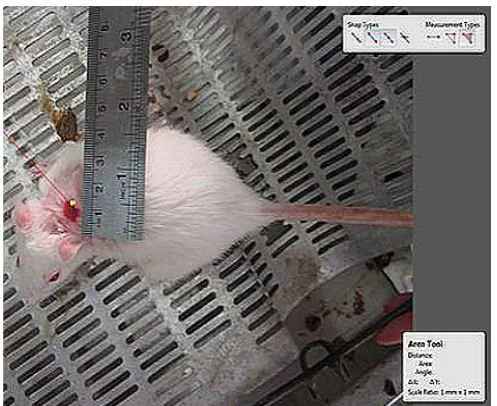
posture on a surgery table. Following shaving and aseptic preparation, a circular excision wound was made by cutting away approximately 300 mm2 full thickness of predetermined area on the anterior-dorsal side of each rat. All the test formulations were applied for 10 days starting from the day of wounding. Wound-healing property was evaluated by wound contraction percentage and wound closure time. Photographs were taken immediately after wounding and on days 6, 9, 12, 15, 18 and 21 post-operation by a digital camera while a ruler was placed near the wounds (Figure 1). The wound areas were analysed by Measuring Tool of Adobe Acrobat 9 Pro Extended software (Adobe Systems Inc, San Jose, CA, USA) and wound contraction percentage was calculated using the following
formula: Percentage of wound contraction = (A0 – At ) / A0 × 100.
Where A0 is the original wound area and At is the wound area at the time of imaging (12). The animals were left in separate cages for four days at room conditions for acclimatisation. Animal houses were in standard environmental conditions of temperature (22 ± 3 °C), humidity (60 ± 5%), and a 12 h light/dark cycle. The animals were maintained on standard pellet diet and tap water. All rats were closely observed for any infection and if they showed signs of infection were separated, excluded from the study and replaced.
Determination of Hydroxyproline Levels On the day 21 after surgery, a piece of skin from the healed wound area was collected and analysed for hydroxyproline content. As a major part of collagen, hydroxyproline has an essential role in collagen stability. The collagen is the major component of extracellular tissue, which gives support and strength. The hydroxyproline contents were estimated using a method
described by others (13). Briefly, tissues were
dried in a hot air oven at 60–70 °C to constant weight and were hydrolysed in 6N HCl at 130 °C for 4 h in sealed tubes. The hydrolysate was neutralised to pH 7.0 and was subjected to chloramine-T oxidation for 20 min. The reaction was terminated by addition of 0.4M perchloric acid and color was developed with the help of Ehrlich reagent at 60 °C and measured at 557 nm using UV-visible spectrophotometer (CamSpec M330, Cambridge CB2 4BG, UK).
Incision Wound Model and Biomechanical Testing
Twenty healthy male Wistar rats weighing approximately 170 g–180 g and six weeks of age were divided into 5 groups (n = 4), randomly. All animals of four groups were anesthetised as mentioned above and a paravertebral long incision of 4 cm length was made through the skin and cutaneous muscle at a distance about 1.5 cm from the middle on right side of the depilated back. After the incision was made, the two ends of the wound were sutured at 0.5 cm intervals with 3/0 nylon. The formulations were applied the same way in the excisional wound model. Ointments were applied once daily for 9 days. On day 9, sutures were removed and a strip of skin, 7 cm long, with the same widths of wound diameter, in the manner that the wound was located at the middle of the strip, was removed by a double-blade scalpel. The skin was then wrapped in Ringer’s soaked gauze, aluminum foils, and plastic bags and kept in -20 °C freezer until mechanical testing. The TA.XTPlus Texture Analyser mechanical test device was used for the assessment (Stable Micro Systems, Surrey GU7 1YL, UK). The samples
were fitted with appropriate clamps, the distance
between the clamps at the start of testing being 4 cm. The strips were loaded with 0 kg–30 kg load cell, with strain rate of 1 cm/min and the load elongation curves were obtained. Yield strength (yield point) (kg), ultimate strength (kg), maximum stored energy (kg/cm), and stiffness (kg/cm) were measured from the load elongation curves.
Statistical Analysis
Normal distributions of data were evaluated by Kruskal-Wallis variance analysis. Multiple comparison tests were used to know differences among experimental groups. One way ANOVA and post hoc Tuky’s test were used in toxicity and mechanical tests. Repeated measures ANOVA and a factorial were used for wound area analyses. SPSS 17 (SPSS Inc., Chicago, IL, USA) was used for statistical analyses. A P-value was set at 0·05.
Results
Acute Toxicity Test Findings
Dermal administration of the pulegone in highest doses, 5000 mg/kg did not produce any mortality and visible signs of toxicity when
observed 24 h after treatment and for further 14 days. Administered orally, the pulegone produced visible signs of toxicity in rats at doses of greater than100 mg/kg and mortality was observed in doses greater than 1600 mg/ kg. The signs included abnormal gait, increased respiration, decreased activity, unresponsive
to writhing test and limb paralysis. Significant
body weight loss was not observed after two weeks of oral and percutaneous administration of pulegone. Pulegone was more toxic through oral route than dermal route.
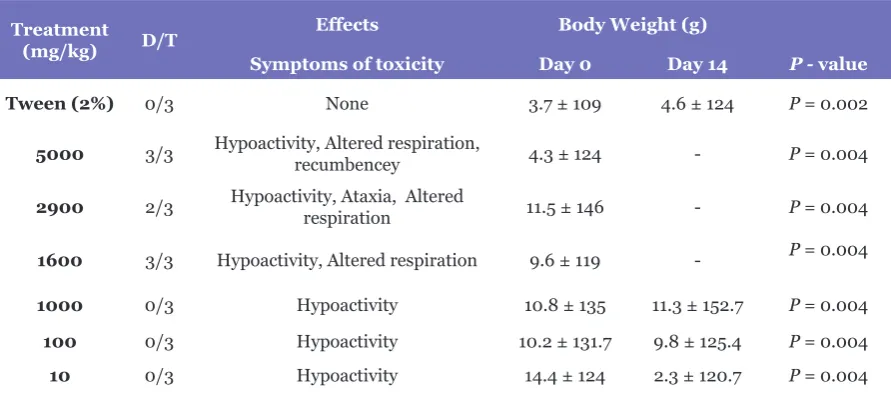
The LD50 was estimated to be 1250 mg/kg for the oral route and greater than 5000 mg/kg in dermal route. Visual inspection, on necropsy, did not reveal any signs of damage to organs (Table 1 and Table 2).
Reduction in Wound Area
Wound contraction percentage in different groups during the course of study is shown in Table 3. The healing rate of 0.5% ointment
treated group was significantly different
compared to other groups (P < 0.05). However,
time had significant effect on wound contraction
of all wounds (P = 0.035) (Figure 2). Hydroxyproline Content of Wound
Proline is hydroxylated to form hydroxyproline after protein synthesis. Hydroxyproline contents in the groups CG, sham surgery, E1, E2 and E3 were found to be 51.25±3.40, 58.41±4.62, 68.59±3.53, 86.32±3.18 and 74.26±4.73 mg g-1, respectively. Hydroxyproline contents were increased
significantly in 0.5% ointment treated group
which implies more collagen deposition compared to other experimental groups (P = 0.001).
Biomechanical Findings
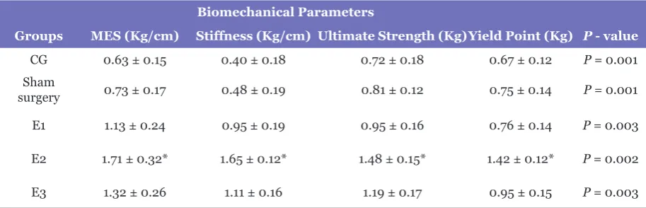
The biomechanical indices, maximum stored energy, stiffness, ultimate strength and yield strength obtained for 0.5% ointment
treated group were significantly higher than
Table 1. Mortality and behavioural changes are shown in acute dermal toxicity of pulegone examined rats. The pulegone dissolved in Tween 80 (2%) was rubbed as single doses in area 5cm × 5cm of the back zone of each rat. All animals were carefully examined for adverse effects, behavioural changes and mortality, for 14 days. Symptoms of toxicity are described for each group
Treatment
(mg/kg) D/T
Effects Body Weight (g)
Symptoms of toxicity Day 0 Day 14 P - value
Tween (2%) 0/3 None 121.3 ± 18.6 137.2 ± 19.3 P = 0.002
5000 0/3 Severe hypoactivity, local redness and congestion 127.3 ± 3.9 137.4 ± 6.2 P = 0.004
2900 0/3 Severe hypoactivity, local redness and congestion 123.2 ± 15.8 138.7 ± 11.3 P = 0.004
1600 0/3 hypoactivity 136.7 ± 10.4 130.3 ± 12.5 P = 0.004
1000 0/3 No clinical signs were observed 129.5 ± 6.9 125.7 ± 14.8 P = 0.002
100 0/3 No clinical signs were observed 110.7 ± 10.4 124.6 ± 15.3 P = 0.002
10 0/3 No clinical signs were observed 133.4 ± 12.1 138.3 ± 13.5 P = 0.002
D/T: dead/treated rats, latency: time to death after the animal was dosed, none: no toxic symptom was observed during the observation period.
Body Wight values are expressed as Mean ± SD. One way ANOVA and post hoc Tuky’s test were used.
Table 2.Mortality and behavioural changes in acute oral toxicity of pulegone are shown in examined rats. The pulegone, dissolved in Tween 80 (2%) was administered as single oral doses to 6 groups of 3 rats each. All animals were carefully examined for adverse effects, behavioural changes and mortality, for 14 days. Symptoms of toxicity are described for each group
Treatment
(mg/kg) D/T
Effects Body Weight (g)
Symptoms of toxicity Day 0 Day 14 P - value
Tween (2%) 0/3 None 3.7 ± 109 4.6 ± 124 P = 0.002
5000 3/3 Hypoactivity, Altered respiration, recumbencey 4.3 ± 124 - P = 0.004
2900 2/3 Hypoactivity, Ataxia, Altered respiration 11.5 ± 146 - P = 0.004
1600 3/3 Hypoactivity, Altered respiration 9.6 ± 119 - P = 0.004
1000 0/3 Hypoactivity 10.8 ± 135 11.3 ± 152.7 P = 0.004
100 0/3 Hypoactivity 10.2 ± 131.7 9.8 ± 125.4 P = 0.004
10 0/3 Hypoactivity 14.4 ± 124 2.3 ± 120.7 P = 0.004
D/T: dead/treated rats, latency: time to death after the animal was dosed, none: no toxic symptom was observed during the observation period.
Table 3. Effect of pulegone on circular excision wound contraction area (mm2) is represented. Values are given as mean ± SD
Wound area in days (mm2)
Groups Day 6 Day 9 Day 12 Day 15 Day 18 Day 21 P - value CG 254.67 ± 0.49 100.78 ± 1.78 85.13 ± 0.26 45.28 ± 1.34 21.43 ± 1.57 8.11 ± 0.21 P = 0.001 Sham
surgery 244.18 ± 0.45 96.55 ± 0.46 82.12 ± 1.39 41.50 ± 0.25 18.10 ± 1.17 4.04 ± 0.24 P = 0.001 E1 254.31 ± 1.37 87.23 ± 1.12 67.62 ± 0.15 25.63 ± 0.39 13.11 ± 0.38 2.75 ± 0.41 P = 0.003 E2 215.09 ± 0.44* 61.52 ± 0.75* 24.12 ± 0.27* 4.40 ± 1.18* 2.50 ± 0.47* 00.35 ± 0.57* P = 0.002 E3 234.60 ± 0.77 76.25 ± 0.49 43.22 ± 0.18 15.14 ± 0.14 7.28 ± 0.47 1.17 ± 0.12 P = 0.003
*: The mean difference is significant at the .05 level vs other experimental groups. Repeated measures ANOVA were used.
Figure 2. The representative photographs of wounds on day 15 show wound area in the experimental groups
Table 4. Biomechanical parameters assessed for each of the experimental groups are represented. Values are given as mean ± SD
Biomechanical Parameters
Groups MES (Kg/cm) Stiffness (Kg/cm) Ultimate Strength (Kg)Yield Point (Kg) P - value CG 0.63 ± 0.15 0.40 ± 0.18 0.72 ± 0.18 0.67 ± 0.12 P = 0.001 Sham
surgery 0.73 ± 0.17 0.48 ± 0.19 0.81 ± 0.12 0.75 ± 0.14 P = 0.001
E1 1.13 ± 0.24 0.95 ± 0.19 0.95 ± 0.16 0.76 ± 0.14 P = 0.003
E2 1.71 ± 0.32* 1.65 ± 0.12* 1.48 ± 0.15* 1.42 ± 0.12* P = 0.002
E3 1.32 ± 0.26 1.11 ± 0.16 1.19 ± 0.17 0.95 ± 0.15 P = 0.003
MES: Maximum Energy Stored. *: The mean difference is significant at the .05 level vs other experimental groups. One way
Discussion
Inflammation, proliferation and tissue
remodeling are three phases of healing process which occur following tissue damages as closely as possible to its natural state. The healing process is activated when platelets come into contact with exposed collagen leading to platelet aggregation and the release of clotting factors
resulting in the deposition of a fibrin clot at the site of injury. The fibrin clot serves as a
provisional matrix and sets the stage for the
subsequent events of healing. Inflammatory cells
also arrive along with the platelets at the injury site providing key signals known as growth
factors. The fibroblast is the connective tissue
cell responsible for collagen deposition required to repair the tissue injury. The collagen is the main constituent of extra cellular tissue, which is responsible for support and strength (14).
Monoterpenes, belonging to a large and diverse group of chemical compounds named ‘terpenes’, represent a group of naturally occurring organic compounds whose basic structure consists of two linked isoprene units, which are formed by a 5-carbon-base (C5) each. They are the most representative molecules constituting 90% of the essential oils and have a great variety of structures with several functions such as antimicrobial, hypotensive,
anti-inflammatory and antipruritic (15–18).
These compounds are inexpensive and have been
widely used in flavourings and fragrances since
the beginning of the 19th century. More recently, in the pharmaceutical industry, they play a great role because of their therapeutic potential (19). Pulegone, a naturally occurring monoterpene ketone, has been reported to be able to reduce the number of writhings induced by acetic acid with the same intensity shown by carvone (20). This monoterpene also reduces the licking time induced by formalin and increases the latency time in hot-plate test with non-participation of the opioid system (21).
Inflammation is the reaction of a normal
living tissue to local injury and it plays an important role in the defense mechanism which helps to protect us from infection or injury (22).
The function of inflammation is to eliminate the
foreign bodies or injurious agents. Furthermore, it removes damaged tissue components, so that the body can begin to heal and recover.
However, if the inflammation is left untreated
or uncontrolled, it will lead to many acute and chronic human diseases (23).
In vitro studies revealed that pulegone is a selective COX-2 inhibitor (24). Although no mechanism of action has been investigated, probably pulegone acts through the inhibition
of the production of inflammatory mediators
from the cascade of cyclooxygenase. It has also been stated that pulegone bears regenerative cell proliferation properties in experimental animals
(25). It seems as a result of anti-inflammatory
and cell proliferation properties of pulegone, the pulegone treated animals showed accelerated wound repair on the present study.
The investigation of cytotoxic activity of pulegone and its metabolites like piperitenone, piperitone, menthofuran and menthone demonstrated their cytotoxic activity against rat at bladder and a rat urothelial cell line (MYP3 cells) (26, 27). The animals in group E2 showed
a significant improvement in wound healing compared to group E3. This significant effect
might be associated with exuberant toxic effects of pulegone with 1g of the pulegone in ointment (group E3) where the toxic effect of the pulegone
dominated its beneficial wound healing activity.
In excisional wound model there was
a significant decrease in wound area. This
indicated improved collagen maturation by increased cross linking. The balance between synthesis and breakdown and so deposition of collagen is important in wound healing and development of wound strength (28).
Mechanical testing is sensitive to changes that occur during the progression of wound healing, and can be used as a tool to measure the quality of healing. One of the most important factors in the healing of wounds is the stimulation of wound strength (29). Wound strength is determined by the amount and quality of newly synthesized and deposited collagen, as well as degradation of preformed collagen (30). Tensile strength, which demonstrates the force per unit of cross-sectional area needed to break the wound, is an important
measure since it reflects the subdermal organisation of the collagen fibers in the newly
deposited collage (29). Tensile strength indicates how much the repaired tissue resists to break under tension and may indicate in part the quality of the repaired tissue (31).
Collagen is one of the major components that is mainly responsible for the mechanical properties of the skin (32). The net amount of wound collagen deposition depends on
collagen turnover and is a reflection of collagen
synthesis minus collagen breakdown (30).
have also been related to mechanical strength
of the skin. Apparently thick collagen fibrils
can resist greater tensile strength as opposed to thin ones (33). Once the skin is injured, the normal collagen will be replaced by scar collagen and the connective tissue will not regain the original highly organised structure of collagen. Thus, the healing skin is weaker and results in lower tensile strength as opposed to the normal skin (34).Mechanical property data provide a clinically relevant and functional assessment of wound healing quality. When compared to other experimental groups, pulegone treated animals
showed a statistically significant difference in
biomechanical parameters.
Hydroxyproline is a major component of the collagen that permits the sharp twisting of the collagen helix. It helps on providing stability to the triple-helical structure of collagen by forming hydrogen bonds. Hydroxyproline is found in few proteins other than collagen. For this reason, hydroxyproline content has been used as an indicator to determine collagen content (14). Increase in hydroxyproline content in pulegone treated groups indicated increased collagen content, since hydroxyproline is the direct estimate of collagen synthesis.
Although the present study showed the promising effect of pulegone on wound healing in rats, data regarding the histological and molecular mechanisms leading to its action remain to be investigated in depth. The authors have not provided the histologic and molecular evidence for the action of pulegone, which may be considered as a limitation of this study.
Conclusions
The pulegone resulted in significant
improvement of full thickness wound healing. Thus, from this study we concluded that pulegone had a reproducible wound healing potential in rats. However, because of toxicity of pulegone further dose-dependent study is needed to determine an appropriate dose that achieve
maximal efficacy in facial nerve transection
models.
Acknowledgements
This work was supported by the Urmia University [1435-94-95]. The authors would like to thank Dr Ali Hassanpour, Department of Mechanical Engineering of Biosystems, Faculty of Agriculture, Urmia University, for mechanical
testings, and Mr Matin, Mr Valinezhad and Mr Ansarinia for their technical expertise and care of animals.
Authors’ Contributions
Conception and design: RM
Analysis and interpretation of the data: RM Drafting of the article: RM
Critical revision of the article for important intellectual content: GJ-A
Final approval of the article: GJ-A
Provision of study materials or patients: GJ-A Statistical expertise: RM
Obtaining of funding: ZC
Administrative, technical, or logistic support: ZC Collection and assembly of data: ZC
Correspondence
Dr Rahim Mohammadi
DVM (Islamic Azad University, Urmia Branch), DVSc (Urmia University)
Assistant Professor of Veterinary Surgery Department Surgery and Diagnostic Imaging, Faculty of Veterinary Medicine, Urmia University, Nazloo Road, Urmia, 57153 1177, Iran.
Tel: +98 441 2770508 Fax: +98 441 277 19 26
E-mail: [email protected]
References
1. Thakur R, Jain N, Pathak R, Sandhu SS. Practices in wound healing studies of plants. Evid Based Complement Alternat Med. 2011;2011:438056. https://doi.org/10.1155/2011/438056
2. Heng MC. Wound healing in adult skin: aiming for perfect regeneration. Int J Dermatol. 2011;50(9):1058–1066. https://doi.org/10.1111/ j.1365-4632.2011.04940.x
3. Nayak BS, Pinto Pereira LM. Catharanthus roseus flower extract has wound-healing activity
in Sprague Dawley rats. BMC Complement Altern Med. 2006;21(6):41. https://doi. org/10.1186/1472-6882-6-41
4. Feedar A. Wound healing alternative in management. 2nd ed. Philadelphia: Davis; 1995.
6. Jalilzadeh-Amin G, Maham M, Dalir-Naghadeh B, Kheiri F. Effects of Mentha longifolia essential oil on ruminal and abomasal longitudinal smooth muscle in sheep. J Essent Oil Res. 2012;24:61–69. https://doi.org/10.1080/10412905.2012.646019
7. Turner GW, Croteau R. Organization of monoterpene biosynthesis in Mentha. Immunocytochemical localizations of geranyl diphosphate synthase, limonene-6-hydroxylase, isopiperitenol dehydrogenase, and pulegone reductase. Plant Physiol. 2004;136(4):4215– 4227. https://doi.org/10.1104/pp.104.050229
8. Flamini G, Cioni PL, Puleio R, Morelli I, Panizzi L. Antimicrobial activity of the essential oil of Calamintha nepeta and its constituent pulegone against bacteria and fungi. Phytother Res. 1999;13(4):349–351. https://doi.org/10.1002/
(SICI)1099-1573(199906)13:4<349::AID-PTR446>3.0.CO;2-Z
9. Kawata J, Kameda M, Miyazawa M. Cyclooxygenase-2 inhibitory effects of monoterpenoids with a p-menthane skeleton. Int J Essen Oil Therapeut. 2008;2:145–148.
10. Guimarães AG, Quintans JSS, Quintans-Júnior LJ. Monoterpenes with analgesic activity-a systematic review. Phytother Res. 2013;27(1):1– 15. https://doi.org/10.1002/ptr.4686
11. Lorke DA. New approach to practical acute toxicity testing. Arch Toxicol. 1983;54(4):275– 287.
12. Reddy BSR, Reddy KK, Naidu VGM, Madhusudhana K, Agwane SB, Ramakrishna S, et al. Evaluation of antimicrobial, antioxidant and wound-healing potentials of Holoptelea integrifolia. J Ethnopharmacol. 2008;115(2):249–256. https://doi.org/10.1016/j. jep.2007.09.031
13. Qiu Z, Kwon AH, Kamiyama Y. Effects of plasma
fibronectin on the healing of full-thickness skin
wounds in streptozotocin-induced diabetic rats. J Surg Res. 2007;138(1):64–70. https://doi. org/10.1016/j.jss.2006.06.034
14. Martin J, Zenilman M, Lazarus GS. Molecular microbiology: new dimensions for cutaneous biology and wound healing. J Invest Dermatol. 2010;130(1):38–48. https://doi.org/10.1038/ jid.2009.221
15. Bakkali F, Averbeck S, Averbeck D, Idaomar M. Biological effects of essential oils- a review. Food Chem Toxicol. 2008;46:446–475. https://doi. org/10.1016/j.fct.2007.09.106
16. Kordali S, Kotan R, Mavi A, Cakir A, Ala A, Yildirim A. Determination of the chemical composition and antioxidant activity of the essential oil of Artemisia dracunculus and of the antifungal and antibacterial activities of Turkish Artemisia absinthium, A. dracunculus, Artemisia santonicum, and Artemisia spicigera essential oils. J Agric Food Chem. 2005;53(24):9452– 9458. https://doi.org/10.1021/jf0516538
17. Bastos JF, Moreira IJ, Ribeiro TP, Medeiros IA, Antoniolli AR, De Sousa DP, et al. Hypotensive and vasorelaxant effects of citronellol, a monoterpene alcohol. Basic Clin Pharmacol Toxicol. 2010;106(4):331–337. https://doi. org/10.1111/j.1742-7843.2009.00492.x
18. Menezes IA, Barreto CM, Antoniolli AR, Santos MR, De Sousa DP. Hypotensive activity of terpenes found in essential oils. Z Naturforsch C. 2010;65(9–10):562–566.
19. Petasch J, Disch E, Markert S, Becher D, Schweder T, Hüttel B, et al. The oxygen-independent metabolism of cyclic monoterpenes in Castellaniella defragrans 65Phen. BMC Microbiol. 2014;14:164. https://doi. org/10.1186/1471-2180-14-164
20. De Sousa DP, Júnior EV, Oliveira FS, Almeida RN, Nunes XP, Barbosa-Filho JM. Antinociceptive activity of structural analogues of rotundifolone: structure–activity relationship. Z Naturforsch C. 2007;62(1–2):39–42.
21. De Sousa DP, Nóbrega FFF, De Lima MRV, Almeida RN. Pharmacological activity of (R)-(+)-pulegone, a chemical constituent of essential oils. Z Naturforsch C. 2011:66(7–8):353–359.
22. Saba ZH, Suzana M, My YA. Honey: food or medicine? Med. Health. 2013;8(1):3–18.
23. Rao CV, Verma AR, Gupta PK, Vijayakumar
M. Anti-inflammatory and antinociceptive
24. Demidova-Rice TN, Hamblin MR, Herman IM. Acute and impaired wound healing: pathophysiology and current methods for drug delivery, Part 1: normal and chronic wounds: biology, causes, and approaches to care. Adv Skin Wound Care. 2012;25(7):304–314. https://doi. org/10.1097/01.ASW.0000416006.55218.d0
25. Cohen, SM. Urinary bladder carcinogenesis. Toxicol. Pathol 1998;26(1):121–127. https://doi. org/10.1177/019262339802600114
26. Yousefbeyk F, Tabaside J, Ostad SN, Salehi Sourmaghi MH, Amin GR. Investigation of chemical composition and cytotoxic activity of aerial parts of Ziziphora clinopodioides Lam. Research Journal of Pharmacognosy (RJP). 2016;3(2):47–51.
27. Da Rocha MS, Dodmane PR, Arnold LL, Pennington KL, Anwar MM, Adams BR, et al. Mode of action of pulegone on the urinary bladder of F344 rats. Toxicol Sci. 2012;128(1):1–8. https://doi.org/10.1093/toxsci/kfs135
28. Dogan S. Demirer I. Kepenekci B. Erkek A. Kiziltay N. Hasirci S, et al. Epidermal growth factor-containing wound closure enhances wound healing in non-diabetic and diabetic rats. Int Wound J. 2009;6(2):107–115. https://doi. org/10.1111/j.1742-481X.2009.00584.x
29. Jimenez PA, Rampy MA. Keratinocytes Growth Factor-2 accelerates wound healing in incisional wounds. J Surg Res. 1999;81(2):238–242. https://doi.org/10.1006/jsre.1998.5501
30. Witte MB, Thornton FJ, Kiyama T, Efron DT, Schulz GS, Moldawer LL, et al. Metalloproteinase inhibitors and wound healing: a novel enhancer of wound strength. Surgery. 1998;124(2):464–470.
31. Rashed AN, Afifi FU, Disi AM. Simple evaluation
of the wound healing activity of a crude extract of Portulaca oleracea L. (growing in Jordan) in Mus musculus JVI-1. J Ethnopharmacol. 2003;88(2):131–136.
32. Oxlund H, Andreassen TT. The roles of hyaluronic acid, collagen and elastin in the mechanical properties of connective tissues. J Anat. 1980;131(Pt 4):611–620.
33. Jorgensen PH, Bang C, Andreassen TT,
Jorgensen KD. Growth hormone influences
collagen deposition and mechanical strength of intact rat skin. A dose-response study. Acta Endocrinologica (Copenh). 1989;120(6):767– 772.