Open Access
Research article
Intra-abdominal pressure in severe acute pancreatitis
Paivi Keskinen*
1, Ari Leppaniemi
2, Ville Pettila
1, Anneli Piilonen
3,
Esko Kemppainen
2and Marja Hynninen
1Address: 1Department of Anesthesiology and Intensive Care Medicine, Meilahti Hospital, Helsinki University Central Hospital, PO Box 340, 00029 HUS, Helsinki, Finland, 2Department of Gastroenterological and General Surgery, Meilahti Hospital, Helsinki University Central Hospital, Helsinki, Finland and 3Department of Radiology, Meilahti Hospital, Helsinki University Central Hospital, Helsinki, Finland
Email: Paivi Keskinen* - paivi.keskinen@helsinki.fi; Ari Leppaniemi - ari.leppaniemi@hus.fi; Ville Pettila - ville.pettila@hus.fi; Anneli Piilonen - anneli.piilonen@hus.fi; Esko Kemppainen - esko.kemppainen@hus.fi; Marja Hynninen - marja.hynninen@hus.fi * Corresponding author
Abstract
Background: Hospital mortality in patients with severe acute pancreatitis (SAP) remains high. Some of these patients develop increased intra-abdominal pressure (IAP) which may contribute to organ dysfunction. The aims of this study were to evaluate the frequency of increased IAP in patients with SAP and to assess the development of organ dysfunction and factors associated with high IAP.
Methods: During 2001–2003 a total of 59 patients with severe acute pancreatitis were treated in the intensive care unit (ICU) of Helsinki University Hospital. IAP was measured by the intravesical route in 37 patients with SAP. Data from these patients were retrospectively reviewed.
Results: Maximal IAP, APACHE II score, maximal SOFA score, maximal creatinine, age and maximal lactate were significantly higher in nonsurvivors. There was a significant correlation of the maximal IAP with the maximal SOFA, APACHE II, maximal creatinine, maximal lactate, base deficit and ICU length of stay. Patients were divided into quartiles according to the maximal IAP. Maximal IAP was 7–14, 15–18, 19–24 and 25–33 mmHg and the hospital mortality rate 10%, 12.5%, 22.2% and 50% in groups 1–4, respectively. A statistically significant difference was seen in the maximal SOFA, ICU length of stay, maximal creatinine and lactate values. The mean ICU-free days in groups 1–4 were 45.7, 38.8, 32.0 and 27.5 days, respectively. The difference between groups 1 and 4 was statistically significant.
Conclusion: In patients with SAP, increased IAP is associated with development of early organ failure reflected in increased mortality and fewer ICU-free days. Frequent measurement of IAP during intensive care is important in optimizing abdominal perfusion pressure and recognizing patients potentially benefitting from decompressive laparotomy.
Background
In spite of advances in the treatment of severe acute pan-creatitis, the hospital mortality rate remains high [1,2]. The major determinants of death are multiple organ
fail-ure (MOF), the extent of necrotic pancreatic parenchyma and the presence of bacterial infection [3]. Recent clinical experience has indicated that some patients dying of "early MOF" might have suffered from untreated abdom-Published: 17 January 2007
World Journal of Emergency Surgery 2007, 2:2 doi:10.1186/1749-7922-2-2
Received: 29 August 2006 Accepted: 17 January 2007
This article is available from: http://www.wjes.org/content/2/1/2
© 2007 Keskinen et al; licensee BioMed Central Ltd.
inal compartment syndrome (ACS). Massive fluid resusci-tation in the early course of the disease combined with the severe inflammatory process in the retroperitoneum could contribute to visceral oedema leading to increased intra-abdominal pressure (IAP). This acute increase of IAP may in the severe cases lead to early organ dysfunction and ACS [4].
In healthy individuals, IAP ranges from 0 to 5 mmHg and varies with the respiratory cycle [5,6]. Because the organs and other contents in abdomen are relatively noncom-pressible, any increase in the volume of the retroperito-neal or abdominal contents increases IAP. New consensus definitions for IAH (intra-abdominal hypertension) and ACS were set by the World Society on the Abdominal Compartment Syndrome (WSACS) [7]. IAH is a sustained increase in IAP above 12 mmHg. ACS is a sustained increase in IAP above 20 mmHg with new onset organ failure with or without a low APP. However, even values of lower than 15 mmHg may cause organ dysfunction [8,9].
The prevalence of IAH in critically ill patients depends on the threshold used. Most commonly, the maximal IAP value instead of median or mean IAP has been used. In patients with severe acute pancreatitis, IAP >25 mmHg was detected in 30% of patients [10]. In a recent study, IAP > 15 mmHg was found in 78% of the patients with severe acute pancreatitis [11]. In a mixed population of ICU patients, prevalence of IAH (cut-off 12 mmHg) was 59%, 8.2% of these patients were classified as having ACS [12]. In another study with mixed ICU population, 32% of the patients had IAH (cut-off 12 mmHg) and 4.2% had ACS on admission [13]. In abdominal surgical patients, incidence of IAH (cut-off 20 mmHg) was 33 to 39% [14,15] and incidence of ACS (cut-off 20–25 mmHg) 2– 36% [15-18].
This was a study on primary IAH and ACS according to the WSACS definitions. The aims of this study were to evalu-ate the degree of increased intra-abdominal pressure (IAP) in a group of patients with severe acute pancreatitis and to assess the development and progression of organ dysfunc-tion and other factors associated with high IAP.
Methods
Patients
ICU computerized database was used to identify all patients with severe acute pancreatitis treated in the ICU of Helsinki University Hospital during 2001–2003. Severe acute pancreatitis was diagnosed, if the patients presented with abdominal pain, increased serum amylases and at least one organ dysfunction. The diagnosis was confirmed by abdominal CT in all but one patient. IAP was measured by the intravesical route in 37 of these 59 patients. IAP
was measured if IAH was clinically suspected due to severe distension of the abdomen combined with a new or a worsening organ failure.
Definitions
Acute Physiology And Chronic Health Evaluation (APACHE) II score [19] and Balthazar classifications [20] were used to assess the severity of pancreatitis. Balthazar classification: Grade A – normal CT, Grade B – focal or dif-fuse enlargement of the pancreas, Grade C – pancreatic gland abnormalities and peripancreatic inflammation, Grade D – fluid collection in a single location, Grade E – two or more collections and/or gas bubbles in or adjacent to pancreas. Sequential Organ Failure Assessment (SOFA) score [21] was calculated daily to assess the extent of organ dysfunction, using the worst value of each day. Based on the highest measured IAP value, the patients were divided into quartiles.
Data collection
Patient data were retrospectively retrieved from the com-puterized patient database and the patient records. Age, gender, height, weight, body mass index (BMI), length of ICU stay, ICU-free days (out of 60 days), length of hospi-tal stay, hospihospi-tal morhospi-tality, medical history, etiology of pancreatitis and type of admission (primary or referral) were recorded. Parameters collected (days 1–14 in ICU) were IAP, lactate, C-reactive protein (CRP) at admission, creatinine, base deficit, need of renal replacement therapy, amount of fluids given, fluid balance, amount of perito-neal fluid (small, moderate or large; evaluated with CT), daily SOFA score, APACHE II score at admission and laparotomies during the ICU stay.
Intra-abdominal pressure
Intra-abdominal pressure was measured through a Foley bladder catheter [22,23]. Intravenous infusion set was connected to normal saline, three-way stopcock and a dis-posable pressure transducer. The urometer was cut near the Foley catheter and the transducer was connected to the urometer by a three-way stopcock. The infusion set was flushed with saline and the pressure transducer was zeroed at the level of symphysis pubis. With the patient in supine position, 50 ml of saline were injected into the bladder and IAP was measured during end expiration. Nowadays minimal instillation volumes (<25 ml) are used for IAP measurement [7,24,25].
Statistical analysis
test statistical significance between multiple groups in continuous variables. Fisher exact test was used in dichot-omous variables. Mann-Whitney test was used to test sig-nificance between two groups. Results are expressed as medians with interquartile (IQ) ranges. ICU-free days out of 60 are expressed as means. A p-value less than 0.05 was considered statistically significant.
Results
The overall hospital mortality rate was 24% (9 out of 37) in our study group. According to the WSACS definition, IAH was found in 84% (31 out of 37) of patients. 17/37 (46%) patients had recurrent IAH. 18/37 (49%) patients had ACS and 7/37 (19%) had recurrent ACS. Table 1 sum-marizes the demographic and clinical data among survi-vors and nonsurvisurvi-vors. The median age of the patients was 46 years (range 21–69 years), the most common etiologi-cal factor in this series was alcohol abuse (84%). Five patients underwent abdominal operations during the two first weeks in the ICU, additional four patients later on. The indication for two of the laparotomies during the study period was ACS. In patient 1 IAP decreased postop-eratively from 33 mmHg to 16–22 mmHg. The urine out-put increased from less than 1 ml/kg/h to more than 2 ml/ kg/h, plasma lactate normalized, and ventilatory function improved. In patient 2 IAP decreased postoperatively from 25 mmHg to 13 mmHg. The urine output increased from less than 0.5 ml/kg/h to 1.5 ml/kg/h and the venti-latory function improved. These effects on IAP were sus-tained in both of these patients over several days. Other reasons for laparotomy included intra-abdominal hemor-rhage, suspected bowel perforation, and verified or sus-pected infection of the peripancreatic necrosis. 43% (16 of 37) of the patients required renal replacement therapy. For those 22 patients in whom the IAP was not measured the mortality rate was 18%.
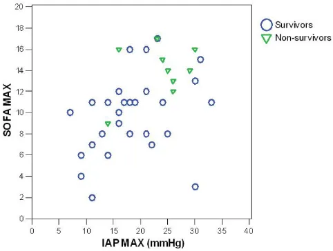
The maximal IAP, APACHE II score at admission, maxi-mal SOFA score, age, maximaxi-mal plasma lactate, maximaxi-mal creatinine and base deficit were significantly higher in the nonsurvivors (Table 2). There was a significant correlation of the maximal IAP with the maximal SOFA score (coeffi-cient 0.49, p = 0.001) (Fig. 1), APACHE II score (0.50, p = 0.001), maximal lactate value (0.46, p = 0.002), base def-icit (0.43, p = 0.008), maximal creatinine (0.56, p < 0.001) and duration of intensive care (0.48, p = 0.001). Maximal IAP did not correlate with the length of hospital stay or body-mass index (Spearman's non-parametric cor-relation).
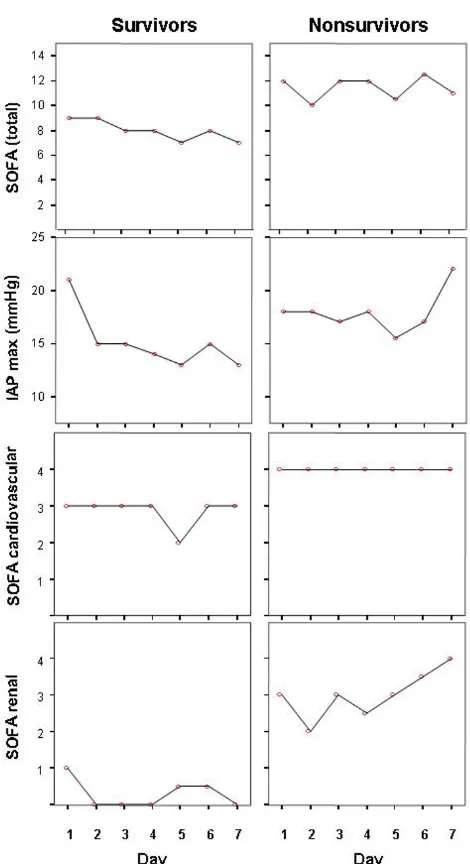
Figure 2 illustrates that nonsurvivors had higher maximal IAP, total SOFA, SOFA cardiovascular and renal scores on ICU-days 1–7 than survivors, whereas there was no signif-icant difference in SOFA respiratory, coagulation, hepatic or neurological score (data not shown). Patients were
divided into quartiles (8–10 patients in each group) according to the maximal IAP measured during days 1–14 in the ICU with maximum IAP values of 7–14, 16–18, 19– 24 and 25–33 mmHg in groups 1 – 4, respectively. The hospital mortality rates in groups 1–4 were 10%, 12.5%, 22.2% and 50%, respectively. A statistically significant dif-ference (Kruskall-Wallis test) between groups was seen in the maximal SOFA score (p = 0.01), maximal creatinine values (p = 0.01), duration of intensive care (p = 0.038) and maximal lactate values (p = 0.039). The difference in mortality rates between groups 1 and 4 was not statisti-cally significant (p = 0.14; Fisher exact test). The mean ICU-free days in groups 1–4 were 45.7, 38.8, 32.0 and 27.5 days, respectively (p = 0.045, Kruskall-Wallis test). The difference in ICU-free days between groups 1 and 4 was statistically significant (p = 0.023, Mann-Whitney test). ROC (receiver operating characteristics) curves for IAP max day 1–7, SOFA day 1 and APACHE II are shown in Figure 3.
Discussion
We found in this study, that high IAP in critically ill patients with acute pancreatitis correlates with the degree of organ dysfunction and length of intensive care.
Table 1: Demographic and clinical data of patients treated for severe acute pancreatitis.
All (%)
37 (100)
Male 33 (89)
Age (years) median, range 46 (21–69)
BMI (kg/m2) median, range 28 (21–42)
Pre-existing diseases
Hypertension 10 (27)
Diabetes 2 (5)
Cardiovascular 3 (8)
Hyperlipidemy 2 (5)
Chronic pancreatitis 2 (5)
Respiratory 2 (5)
Renal 2 (5)
Psychiatric 4 (11)
Etiology
Alcohol 31 (84)
Biliary 6 (16)
Amount of peritoneal fluid in CT
small 13 (35)
moderate 19 (51)
large 4 (11)
Balthazar classification
A, B 0 (0)
C 1 (3)
D 1 (3)
E 34 (92)
no CT 1 (3)
Primary admission 4 (11)
Increased IAP has deleterious effects on several organ sys-tems. Cardiovascular effects include decrease in cardiac output, ventricular end-diastolic volume, preload and venous return, and increase in afterload and intrathoracic pressure [14,26]. Respiratory failure is caused by the ele-vation of the diaphragm leading to a decline in lung and chest wall compliance, decrease in functional residual capacity, total lung capacity and residual volume. Ventila-tion-perfusion mismatch leads to hypoxia, hypercapnia and need of mechanical ventilation. Renal dysfunction is probably caused by a decrease in renal perfusion pressure, the filtration gradient and renal blood flow. Splanchnic perfusion may diminish due to a decrease in cardiac out-put or a direct mechanical compression of the splanchnic bed. Increased concentrations of vasopressin may also play a role in the development of splanchnic ischemia [27-31]. Several scoring systems such as APACHE II [19]
and SAPS [32] have been developed to predict outcome of critically ill patients. However, IAP is not included in any of these.
In earlier studies, an increase in IAP has been shown to be associated with increased mortality in surgical and trauma patients [17,18,29,30]. In trauma patients and liver recip-ients, acute ACS was associated with multiorgan failure and increased mortality [33-35]. In a recent multicenter study, the prevalence of IAH in critically ill patients was more than 50% [12]. In another study in a mixed ICU population, IAH during intensive care was an independ-ent outcome predictor [13].
In our study, the hospital mortality rate showed an increasing trend from 10% to 50% with the maximal IAP increasing from 7–14 to 25–33 mmHg, respectively. The maximal IAP correlated with the highest SOFA score, APACHE II-score on admission, maximal lactate and cre-atinine values, base deficit and the duration of intensive care. For quartiles divided by maximal IAP, the mean ICU-free days significantly decreased with increasing maximal IAP values. In a recent study, where IAP was measured in patients with SAP only when IAH was clinically suspected, the incidence of IAH was 78%. This is in agreement with our study where the incidence of IAH was 84%. In the cur-rent study the IAP showed an increasing trend during the first week in the ICU in non-survivors, whereas it decreased in survivors during the same time period. In contrast, SOFA score remained relatively unchanged in non-survivors. This may indicate that IAP could be a sen-sitive indicator of poor prognosis in patients with SAP. However, a larger study to confirm this finding is needed.
As a limitation to this study IAP was not measured in all patients with SAP in our ICU during the study period. Also, the patient number was not large enough to com-pare the predictive value of different factors on patient outcome. However, the adverse effects of high IAP on
dif-Correlation of maximal intra-abdominal pressure (IAP) with maximal Sequential Organ Failure Assessment (SOFA) score in survivors and nonsurvivors with severe acute pancreatitis
Figure 1
Correlation of maximal intra-abdominal pressure (IAP) with maximal Sequential Organ Failure Assessment (SOFA) score in survivors and nonsurvivors with severe acute pancreatitis.
Table 2: ICU data from survivors and nonsurvivors of severe acute pancreatitis.
Nonsurvivors Survivors
Median (IQ range) Median (IQ range) p value
IAP max (mmHg) 25 (19.5–27.5) 18 (13.3–22.8) 0.043
SOFA max 14 (12.5–16) 10.5 (7.3–11.8) 0.003
ICU stay (days) 27 (7.0–54.0) 15.5 (7.3–20.8) 0.257
Hospital stay (days) 28 (9.5–107.5) 26 (20.0–37.5) 0.986
CRP at admission (mg/l) 293 (212–385) 316 (246–378) 0.671
APACHE II 19 (17.0–22.5) 13 (10.0–17.0) 0.001
Lactate max (mmol/l) 2.7 (2.1–7.1) 1.5 (1.3–2.1) 0.006
BE min (mmol/l) -10.5 (-13.2-(-8.0)) -1.3 (-6.6-(-1.3)) <0.001
Creatinine max (mmol/l) 338 (181.5–547) 140.5 (67.5–280.3) 0.020
ferent organ systems are fairly well documented and IAH may be a contributing factor to worsening organ function (SOFA score) in patients with SAP.
For patients with severe acute pancreatitis IAH could be especially deleterious because increased IAP in animal studies has been associated with bacterial translocation [36,37]. General splanchnic hypoperfusion and decreased
blood flow to pancreas together with bacterial transloca-tion may predispose the patient to infected necrosis and poor outcome [31]. However, the role and the treatment of IAH in severe acute pancreatitis still remains to be elu-cidated. Recently published international recommenda-tions on the management of severe acute pancreatitis do not specifically address the management of IAH or ACS [38].
Once ACS is recognized, prompt treatment with decom-pressive laparotomy seems to be the best option although the exact indications, threshold IAP values and the most appropriate technique need further research. It is even more crucial in view of the considerable morbidity associ-ated with the procedure itself, especially if the fascial clo-sure is impossible leading to an open abdomen with significant long-term morbidity and need for reconstruc-tive surgery of the abdominal wall later on. As shown in selected trauma and other surgical patients, however, the risk of organ dysfunction can be decreased with timely decompressive laparotomy in patients not responding to nonoperative management of severe IAH [18,39,40]. The same effect can be expected in patient with severe acute pancreatitis.
Conclusion
In patients with severe acute pancreatitis, increased IAP is associated with development of early organ failure and fewer ICU-free days. Frequent measurement of IAP during intensive care in patients with severe acute pancreatitis could be important in optimizing abdominal perfusion pressure and recognizing patients potentially benefiting from early decompressive laparotomy.
ROC curves for IAP max day 1–7, APACHE II and day 1 SOFA points
Figure 3
ROC curves for IAP max day 1–7, APACHE II and day 1 SOFA points. AUC: area under curve.
Maximal intra-abdominal pressure (IAP) values, total Sequen-tial Organ Failure Assessment (SOFA) score, cardiovascular and renal SOFA scores during ICU-days 1–7 in the survivors and nonsurvivors of severe acute pancreatitis
Figure 2
Competing interests
The authors declare that they have no competing interests.
Authors' contributions
PK: Acquisition, analysis and interpretation of data, draft-ing the manuscript
AL: Interpretation of data, revising the manuscript criti-cally
VP: Analysis and interpretation of data, revising the man-uscript critically
AP: Analysis of data
EK: Revising the manuscript critically
MH: Analysis and interpretation of data, revising the man-uscript critically
All authors read and approved the final manuscript
References
1. Mayerle J, Hlouschek V, Lerch MM: Current management of acute pancreatitis. Nat Clin Pract Gastroenterol Hepatol 2005,
2:473-483.
2. Buter A, Imrie CW, Carter CR, Evans S, McKay CJ: Dynamic nature of early organ dysfunction determines outcome in acute pancreatitis. Br J Surg 2002, 89:298-302.
3. Garg PK, Madan K, Pande GK, Khanna S, Sathyanarayan G, Bohidar NP, Tandon RK: Association of extent and infection of pancre-atic necrosis with organ failure and death in acute necrotiz-ing pancreatitis. Clin Gastroenterol Hepatol 2005, 3:159-166. 4. Wilmer A: ICU management of severe acute pancreatitis. Eur
J Intern Med 2004, 15:274-280.
5. Sanchez NC, Tenofsky PL, Dort JM, Shen LY, Helmer SD, Smith RS:
What is normal intra-abdominal pressure? Am Surg 2001,
67:243-248.
6. Ivatury RRCML Malbrain ML, Sugrue M: Abdominal compartment syndrome. Georgetown, Landes Bioscience; 2006:308.
7. Malbrain ML, Cheatham ML, Kirkpatrick A, Sugrue M, Parr M, De Waele J, Balogh Z, Leppaniemi A, Olvera C, Ivatury R, D'Amours S, Wendon J, Hillman K, Johansson K, Kolkman K, Wilmer A: Results from the International Conference of Experts on Intra-abdominal Hypertension and Abdominal Compartment Syndrome. I. Definitions. Intensive Care Med 2006, 32:1722-1732. 8. Schwarte LA, Scheeren TW, Lorenz C, De Bruyne F, Fournell A:
Moderate increase in intraabdominal pressure attenuates gastric mucosal oxygen saturation in patients undergoing laparoscopy. Anesthesiology 2004, 100:1081-1087.
9. Malbrain ML: Is it wise not to think about intraabdominal hypertension in the ICU? Curr Opin Crit Care 2004, 10:132-145. 10. Pupelis G, Austrums E, Snippe K, Berzins M: Clinical significance of
increased intraabdominal pressure in severe acute pancrea-titis. Acta Chir Belg 2002, 102:71-74.
11. De Waele JJ, Hoste E, Blot SI, Decruyenaere J, Colardyn F: Intra-abdominal hypertension in patients with severe acute pan-creatitis. Crit Care 2005, 9:R452-7.
12. Malbrain ML, Chiumello D, Pelosi P, Wilmer A, Brienza N, Malcangi V, Bihari D, Innes R, Cohen J, Singer P, Japiassu A, Kurtop E, De Keu-lenaer BL, Daelemans R, Del Turco M, Cosimini P, Ranieri M, Jacquet L, Laterre PF, Gattinoni L: Prevalence of intra-abdominal hyper-tension in critically ill patients: a multicentre epidemiologi-cal study. Intensive Care Med 2004, 30:822-829.
13. Malbrain ML, Chiumello D, Pelosi P, Bihari D, Innes R, Ranieri VM, Del Turco M, Wilmer A, Brienza N, Malcangi V, Cohen J, Japiassu A, De Keulenaer BL, Daelemans R, Jacquet L, Laterre PF, Frank G, de Souza P, Cesana B, Gattinoni L: Incidence and prognosis of
intraab-dominal hypertension in a mixed population of critically ill patients: A multiple-center epidemiological study. Crit Care Med 2005, 33:315-322.
14. Sugrue M, Jones F, Lee A, Buist MD, Deane S, Bauman A, Hillman K:
Intraabdominal pressure and gastric intramucosal pH: is there an association? World J Surg 1996, 20:988-991.
15. Balogh Z, McKinley BA, Cocanour CS, Kozar RA, Valdivia A, Sailors RM, Moore FA: Supranormal trauma resuscitation causes more cases of abdominal compartment syndrome. Arch Surg
2003, 138:637-42; discussion 642-3.
16. Malbrain ML: Abdominal pressure in the critically ill: measure-ment and clinical relevance. Intensive Care Med 1999,
25:1453-1458.
17. Raeburn CD, Moore EE, Biffl WL, Johnson JL, Meldrum DR, Offner PJ, Franciose RJ, Burch JM: The abdominal compartment syn-drome is a morbid complication of postinjury damage con-trol surgery. Am J Surg 2001, 182:542-546.
18. Ertel W, Oberholzer A, Platz A, Stocker R, Trentz O: Incidence and clinical pattern of the abdominal compartment syndrome after "damage-control" laparotomy in 311 patients with severe abdominal and/or pelvic trauma. Crit Care Med 2000,
28:1747-1753.
19. Knaus WA, Draper EA, Wagner DP, Zimmerman JE: APACHE II: a severity of disease classification system. Crit Care Med 1985,
13:818-829.
20. Balthazar EJ, Ranson JH, Naidich DP, Megibow AJ, Caccavale R, Cooper MM: Acute pancreatitis: prognostic value of CT. Radi-ology 1985, 156:767-772.
21. Vincent JL, Moreno R, Takala J, Willatts S, De Mendonca A, Bruining H, Reinhart CK, Suter PM, Thijs LG: The SOFA (Sepsis-related Organ Failure Assessment) score to describe organ dysfunc-tion/failure. On behalf of the Working Group on Sepsis-Related Problems of the European Society of Intensive Care Medicine. Intensive Care Med 1996, 22:707-710.
22. Cheatham ML, Safcsak K: Intraabdominal pressure: a revised method for measurement. J Am Coll Surg 1998, 186:594-595. 23. Kron IL, Harman PK, Nolan SP: The measurement of
intra-abdominal pressure as a criterion for intra-abdominal re-explora-tion. Ann Surg 1984, 199:28-30.
24. Malbrain ML, Deeren DH: Effect of bladder volume on meas-ured intravesical pressure: a prospective cohort study. Crit Care 2006, 10:R98.
25. De Waele J, Pletinckx P, Blot S, Hoste E: Saline volume in trans-vesical intra-abdominal pressure measurement: enough is enough. Intensive Care Med 2006, 32:455-459.
26. Bloomfield GL, Ridings PC, Blocher CR, Marmarou A, Sugerman HJ:
A proposed relationship between increased intra-abdomi-nal, intrathoracic, and intracranial pressure. Crit Care Med
1997, 25:496-503.
27. Walker J, Criddle LM: Pathophysiology and management of abdominal compartment syndrome. Am J Crit Care 2003,
12:367-71; quiz 372-3.
28. Hunter JD, Damani Z: Intra-abdominal hypertension and the abdominal compartment syndrome. Anaesthesia 2004,
59:899-907.
29. McNelis J, Soffer S, Marini CP, Jurkiewicz A, Ritter G, Simms HH, Nathan I: Abdominal compartment syndrome in the surgical intensive care unit. Am Surg 2002, 68:18-23.
30. McNelis J, Marini CP, Simms HH: Abdominal compartment syn-drome: clinical manifestations and predictive factors. Curr Opin Crit Care 2003, 9:133-136.
31. Malbrain ML, Deeren D, De Potter TJ: Intra-abdominal hyperten-sion in the critically ill: it is time to pay attention. Curr Opin Crit Care 2005, 11:156-171.
32. Le Gall JR, Lemeshow S, Saulnier F: A new Simplified Acute Phys-iology Score (SAPS II) based on a European/North Ameri-can multicenter study. Jama 1993, 270:2957-2963.
33. Balogh Z, McKinley BA, Holcomb JB, Miller CC, Cocanour CS, Kozar RA, Valdivia A, Ware DN, Moore FA: Both primary and second-ary abdominal compartment syndrome can be predicted early and are harbingers of multiple organ failure. J Trauma
2003, 54:848-59; discussion 859-61.
34. Biancofiore G, Bindi ML, Romanelli AM, Bisa M, Boldrini A, Consani G, Filipponi F, Mosca F: Postoperative intra-abdominal pressure and renal function after liver transplantation. Arch Surg 2003,
Publish with BioMed Central and every scientist can read your work free of charge "BioMed Central will be the most significant development for disseminating the results of biomedical researc h in our lifetime."
Sir Paul Nurse, Cancer Research UK
Your research papers will be:
available free of charge to the entire biomedical community
peer reviewed and published immediately upon acceptance
cited in PubMed and archived on PubMed Central
yours — you keep the copyright
Submit your manuscript here:
http://www.biomedcentral.com/info/publishing_adv.asp
BioMedcentral 35. Biancofiore G, Bindi ML, Romanelli AM, Boldrini A, Consani G, Bisa
M, Filipponi F, Vagelli A, Mosca F: Intra-abdominal pressure mon-itoring in liver transplant recipients: a prospective study.
Intensive Care Med 2003, 29:30-36.
36. Gargiulo NJ 3rd, Simon RJ, Leon W, Machiedo GW: Hemorrhage exacerbates bacterial translocation at low levels of intra-abdominal pressure. Arch Surg 1998, 133:1351-1355.
37. Diebel LN, Dulchavsky SA, Brown WJ: Splanchnic ischemia and bacterial translocation in the abdominal compartment syn-drome. J Trauma 1997, 43:852-855.
38. Nathens AB, Curtis JR, Beale RJ, Cook DJ, Moreno RP, Romand JA, Skerrett SJ, Stapleton RD, Ware LB, Waldmann CS: Management of the critically ill patient with severe acute pancreatitis. Crit Care Med 2004, 32:2524-2536.
39. Sugrue M, Jones F, Janjua KJ, Deane SA, Bristow P, Hillman K: Tem-porary abdominal closure: a prospective evaluation of its effects on renal and respiratory physiology. J Trauma 1998,
45:914-921.
40. Meldrum DR, Moore FA, Moore EE, Franciose RJ, Sauaia A, Burch JM:
Prospective characterization and selective management of the abdominal compartment syndrome. Am J Surg 1997,