C A S E R E P O R T
Open Access
Successful pre-emptive kidney
transplantation in a cystinuria patient with
nephrolithiasis-related end-stage renal
disease
Masatoshi Matsunami
1*, Kazuya Kinoshita
1, Kiho Tanaka
1, Yuki Nakamura
1, Kenichi Ohashi
3,5, Takeshi Fujii
3,
Yoshifumi Ubara
2,4and Yasuo Ishii
1Abstract
Background:Cystinuria is a rare autosomal recessive metabolic disorder that affects renal and intestinal cystine transport. Cystine stones are found in only 1–2% of all stone formers. Patients with cystinuria are at high risk for nephrolithiasis and subsequent morbidity. In spite of the various medical and surgical treatments that are currently available for cystinuria, some patients gradually develop kidney failure, with only a few reported cases regarding kidney transplantation (KTx) to treat end-stage renal disease (ESRD) secondary to cystinuria. Cystinuria is likely not to be a systemic disease; thus, renal replacement with transplantation seems a good therapeutic option for ESRD. However, few cystinuria patients have undergone KTx due to ESRD.
Case presentation:We herein describe the case of a 49-year-old man with cystinuria, frequent stone events, and ESRD who underwent pre-emptive ABO-incompatible kidney transplantation. At 2 years and 6 months post-transplantation, the patient remains asymptomatic with no prophylactic therapy for cystinuria, and the allograft function has been preserved without evidence of rejection.
Conclusions:In conclusion, a cystinuria patient with nephrolithiasis-related ESRD was successfully treated by transplantation. Although additional cases are required to confirm the efficacy of this approach, renal replacement may be useful for treating ESRD in patients with rare hereditary forms of kidney stone disease.
Keywords: Cystinuria, Nephrolithiasis, Kidney transplantation, Pre-emptive, ESRD
Background
Cystinuria is a rare, autosomal recessive hereditary disease that was first described by Archibald Garrod in 1908 [1]. It is also known as the most frequent monogenic cause of renal stones. The condition is present in approximately 1% of adults and 8% of children with nephrolithiasis. Glo-bally, the average prevalence of cystinuria is approximately 1 per 7000 births; however, the prevalence ranges and widespread variation are observed [2, 3]. This disease is caused by the impairment of the epithelial transport of di-basic amino acids (cystine, ornithine, arginine, and lysine)
in the renal proximal tubule and small intestine [2]. Be-cause of its poor solubility at a typical urine pH, cystine alone leads to urinary precipitation and recurrent nephro-lithiasis that can cause obstruction, infection, and, ultim-ately, chronic kidney disease (CKD) [2, 4]. Usually, the diagnosis is based on the detection of urinary cystine crys-tals, a stone analysis, or increased urinary cystine levels. In some patients, stones develop into staghorn calculi [3].
Preventive medical managements include hydration (high fluid intake) and urinary alkalinization using orally administered potassium citrate. If hydration and urinary alkalinization fail to prevent cystine stone recurrence, the next step in the treatment should be the introduc-tion of chelaintroduc-tion or anti-urolithic therapy (such as tio-pronin) to reduce the urinary concentration of cystine
© The Author(s). 2019Open AccessThis article is distributed under the terms of the Creative Commons Attribution 4.0 International License (http://creativecommons.org/licenses/by/4.0/), which permits unrestricted use, distribution, and reproduction in any medium, provided you give appropriate credit to the original author(s) and the source, provide a link to the Creative Commons license, and indicate if changes were made. The Creative Commons Public Domain Dedication waiver (http://creativecommons.org/publicdomain/zero/1.0/) applies to the data made available in this article, unless otherwise stated. * Correspondence:matsunami-m@toranomon.gr.jp
1Department of Surgery, Nephrology Center, Toranomon Hospital, 2-2-2
Toranomon, Minato-ku, Tokyo 105-8470, Japan
[3]. In cases of stone formation, large stones that are as-sociated with pain, infection, or symptoms of obstruc-tion require surgical intervenobstruc-tion, such as extracorporeal shockwave lithotripsy (ESWL), percutaneous nephro-lithotomy (PCNL), or endoscopic retrograde techniques [2]. In contrast, smaller asymptomatic stones may be monitored with close ultrasonographic follow-up [2].
End-stage renal disease (ESRD) seems to be an un-common condition in cystinuria patients, and few stud-ies have evaluated the comorbiditstud-ies associated with cystinuria [4, 5]. Our search of the PubMed database for English studies on successful kidney transplantation (KTx) for the treatment of ESRD secondary to cystinuria revealed only four previous reports [6–9]. The most re-cent case, that of a male aged in his 20s with bilateral staghorn calculi, was reported in 1993. Despite bilateral pyelolithotomy, the patient developed ESRD requiring renal replacement therapy (RRT) and eventually under-went KTx [8].
Thus, it is likely that cystinuria is not a systemic dis-ease, making renal replacement with transplantation seems a good therapeutic option for ESRD due to uria. We herein report a case in which an adult cystin-uria patient with recurrent nephrolithiasis leading to ESRD was successfully treated by KTx.
Case presentation
The patient was a 49-year-old man from Japan with a life-long and complicated history of cystinuria. He was diag-nosed with cystinuria based on a stone analysis shortly after birth. His medical history was notable for multiple episodes of renal stones. He had no known family history of nephrolithiasis. At 1 year of age, he developed right-sided ureteric calculi and ureterolithotomy was per-formed. At 6 years of age, he suffered from left-sided ur-eteric calculi, and ureterolithotomy was performed as well. Despite the initiation of conservative treatment with potassium citrate and tiopronin, disease remission was not achieved. His next symptomatic stone event occurred at 23 years of age, when he was diagnosed with bilateral
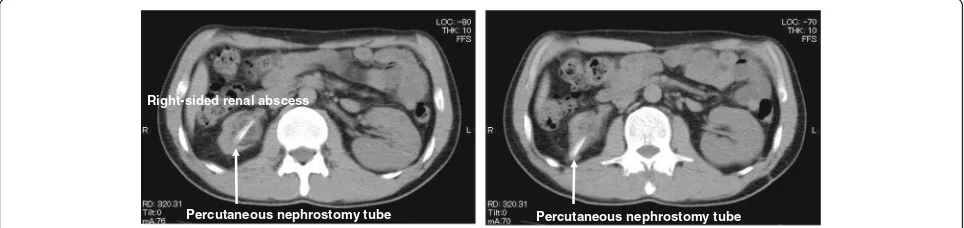
staghorn calculi. At this point, unfortunately, his right-sided kidney was already non-functioning. At 31 years of age, he had recurrent left-sided renal calculi and required PCNL for stone removal. At 39 years of age, the patient had an episode of right-sided renal abscess and was treated with intravenous antibiotic therapy with percutan-eous nephrostomy (Fig. 1). His serum creatinine (Cr) gradually increased to approximately 1.8 mg/dL. The pa-tient was referred to our hospital for further investigation. Since there was a possibility of renal abscess recurrence, right-sided nephrectomy was eventually performed. The isolated kidney with obstructive pyelonephritis showed diffuse thinning of the renal parenchyma, interstitial fibro-sis, hyalinization, and obscuring of the corticomedullary junction (Fig. 2). Thereafter, his renal function grad-ually declined and progressed to ESRD.
At 47 years of age, RRT was required because of fre-quent episodes of calculi, such as ureterolithiasis and nephrolithiasis, which ultimately resulted in ESRD. Both of the patient’s parents carried a risk of having heterozy-gous mutations and were excluded as donors. However, his wife, a 45-year-old woman, volunteered to donate her kidney to him, so we decided to undergo pre-emptive living donor KTx.
The proposed transplant was ABO incompatible, from a donor with blood type A to a recipient with blood type B. At 2 weeks prior to transplantation, the administra-tion of mycophenolate mofetil (MMF, 500 mg/day) and the anti-CD20 monoclonal antibody rituximab (1 dose of 200 mg) was initiated according to our pre-transplantation regimen (Fig.3).
Preoperatively, tacrolimus (TAC) and methylprednisolone (mPSL) were also administered from 6 days before trans-plantation. TAC was started at a dose of 0.1 mg/kg/day and then increased to 0.15 mg/kg/day. Following 2 weeks of desensitization therapy, the patient underwent two sessions of hemodialysis (HD) and one session of double filtration plasmapheresis (DFPP) before transplantation.
In addition, induction therapy with intravenous methyl-prednisolone (mPSL [500 mg]) and basiliximab (1 dose of 20 mg) was administered on the day of transplantation.
Percutaneous nephrostomy tube Percutaneous nephrostomy tube Right-sided renal abscess
The renal graft was transplanted without incident into the left iliac fossa (because of a previous operation at the right iliac fossa). Subsequently, the graft became pink and the urine was immediately produced. The post-transplantation immunosuppression protocol consisted of TAC, MMF (1500 mg/day), and mPSL. Basiliximab was also administered 4 days after transplantation. The TAC trough level was maintained at 8–12 ng/mL for the first few weeks after transplantation. mPSL was gradually tapered to 5 mg by the end of the post-transplantation period and switched to an oral formulation (Fig.3).
Notably, the patient’s Cr levels decreased after trans-plantation and are now maintained at 1.2 mg/dL under immunosuppression with a triple-drug regimen (TAC,
MMF, and mPSL) (Fig. 3). On post-operative days 180 and 544, a urinary amino acid analysis was performed and revealed that the patient’s urinary amino acid levels remained within the normal ranges.
At present, 2 years and 6 months after KTx, the patient remains asymptomatic with no prophylactic therapy for cystinuria, and the allograft function has been preserved without evidence of rejection. On magnetic resonance angiography (MRA), there was no transplant renal artery stenosis, and the renal blood flow was maintained (Fig.4). In addition, the hematologic follow-up has not demon-strated any significant adverse effects of immunosuppres-sion (such as dyslipidemia or myelosuppresimmunosuppres-sion) or cytomegalovirus (CMV) infection events.
Tubular atrophywith thyroidization
Strong hyalinization of glomeruli (circle)
20x, HE-staining
a
b
Fig. 2The gross and microscopic observation of the isolated polycystic kidney.aThe gross specimen showed a massively enlarged kidney with obstructive pyelonephritis.bMicroscopic (histologic) observation showed wide interstitial fibrosis, tubular thyroidization, and tubular atrophy. Most glomeruli were hyalinized and collapsed
0 1 0 0 2 0 0 3 0 0 4 0 0 5 0 0 6 0 0 7 0 0 8 0 0 0
2 4 6 8 1 0
d a y s a fte r tra n s p la n ta tio n C r (m g /d L )
0 2 0 4 0 6 0
m P S L (m g/day )
0 5 0 0 1 0 0 0 1 5 0 0
M M F (m g /d a y )
0 5 1 0 1 5
TA C (m g/day )
1500 1250
KTx HD (day-2) HD+DFPP (day-1)
Rituximab 200mg (day -13) Basiliximab 20mg (day 0, 4) 4-15
2 4
5-60 mPSL500mg (day 0)
3.5
Urinary amino acid analyses
HD: hemodialysis
DFPP: double filtration plasmapheresis
TAC: tacrolimus
MMF: mycophenolate mofetil
mPSL: methylprednisolone
day 180 day 544
Discussion and conclusions
We presented the case of a cystinuria patient with nephrolithiasis-related ESRD who was followed for 2 years and 6 months after pre-emptive KTx. An accur-ate and early diagnosis of cystinuria is important for the long-term management of patients. In this case, the diagnosis of cystinuria was made based on a stone ana-lysis and no genetic anaana-lysis was performed. The condi-tion was characterized by lifelong, recurrent stone symptoms that were difficult to manage, both medically and surgically.
Patients with rare hereditary forms of kidney stone dis-eases, including primary hyperoxaluria, cystinuria, Dent disease, and adenine phosphoribosyltransferase deficiency, experience recurring stones—often starting in child-hood—and are at high risk for CKD [5]. ESRD is common in primary hyperoxaluria, cystinuria, Dent disease, and ad-enine phosphoribosyltransferase deficiency. However, the CKD in cystinuria is usually less aggressive [5].
Recently, Prot-Bertoye et al. investigated CKD and its risk factors among a large series of French patients with cystinuria. This study showed that the prevalence of CKD in cystinuria patients was (26.7%), but that only 1.1% (5 of 442) of cystinuria patients developed ESRD requiring RRT [4]. Among these CKD patients with cystinuria, the preva-lence of hypertension (28.6%) was high, and hypertension was found to be significantly associated with male sex, age, and CKD [4]. In this case, the cystinuria patient
underwent nephrectomy, which has the potential to lead to ESRD. In addition to CKD, he also suffered from hyper-tension. Following transplantation, a blood pressure of 120/80–135/85 mmHg has been maintained on Nifedipine (20 mg, once daily).
Kidney transplantation is the preferred treatment for ESRD. Our search of the literature only identified four cases in which KTx was performed for the treatment of ESRD secondary to cystinuria [6–9]. Among them, only one case resulted in graft failure 10 days after KTx due to acute rejection, not due to cystinuria, and the trans-planted kidneys functioned well without recurrence of cystinuria for the remaining three cases (Table1). These findings suggest that cystinuria does not develop in the transplanted kidneys with intact cystine transport.
In addition, as Tuso et al. described, hereditary kidney stone disease such as cystinuria is likely not to be a sys-temic disease, but causes kidney dysfunction [8]. Thus, we hypothesized that transplantation could be used to treat the patient’s ESRD. Theoretically, there should be no recurrence of cystinuria, and in this case, the func-tion of the allograft was nearly normal with normal urin-ary amino acid levels maintained for 2 years and 6 months after transplantation.
Meanwhile, since cystinuria is a hereditary disease, do-nors need to be carefully selected. In this case, we were not able to conduct genetic analysis because the patient’s parents and wife did not consent. Despite the donor be-ing the patient’s wife, the patient is currently doing well with no subjective symptoms after transplantation.
According to recent studies, pre-emptive transplant-ation has many benefits, including improved patient and graft survival, and a less delayed graft function in compari-son to transplantation after dialysis [10]. Other outcomes are improved, including a reduced overall cost of care, and improved patient employment status [11]. Despite the clearly defined benefits, the identification of a living donor is essential for successful pre-emptive transplantation. In Japan, patients can find the process of asking family mem-bers to donate a kidney to be daunting. Hence, the neph-rologist or transplant surgeon should provide education on the donation process and the general safety of dona-tion. If potential donors move through the process smoothly, then the likelihood of transplantation increases. In Japan, the rate of pre-emptive transplantation from Aorta
Transplanted kidney Transplant renal artery
Left internal iliac artery
Left external iliac artery
Fig. 4MRA coronal image of a transplanted kidney. Renal artery anastomosis to the left external iliac artery showed no anastomotic stenosis
Table 1Clinical characteristics of recipients with cystinuria after kidney transplantation
Patient Underlying disease Age (years) Sex KTx Cystinuria recurrence Follow-up Reference
Case 1 Cystinuria 28 M NA No 1 year and 8 months Kelly et al. [6]
Case 2 Cystinuria 38 M DDKT NA Rejection (POD 10) Hoitsma et al. [7]
Case 3 Cystinuria 20s M LDKT No 3 years and 6 months Tuso et al. [8]
Case 4 Cystinuria 46 F DDKT No NA Krizek et al. [9]
living donors has increased year by year and reached ap-proximately 25.5% in 2013 [10,12].
In conclusion, based on the excellent results that were observed, nephrolithiasis-related ESRD in a cys-tinuria patient was considered to have been successfully treated by transplantation. Although continued urinary amino acid analyses and long-term follow-up are needed to accurately determine the validity of KTx as a treatment for cystinuria, renal replacement may poten-tially be useful in patients who have rare hereditary forms of kidney stone diseases, including cystinuria. We hope that this case will be helpful to others facing the problem of deciding whether or not to attempt transplantation for the treatment of patients with a his-tory of ESRD secondary to cystinuria. Furthermore, the accumulation of new reports in which good outcomes are achieved may help to establish KTx as an acceptable therapeutic option.
Abbreviations
CKD:Chronic kidney disease; ESRD: End-stage renal disease; KTx: Kidney transplantation; RRT: Renal replacement therapy
Acknowledgements
The authors would like to thank Dr. Atsushi Kato and Dr. Hideshi lshizuka for their careful patient management at Saitama Shakaihoken Hospital (current JCHO Saitama Medical Center) in Saitama, Japan.
Authors’contributions
MM designed and wrote the manuscript. KK, KT, and YN treated the patient. KO and TF supplied the pathology reports. YU and YI supervised the transplant program and corrected the manuscript. All authors read and approved the final manuscript.
Funding
No funding was obtained for this study.
Availability of data and materials
All data supporting our findings are contained within the manuscript.
Ethics approval and consent to participate
Not applicable.
Consent for publication
Written informed consent was obtained from the patient to publish this case report and any accompanying images. A copy of the written consent form is available for review by the editor of this journal.
Competing interests
The authors declare that they have no competing interests.
Author details
1Department of Surgery, Nephrology Center, Toranomon Hospital, 2-2-2
Toranomon, Minato-ku, Tokyo 105-8470, Japan.2Nephrology Center, Toranomon Hospital, Tokyo, Japan.3Department of Pathology, Toranomon Hospital, Tokyo, Japan.4Okinaka Memorial Institute for Medical Research, Tokyo, Japan.5Department of Pathology, Yokohama City University Graduate School of Medicine, Yokohama, Japan.
Received: 11 March 2019 Accepted: 25 June 2019
References
1. Garrod A. The Croonian lectures on inborn errors of metabolism. Lancet. 1908;172(4427):1–7.
2. Chillaron J, Font-Llitjos M, Fort J, Zorzano A, Goldfarb DS, Nunes V, Palacin M. Pathophysiology and treatment of cystinuria. Nat Rev Nephrol. 2010;6(7):424–34.
3. Cochat P, Pichault V, Bacchetta J, Dubourg L, Sabot JF, Saban C, Daudon M, Liutkus A. Nephrolithiasis related to inborn metabolic diseases. Pediatr Nephrol. 2010;25(3):415–24.
4. Prot-Bertoye C, Lebbah S, Daudon M, Tostivint I, Bataille P, Bridoux F, Brignon P, Choquenet C, Cochat P, Combe C, et al. CKD and its risk factors among patients with cystinuria. Clin J Am Soc Nephrol. 2015;10(5):842–51. 5. Rule AD, Krambeck AE, Lieske JC. Chronic kidney disease in kidney stone
formers. Clin J Am Soc Nephrol. 2011;6(8):2069–75.
6. Kelly S, Nolan EP. Excretory rates in posttransplant cystinuric patient. Jama. 1978;239(12):1132.
7. Hoitsma AJ, Koene RA, Trijbels FJ, Monnens LA. Disappearance of cystinuria after renal transplantation. Jama. 1983;250(5):615.
8. Tuso P, Barnett M, Yasunaga C, Nortman D. Cystinuria and renal transplantation. Nephron. 1993;63(4):478.
9. Krizek V, Erben J, Lazne M, Navratil P, Svab J. Disappearance of cystinuria after kidney transplantation. Br J Urol. 1983;55(5):575.
10. Goto N, Okada M, Yamamoto T, Tsujita M, Hiramitsu T, Narumi S, Katayama A, Kobayashi T, Uchida K, Watarai Y. Association of dialysis duration with outcomes after transplantation in a japanese cohort. Clin J Am Soc Nephrol. 2016;11(3):497–504.
11. Fishbane S, Nair V. Opportunities for increasing the rate of preemptive kidney transplantation. Clin J Am Soc Nephrol. 2018;13(8):1280–2. 12. Yagisawa T, Mieno M, Yoshimura N, Yuzawa K, Takahara S. Current status of
kidney transplantation in Japan in 2015: the data of the Kidney Transplant Registry Committee, Japanese Society for Clinical Renal Transplantation and the Japan Society for Transplantation. Ren Replace Ther. 2016;2(1):68.
Publisher’s Note