R E S E A R C H
Open Access
Possible effects of
EXT2
on mesenchymal
differentiation - lessons from the zebrafish
Malgorzata I Wiweger
1,2, Carlos E de Andrea
1,3, Karel W F Scheepstra
1, Zhe Zhao
1,4and Pancras C W Hogendoorn
1*Abstract
Background:Mutations in theEXTgenes disrupt polymerisation of heparan sulphates (HS) and lead to the development of osteochondroma, an isolated/sporadic- or a multifocal/hereditary cartilaginous bone tumour. Zebrafish (Danio rerio) is a very powerful animal model which has shown to present the same cartilage phenotype that is commonly seen in mice model and patients with the rare hereditary syndrome, Multiple Osteochondroma (MO).
Methods:Zebrafishdackel (dak)mutant that carries a nonsense mutation in theext2gene was used in this study. A panel of molecular, morphological and biochemical analyses was used to assess at what step bone formation is affected and what mechanisms underlie changes in the bone formation in theext2mutant.
Results:During bone development in theext2−/−zebrafish, chondrocytes fail to undergo terminal differentiation; and pre-osteoblasts do not differentiate toward osteoblasts. This inadequate osteogenesis coincides with increased deposition of lipids/fats along/in the vessels and premature adipocyte differentiation as shown by biochemical and molecular markers. Also, theext2-null fish have a muscle phenotype, i.e. muscles are shorter and thicker. These changes coexist with misshapen bones. Normal expression ofrunx2together with impaired expression ofosterix and its master regulator -xbp1suggest that unfolded protein responses might play a role in MO pathogenesis.
Conclusions:Heparan sulphates are required for terminal differentiation of the cartilaginous template and consecutive formation of a scaffold that is needed for further bone development. HS are also needed for
mesenchymal cell differentiation. At least one copy ofext2is needed to maintain the balance between bone and fat lineages, but homozygous loss of theext2function leads to an imbalance between cartilage, bone and fat lineages. Normal expression ofrunx2and impaired expression ofosterixin theext2−/−fish indicate that HS are required by osteoblast precursors for their further differentiation towards osteoblastic lineage. Lower expression of xbp1,a master regulator ofosterix,suggests that HS affect the‘unfolded protein response’, a pathway that is known to control bone formation and lipid metabolism. Our observations in theext2-null fish might explain the
musculoskeletal defects that are often observed in MO patients.
Keywords:Zebrafish, Heparan sulphate, Bone, Fat, Osteochondroma, Exostosis, MHE/HME, Osteoblasts, Differentiation, Bone tumour
* Correspondence:[email protected] 1
Department of Pathology, Leiden University Medical Center, Leiden, The Netherlands
Full list of author information is available at the end of the article
Introduction
Bone formation and homeostasis are complex processes in which many cell types and various signalling pathways are involved. Chondrocytes and osteoblasts originate from the same precursors - mesenchymal stem cells, which can also differentiate towards adipocytes, fibro-blasts, myoblasts and epithelial cells. The osteoblast lineage is under strict control of RUNT-RELATED TRANSCRIPTION FACTOR 2 (RUNX2) and its down-stream target - SP7/OSTERIX, which can work in a Runx-dependent and/or an independent manner. Runx2 andOsterixmRNA are expressed in the immature chon-dro/osteoprogenitor cells and osteoblasts only [1] and al-terations in the expression of either of the two genes affect bone development. Runx- and Osterix-null mice have normal cartilage but their reduced or absent ex-pression of type I collagen, bone sialoprotein, osteonec-tin, osteoponosteonec-tin, and osteocalcin indicate that bone development is blocked at the step of pre-osteoblast to osteoblast differentiation [2]. Mammalian cells treated with an Osterix inhibitor, dexamethasone, enter an adipogenic- instead of osteoblastic lineage [3]. This im-balance between bone and fat is a known phenomenon. For example, it has been shown that knockout mice, which are heterozygous for Peroxisome proliferator-activated receptorγ (PPARγ have impaired adipogenesis, coinciding with an increased osteoblast number [4]. Other signalling molecules such as wingless (Wnt), bone morphogenic protein (BMP), and hedgehog were also shown to trigger the switch between different lineages including a bone-to-fat change. Remarkably, in all of these pathways, receptor-ligand binding and gradient formation is dependent on heparan sulphates (HS).
Heparan sulphate (HS) are glycosaminoglycans, heavily sulphated linear polysaccharides, that are present in all type of cells. Once they become attached to a core pro-tein they form proteoglycans. The biosynthesis of HS take place in the Golgi apparatus and endoplasmic reticulum, where the elongation of glycosaminoglycan chains is maintained by type II glycosyltransferases encoded by the EXOSTOSINs genes, EXT1 and EXT2 [5]. Several genes are involved in the biosynthesis and degradation of HS, and mutations affecting the HS pro-duction have serious consequences. Abnormal accumu-lation of HS, due to its impaired degradation, causes mucopolysaccharidosis, a progressive disorder affecting mental and physical abilities, causing damage to various organs and leading to premature death. Patients with mucopolysaccharidosis often display skeletal abnormal-ities such as short stature or abnormal bone density [6,7]. Decreased levels of HS due to mutations inEXT1 or EXT2also lead to a skeletal abnormality resulting in one of the most common benign bone tumours in young adults – osteochondroma [8]. The hereditary form of
osteochondroma, multiple osteochondromas (MO; pre-viously named multiple hereditary exostosis, MHE or hereditary multiple exostosis, HME), is a syndrome that is characterized by the development of multiple tumours (osteochondromas) at different sites of the endochondral skeleton [9]. MO is also associated with various other skeletal and non-skeletal phenotypes such as short stat-ure, bone bowing (Figure 1), impingement of tendons, muscles or nerves as well as low bone density, lipid de-position within osteochondromas, pain and scarring [9-13].
Several mice models have been developed to study the role ofEXT1orEXT2in bone and osteochondroma for-mation [14]. Zebrafish (Danio rerio) have also been shown to be a powerful animal model with morpho-logical and developmental pathways comparable to those seen in humans [15]. We use zebrafishdackel (dak) mu-tants that carry a nonsense mutation in the ext2, gene
which is 84.7% identical (at protein level) with human EXT2[16]. The ext2−/−fish have been used as a model for MO. They have shown to mimic the cartilage pheno-type (organization and behaviour) that is common to all models and the dental phenotype present in a number of patients but never described in mice [17-20].
In this study we show that bone development in the zebrafish ext2−/− mutant is affected at two levels/stages in osteogenesis. Firstly development of the scaffold that is needed for osteoblasts to generate the bone is de-layed/absent because chondrocytes fail to undergo ter-minal differentiation. Secondly, bone formation fails to progress from pre-osteoblasts towards osteoblasts and this change coexists with abnormal lipid depositions and premature adipocyte differentiation. Compounds stimu-lating fat-to-bone shift, GW9662 and purmorphamine, stimulate bone development in WT and ext2 heterozy-gote but do not rescue theext2-null bones. Reduced ex-pression of xbp1, the master regulator of osterix, suggests that unfolded protein responses might play an important role in MO pathogenesis. Beside the “low bone-high fat phenotype”, the ext2-null fish also have a muscle phenotype, i.e. muscles are shorter and thicker, and therefore might have different mechanical proper-ties. Bone bowing, weak muscles and muscle fatigue are often observed in MO patients. Based on our findings in the fish model we speculate that bone bowing may occur as a result of weaker“fat bones”being distorted by mus-cles (with different mechanical properties). In support of this concept misshaped clavicles and bowed Meckel’s are a very frequent phenomenon in the ext2-null mutant fish (data not shown).
Materials and methods Animals
All experiments on zebrafish were performed in accord-ance with national and institutional guidelines for the care and use of laboratory animals. Zebrafish (Danio rerioH.) AB, golden and albino strains were used as wild type (WT) lines. Homozygote dackel (dak, ext2to273b), knypek (kny, gpcu34.8), pinscher (pic, slc35b214MX), hi307 (β3gat3hi307) and hi954 (uxs1hi954) mutants were obtained in natural crosses and staged according to Kimmel et al. [21]. The dakmutant was also kept in aTg(osteix:GFP)background [22]. Unless stated otherwise, embryos were anaesthetized in tricane, fixed in 4% paraformaldehyde, dehydrated in a series of methanol dilutions and stored at−20°C.
Bones were stained with Alizarin red as described pre-viously [17]. Lipid deposits were visualized with Oil red O as described by Li and co-authors [23].
Drug treatment
Groups of 50 eggs were placed in a Petri dish with 20 ml E3 medium. Prior to treatment fish larvae were manually
decorionated. GW9662 (Sigma) at concentration of 5-20μM and/or purmorphamine (Calbiochem) at concen-trations of 2,5-20 μM were added at 48, 60, 72, and 96 hours post fertilization (hpf ) directly into E3 in which larvae were grown. 2-40 μM SB431542 (Tocris Bio-trend), 0.2-4 μM dorsomorphin, 1-10 ng/ml TGF-β3 (Oncogene Sci.), or 10-2500 ng/ml BMP6 (a gift from Dr. K. Sampath, Curis, Cambridge, MA) were added into E3 from 48 hpf. For control, equal volume of DMSO (solvent) was added. In case of TGF-β3 and BMP6 acti-vators, as a solvent and control, 4 mM HCl and 0.1% BSA were used. At 6 days post fertilization (dpf ) fish were analysed forosterixexpression (fish with transgenic osterix:GPP in the background) and for bone calcifica-tion (Alizarin red).
In situ hybridization and immunohistochemistry
Whole mount mRNA in situ was done accordingly to Thiesse 2008 [24] using: fabp11a, fabp11b and pparg rybo-probes. For amplification of the probe templates fol-lowing primers were used: fabp11a_F 5′-GATCAAATC TCAATTTACAGCTGTTG-3′,fabp11a_R + T75′-TAAT ACGACTCACTATAGGGTTCAAAGCACCATAAAGAC TGATAAT-3′, fabp11b_F 5′-AACACTTTGTGCTATT ATCTGTC-3′, fabp11b_R + T7 5′-TAATACGACTCACT ATAGGGCCATCCGCAAGGCTCATAG-3′, pparg_F2 5′-TGCAGAGAACAGCGTTTCAT-3′ and pparg_R1 + T7 5′-TAATACGACTCACTATAGGGCACTTCGATGA CCCCGTACT-3′. Whole mount immunostaining on zeb-rafish embryos was performed as described previously [17] using as a primary antibodies from the Developmental Studies Hybridoma Bank: anti-MF-20 for muscles and col-lagen II for cartilage, both in dilution of 1:250. For light microscopy, the anti-Digoxigenin-AP, Fab fragments (Roche) at 1:4000 or anti-mouse AP (Sigma) at 1:500 followed by BCIP/NBT (Sigma) were used to detect the signal. For confocal microscopy, Alexa 488 and 546 were used as the secondary antibody in dilution 1:200. Each ex-periment was repeated at least three times. Morphological evaluation was then performed by comparing of the ext2 homozygote mutant with its normal counterpart.
Quantitative RT-PCR
that the amplicons were 100–150 bp, spanning at least one intron. Tm was set at 60 ± 1°C. Quantitative real time PCR was carried out in BioRad iCycler system with SYBR Green SuperMix (BioRad), and was ana-lysed with iCycler IQ (40 cycles, 1 min 95°C for denaturation and 1 min 60°C for annealing and elongation). All the samples were examined in dupli-cate or triplidupli-cate, and the expression of each marker was normalized toslc25a5level.slc25a5is one of a few house-keeping genes which, accordingly to our array data, is not differentially regulated in the ext2−/− fish (unpublished).
Lipid analysis
For Oil red O stain, 6 days old fish were anesthetized in tricane and fixed in 4% paraformaldehyde for 1–3 hours at room temperature prior to 10 minutes incubation with the dye. After staining, fish were washed twice in PBS and sorted by phenotype to homozygote mutant and siblings. Oil red O was extracted from a group of 10 phenotyped fish by over-night incubation in 100% methanol and quantified by measuring absorbance at 518 nm.
For TLC analysis, 6 days old fish were anaesthetized in tricane, sorted in groups of 20 fish. WT, siblings or
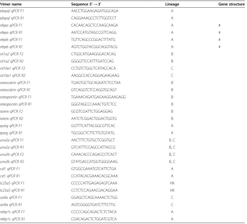
Table 1 Sequences of primers used for quantitative PCR
Primer name Sequence 5′→3′ Lineage Gene structure
adopql qPCR F1 AACCTGGAAGAGATGGCAGA A
adopql qPCR R1 CAGGAAAGCCTCTTGGTCCT A
cebpa qPCR F1 CACAACAGCTCCAAGCAAGA A #
cebpa qPCR R1 AATCCATGTAGCCGTTCAGG A #
cebpb qPCR F1 TGTTCAGCCCGGACTTTATG A #
cebpb qPCR R1 AGTCTGGTACGGCAGGTACG A #
col1a2 qPCR F2 CTGGCATGAAGGGACACAG B
col1a2 qPCR R2 GGGGTTCCATTTGATCCAG B
col10a1 qPCR F2 CCTGTCTGGCTCATACCACA C
col10a1 qPCR R2 AAGGCCACCAGGAGAAGAAG C
osteocalcin qPCR F1 TGAGTGCTGCAGAATCTCCTAA B
osteocalcin qPCR R1 GTCAGGTCTCCAGGTGCAGT B
osteopontin qPCR F1 TGAAACAGATGAGAAGGAAGAGG B
osteopontin qPCR R1 GGGTAGCCCAAACTGTCTCC B
osterix qPCR F2 GCGTCGATTCTGGAGGAG B
osterix qPCR R2 AATCTCGGACTGGACTGGTG B
pparg qPCR F1 GGTTTCATTACGGCGTTCAC A
pparg qPCR R1 TGCGGCTCTTCTTGTGTATG A
runx2a qPCR F1 AACTTTCTGTGCTCGGTGCT B, C
runx2a qPCR R1 GTCATTTCCAGCCATTACCG B, C
runx2b qPCR F2 CAAACACCCAGACCCTCACT B, C
runx2b qPCR R2 GTATGACCATGGTGGGGAAG B, C
scd1 qPCR F1 GTGGCGAAATGTCATTCTGA A
scd1 qPCR R1 CCATACACGAAACACGCAAA A
slc25a5 qPCR F1 CCCCCATTGAGAGAGTCAAA HK
slc25a5 qPCR R1 CCTCTCCAGAACGACAGGAA HK
sox9a qPCR F1 GGAGCTCAGCAAAACTCTGG C
sox9a qPCR R1 AGTCGGGGTGATCTTTCTTG C
srebp1c qPCR F1 CCCCCAGCAGACTCTCTACA A
srebp1c qPCR R1 CGACAGACTCTGGATCGTCA A
A, adipocyte-specific gene; B, bone specific gene; C, cartilage specific gene; HK, house keeping gene. #, gene with one exon.
homozygote mutants were ground with a plastic pestle in a mixture of chloroform:methanol (2:1, v/v) and incu-bated at room temperature for 15 minutes. To 1 ml of extract 300μl of water was added. Samples were quickly vortexed at 2000 rpm for 5 minutes. Bottom phase was washed twice with 0.5 ml of water to be finally reduced in a speed-vacuum. Concentrated lipid extracts were spotted on a Silica gel 60 TLC plate (Merck). Plates were developed in a mixture of chloroform–ethanol–water– triethylamine (30:35:7:35, v/v/v/v), sprayed with primu-line and viewed under ultraviolet light.
Statistical analysis
Data are given as mean ± standard error of mean (SEM). One sample t-test for comparing column means to a hypothetical value or two samples unpaired Student’s test for comparison of two groups were used to deter-mine statistical significance and described as * for p < 0.05, ** for p < 0.005 and *** for p < 0.001.
Results
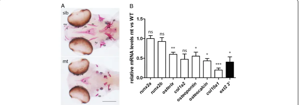
Impaired bone development in theext2−/−fish has been described previously [17-19]. In order to identify at what step bone formation is affected and what mechanisms underlie changes in theext2mutant, we examined the ex-pression of various bone molecular markers in theext2−/− fish and compared it with its siblings (Figure 2).
The importance of HS for pre-osteoblast differentiation
Our previous mRNAin situ analyses demonstrated nor-mal expression patterns of collagen2, sox9a, and chon-dromodulin in the ext2−/− fish, but did not give good
estimates of the expression levels of these molecules [17,18]. In this work, using real time PCR, we confirm that in the homozygote ext2 mutants, the expression levels of early skeletal markers such as runx2are main-tained at wild-type-levels whereas late skeletal markers such as osterix, collagen1a1, osteopontin and osteocal-cinare approximately 2-fold down-regulated and colla-gen 10a1 shows even greater reduction (Figure 2B). Gene expression data indicate that HS are needed by chondrocytes for terminal differentiation for providing a scaffold for developing bone, and for maintenance of the osteochondroprogenitors/preosteoblasts to osteo-blastic lineage.
Bone loss coincides with elevated lipid levels, premature adipocyte differentiation and misshapen musculature in theext2−/−fish
Mesenchymal precursors can differentiate toward skeletal-forming cells (osteoblasts and/or chondroblasts) and/or other lineages such as myoblasts and adipocytes [27]. Al-though differentiation of each lineage is controlled by multiple factors including HS-dependent hedgehog, Wnt or BMP, a switch in the fate of single or multiple lineages can be trigged relatively easily. Thus, we assessed whether diminished bone development in the ext2−/−fish is com-pensated with gain of other lineage(s).
The whole mount MF-20-immunohistology revealed no obvious differences in the musculature between het-erozygous ext2 mutant and its wild type siblings (data not shown). However, the craniofacial muscles in the ext2−/−fish were shorter, broader and fitted the missha-pen cartilaginous skeleton (Figure 3). Moreover, some
muscles such as the hh were absent, whereas extra de-position of muscles was observed around ext2−/− heart (Figure 3, Table 2, Additional file 1).
Oil red O, a stain for neutral triglycerides, lipids and some lipoproteins, highlighted blood vessels, heart, tec-tum, guts, swim bladder and the remains of yolk in all fish (Figure 4A). In theext2−/−fish, the staining was in-tense and abnormally high lipid accumulation was ob-served. Especially, deposits in the vasculature were more pronounced (Figure 4A). Staining at the position of missing bones could be observed in some larvae. Signifi-cantly stronger (P < 0.001) Oil red O stain in theext2−/− fish coincided with over two fold overexpression of pparg(Figure 4).Other adipogenic markers such ascebp, srebp1cand scd1were expressed at levels similar to wild type (Figure 4C). Despite intense staining, abnormal
accumulation of lipids and overexpression ofpparg, TLC analysis of lipid extracts did not reveal any changes in the profiles from wild type and ext2−/− fish (data not shown).
Zebrafish adipocytes start to form by 8dpf and only upon feeding [29]. Interestingly, in the ext2−/−fish, the mRNA in situ hybridization showed that fabp11a- ex-pressing cells are present in unfed larvae already at 5dpf (Figure 4D).
Bone-to-fat switch in proteoglycan mutants
Observing a disturbance in the differentiation of mesen-chymal cell lineages, we wonder if this is specific to the ext2mutant, or to proteoglycan deficiencies in common. Using a panel of mutants described in previous studies [18,20], we found that the hi954 (uxs1) mutant lacking various proteoglycans and with a mild bone phenotype did not show any alteration in lipid deposition as judged by Oil red O (Figure 4B). Significantly increased (P < 0.005) levels of lipids were detected in the knypek (kny, gpc4−/−) mutant, which lacks only a portion of HS and has a mild bone phenotype (Figure 4B and Additional file 2) [18,20]. Interestingly, the pinscher (pic/slc35b2) mutant, which fails to sulphate different molecules (in-cluding HS) and has a stronger bone phenotype [17,18], only showed a very small, but statistically significant in-crease in lipid levels (P < 0.05).
Can PPARG inhibition rescue bone formation in the
ext2−/−homozygote mutant?
Several drugs are known to affect lipid metabolism and influence the bone-to-fat balance. Although it is unlikely to expect a strong effect on total lipid levels in the early stages of zebrafish development where the majority of lipids come from yolk, application of GW9662, the an-tagonist of PPARG, was shown to enhance bone differ-entiation in zebrafish larvae [30]. As expected, we found that treatment with 15μM GW9662 added at 60hpf did not have any significant effect on lipid levels (Figure 5A) but did enhance formation of cartilage and dermal bones in wild type and in the ext2 heterozygous mutant (Figure 5B). In theext2−/−fish, with the same treatment, enhanced GFP expression was noted in tg(osterix:gfp) larvae (data not shown) with improved ossification of the previously existing bones. Bones that normally do not develop inext2−/−mutants, responded only partially to the treatment with rescue and stimulated ossification being observed in only some of the dermal bones; the ext2−/−-cartilage bones were not rescued by this treatment (Figure 5B). Similar effects were seen upon≥7.5μM pur-morphamine treatment, which should stimulate a fat-to-bone switch by activating hedgehog signalling (Figure 5). Furthermore, we tested involvement of other signaling pathways (HS-dependent) which stimulate bone-to-fat
Figure 3Homozygousext2mutant displays musco-skeletal phenotype.Whole mount immunolocalisation at 4dpf using MF-20 antibody for muscles (green) and collagen II for cartilage (red) shows thicker and shorter muscles fitting the malformed cartilaginous skeleton in theext2−/−fish. Muscles:intermandibularis anterior (ima),
intermandibularis posterior (imp), adductor mandibulae (am),
change. Treatment with BMP6 (an activator of BMP pathway) or dorsomorphine (an inhibitor of BMP) did not show significant effect at any time point on the craniofacial ext2−/− bones and TGF-β activator (TGF-β3 ligand) only partially stimulated dermal bones (data not shown).
Is Ira1/Xbp1 pathway involved in the bone/lipid phenotype of theext2−/−fish?
Recently, Xbp1 was shown to regulate osteoblast dif-ferentiation in a Runx2 independent manner [31]. Since in the ext2−/− fish the levels of runx2 tran-script were normal while osterix levels were reduced, we wondered if the unfolded protein response is affected by the lack of HS. We found that heterozy-gotes maintained WT-levels of ern1 and xpb1. In the ext2−/− mutant, the expression of ern1 was only slightly downregulated (2ΔΔCt ext2−/−/WT = 0,71), but the expression of its downstream target, the xbp1, was reduced to 0,64.
Discussion
Abnormal lipid deposition coinciding with impaired bone formation is not common to all types of proteogly-can deficiencies (see Additional file 2). b3gat3- and uxs1-homozygote mutants, that are upstream of ext2in the biosynthesis pathway and lack heparan and chondro-itin sulphates, have a very mild bone phenotype and do not show increased lipid deposition (this work and data not shown). Interestingly, thefam20band xylt1mutants downstream of uxs1 and upstream of b3gat3 and ext2 were shown to have enhanced bone ossification [32]. Unfortunately nothing is known aboutfam20band xylt1 lipid metabolism. The ext2−/−and gpc4−/−, two mutants with reduced HS-levels only, have high lipid content; but only the ext2 mutants have severely reduced bone for-mation, while thegpc4-null fish have very mild bone im-pairment. The slc35b2 homozygote mutant, which has diminished levels of all sulphated proteoglycans, has an even more severe bone phenotype than the ext2−/−fish and show only very mild enhancement of lipid depos-ition. Why different proteoglycan deficiencies have such
Table 2 Cranial muscles in zebrafish head
3dpf 4dpf 5dpf
Region Time Muscle: Wild type ext2 (−/−) Wild type ext2 (−/−) Wild type ext2 (−/−)
M 62 Intermandibularis anterior ima x x* x x x x
H 58 Interhyoideus ih x x x x x x
B 62 Tranversus ventralis tv x x x x x x
M 62 Intermandibularis posterior imp x x x x x x
H 58 Hyohyoideus hh x a x a x a
B 85 Rectus ventralis rv na na na na na na
B 85 Rectus communis rc na na na na na na
M 53 Adductor mandibulae am x x x x x x
M 62 Levator arcus palatini lap x x x x x x
M 62 Dilator operculi do x x x x x x
H 68 Adductor hyoideus ah x x x x x x
H 68 Adductor operculi ao x x* x x* x x*
B 72 Dorsal pharyngeal wall dpw na na na na na na
H 85 Levator operculi lo na na na na na na
E 62 Inferior oblique io na na na na na na
E 58 Inferior rectus ir na na na na na na
E 58 Lateral rectus lr na na na na na na
E 53 Medial rectus mr na na na na na na
B 53 Sternohyoideus sh x xx x xx x xx
E 58 Superior oblique so na na na na na na
E 58 Superior rectus sr na na na na na na
D 72 Protractor pectoralis pp a x* x x x x
different effects on bone and lipid metabolisms is not clear. Holmborn and coauthors [33] showed that, in the ext2 homozygote mutant, the remaining HS are over-sulphated which changes their properties (i.e. increase occurrence of protein-interacting domains). Although, heparin, a highly sulphated glycosaminoglycan and a po-tent anticoagulant, which is often used in clinical practice, negatively affects bone density and is known to increase lipid deposition in sera, the role of over-sulphation of (proteo-)glycans would need to be confirmed.
Craniofacial skeletal development in zebrafish is of mixed origin being derived from cranial neural crest and/or mesoderm [34]. The presence of one functional copy of theext2gene is sufficient for the maintenance of normal differentiation of chondrocytes, osteoblasts and other mesenchyme-derived cells. Reduction in HS-levels in theext2−/−larvae clearly affects skeletal development. Loss of bones cannot be linked specifically to one type of precursor cell as both neural crest- and mesoderm-derived structures are affected. Despite their origin, two
populations of osteoblasts with different sensitivity to hedgehog signalling have been described in zebrafish [35]. As no defects in the hedgehog signalling were found in the craniofacial skeleton of theext2−/−fish, it is unlikely that bone defects could be linked to a specific type of hedgehog-sensitive osteoblast. However, it is pos-sible that there are multiple types of osteoblasts existing in fish, differing in their sensitivity for HS.
Bone homeostasis depends on the balance between osteoblastic and osteoclastic activity. Lipids are known to attract osteoclasts while suppressing osteoblastogene-sis (for review see [36]). Unfortunately, we were not able to test this in zebrafish as the first osteoclasts develop by 16 dpf, beyond the time of premature death of theext2−/− fish. Nevertheless, observations from patient material sug-gest that indeed both osteoblasts and osteoclasts are af-fected by HS-deficiencies [10,26] or by HS abnormal accumulation [7] and, in both cases, bone mineral density is altered. Osteoblasts and adipocytes might not be the only lineages affected by imbalanced HS. EXT1-null
Figure 4Increased lipid levels in theext2−/−fish coincide with decreased bone formation. A, Oil red O stain (ORO) in fish at 6dpf;
embryonic stem cells also appear to have impaired differ-entiation hematopoietic lineages [37], while osteochondro-mas exhibit impaired vascularisation [38].
Fatty acids, when not stored in adipocytes, accumulate into the circulation [39]. Although premature adipocyte-like cells were detected in the ext2−/− fish it is unlikely that they would be able to store all the lipids as cytoplas-mic droplets. Therefore, Oil red O stain in vasculature could reflect only a surplus of fatty acids/lipids. How-ever, it is also possible that mutation in the ext2 gene leads to an abnormal intravascular accumulation of lipids. The changes in bones and fat that we have de-scribed in fish were a characteristic of an organism homozygous for a mutation in theext2gene in all cells. Since MO patients are mostly heterozygous for a muta-tion inEXTthey should have very mild (if any) systemic phenotype. However, if findings from this fish model are true for humans, strong focal changes should be ex-pected at the site where loss of heterozygosity/haplo-insufficiency occured. Not much is known about lipid metabolism in patients with MO. Lemos and co-authors [10] reported lower bone mineral density of femoral neck and lumbar spine in MO patients near osteochon-dromas. In addition, single reports described deposition of fat within the cartilaginous cap of osteochondromas [11] and development of lipoma, a benign bone tumour, or fat-pads in association with osteochondromas [40,41]. These finding might have been coincidental in MO but increased lipid levels often remain asymptomatic. In light of our findings in the fish model on the bone-fat imbalance the status of lipids in human MO seems worth investigating.
Humans, mice and fish with MO are often short in stature and have bowed bones. Recently, Jones and co-authors [42] demonstrated that osteochondroma grow-ing on account of deranged bone growth is apparent only in some individuals and other mechanisms must contribute to the short bone phenotype. Also bone bow-ing does not always require osteochondroma formation to generate the observed anatomical changes (K. Jones, University of Utah School of Medicine, personal com-munication). The presence of muscle phenotype needs to be confirmed in non-fish MO. Further work will show how (if ) muscles with different mechanical properties contribute to the formation of shorter and bowed bones in patients.
Conclusions
Our data indicated that HS have multiple functions dur-ing endochondral bone development. First of all, HS are required for terminal differentiation of the cartilaginous template and consecutive formation of a scaffold that is needed for further bone development. Secondly, normal expression of runx2 and impaired expression of osterix
in the ext2−/− fish indicated that HS are required by osteoblast precursors for their further differentiation within the osteoblastic lineage. Furthermore, the in-creased lipid deposition in the ext2−/− fish suggest that HS are involved in determining the cell lineage when mesenchymal precursor cell differentiates into bones and/or fat. PCR analyses confirm the increase in the ex-pression of lipid markers and down-regulation of early skeletal markers. It still remains to be established how HS are involved in this shift, but lower expression of xbp1, a master regulator of osterix, suggests that HS affect the unfolded protein response, a pathway which is known to control bone formation and lipid metabolism.
Supporting data
The data sets supporting the results of this article are in-cluded within the article and its additional files.
Additional files
Additional file 1:Muscle phenotype in theext2−/−fish.Muscles were detected with MF-20 antibody. Scale = 0.1 mm.
Additional file 2:Information about affected proteoglycans and the bone- and fat phenotypes of mutants used in this study.
Proteoglycans (PGs), heparan sulphate (HS), dermatan sulphate (DS), chondroitin sulphate (CS). keratan sulphate (KS) proteoglycans are the forth group of proteoglycan that is defected theslc35b2−/−mutant.
Abbreviations
AP:Alkaline phosphatase;b3gat3:Beta-1,3-glucuronyltransferase 3; BCIP/ NBT: 5-bromo-4-chloro-3-indolyl-phosphate/nitro blue tetrazolium; BMP: Bone morphogenetic proteins; BSA: Bovine serum albumine;
dak:dackel; Dpf: Days post fertilization;ext1:exostosin 1;ext2:exostosin 2; HS: Heparan sulphates; HSPG: Heparan sulphate proteoglycan; Hpf: Hours post fertilization;kny:knypek; GFP: Green fluorescence protein; MO: Multiple osteochondromas;pic:pinscher;pparg:peroxisome proliferator-activated receptor gamma;runx2:runt-related transcription factor 2;slc35b2:transport of adenosine 3′-phospho 5′- phosphosulfate (PAPS); TCL: Thin layer chromatography; TGF: Transforming/tumour growth factor;uxs1:
UDP-glucuronic acid decarboxylase 1;xbp1:x-box binding protein 1.
Competing interests
All authors declare that they have no competing interests.
Authors’contributions
MIW, CEdA, KWFS, ZZ and PCWH designed this study, analyzed interpreted data; MIW, CEdA and KWFS collected data and generated figures. All authors were involved in writing the paper and had final approval of the submitted and published versions.
Acknowledgments
We thank Marcel Winter for the assistance with RT-PCR, Magnus Palmblad for shearing the lipid standards, Prof. van den Maren and his group for help with lipid analysis, Prof. Schulte-Merker for theTg(osterix:gfp)zebrafish line, Dr. Mikel San Julian for sharing pictures of bowed bones, and Dr J.F. Graadt van Roggen for critical comments on the manuscript. The MF 20 antibody developed by D. Fischman was obtained from the Developmental Studies Hybridoma Bank (the auspices of the NICHD and maintained by The University of Iowa, Department of Biology, Iowa City, IA 52242). This work was supported by the European network of excellence EuroBoNeT [grant number 018814 (LSHC-CT-2006-018814)].
Author details
1
Department of Pathology, Leiden University Medical Center, Leiden, The Netherlands.2Current address: Zebrafish Core Facility, International Institute of Molecular and Cell Biology, Warsaw, Poland.3Department of Histology and Pathology, University of Navarra, Pamplona, Spain.4Nuffield Department of Medicine, Ludwig Institute for Cancer Research, University of Oxford, Oxford, UK.
Received: 18 October 2013 Accepted: 10 February 2014 Published: 14 March 2014
References
1. Kaback LA, Soung DY, Naik A, Smith N, Schwarz EM, O’Keefe RJ, Drissi H:
Osterix/Sp7 regulates mesenchymal stem cell mediated endochondral ossification.J Cell Physiol2008,214:173–182.
2. Nakashima K, Zhou X, Kunkel G, Zhang Z, Deng JM, Behringer RR, de Crombrugghe B:The novel zinc finger-containing transcription factor osterix is required for osteoblast differentiation and bone formation.Cell2002,108:17–29. 3. Mikami Y, Lee M, Irie S, Honda MJ:Dexamethasone modulates
osteogenesis and adipogenesis with regulation of osterix expression in rat calvaria-derived cells.J Cell Physiol2011,226:739–748.
4. Akune T, Ohba S, Kamekura S, Yamaguchi M, Chung UI, Kubota N, Terauchi Y, Harada Y, Azuma Y, Nakamura K, Kadowaki T, Kawaguchi H:PPARgamma insufficiency enhances osteogenesis through osteoblast formation from bone marrow progenitors.J Clin Invest2004,113:846–855.
5. McCormick C, Leduc Y, Martindale D, Mattison K, Esford LE, Dyer AP, Tufaro F:The putative tumour suppressor EXT1 alters the expression of cell-surface heparan sulfate.Nat Genet1998,19:158–161.
6. Fung EB, Johnson JA, Madden J, Kim T, Harmatz P:Bone density assessment in patients with mucopolysaccharidosis: a preliminary report from patients with MPS II and VI.J Pediatr Rehabil Med2010,3:13–23. 7. Rigante D, Caradonna P:Secondary skeletal involvement in Sanfilippo
syndrome.QJM2004,97:205–209.
8. Bovée JVMG, Heymann D, Wuyts W:Osteochondroma.InWHO Classification of Tumours of Soft Tissue and Bone.Edited by Fletcher CDM, Bridge JA, Hogendoorn PCW, Mertens F. Lyon: IARC; 2013:250–251. 9. Wuyts W, Bovée JVMG, Hogendoorn PCW:Multiple Osteochondromas.In
WHO Classification of Tumours of Soft Tissue and Bone.Edited by Fletcher CDM, Bridge JA, Hogendoorn PCW, Mertens F. Lyon: IARC; 2013:384–385. 10. Lemos MC, Kotanko P, Christie PT, Harding B, Javor T, Smith C, Eastell R,
Thakker RV:A novelEXT1splice site mutation in a kindred with hereditary multiple exostosis and osteoporosis.J Clin Endocrinol Metab
2005,90:5386–5392.
11. Schick F, Duda SH, Lutz O, Claussen CD:Lipids in bone tumors assessed by magnetic resonance: chemical shift imaging and proton spectroscopy in vivo.Anticancer Res1996,16:1569–1574.
12. Darilek S, Wicklund C, Novy D, Scott A, Gambello M, Johnston D, Hecht J:
Hereditary multiple exostosis and pain.J Pediatr Orthop2005,25:369–376. 13. Hosalkar H, Greenberg J, Gaugler RL, Garg S, Dormans JP:Abnormal scarring
with keloid formation after osteochondroma excision in children with multiple hereditary exostoses.J Pediatr Orthop2007,27:333–337. 14. Huegel J, Sgariglia F, Enomoto-Iwamoto M, Koyama E, Dormans JP, Pacifici
M:Heparan sulfate in skeletal development, growth, and pathology: the case of hereditary multiple exostoses.Dev Dyn2013,242:1021–1032. 15. Dooley K, Zon L:Zebrafish: a model system for the study of human
disease.Curr Opin Genet Dev2000,10:252–256.
16. Lee J-S, der HS V, Rusch MA, Stringer SE, Stickney HL, Talbot WS, Geisler R, Nüsslein-Volhard C, Selleck SB, Chien CB, Roehl H:Axon sorting in the optic tract requires HSPG synthesis byext2 (dackel)andextl3 (boxer).Neuron
2004,44:947–960.
17. Clément A, Wiweger M, von der Hardt S, Rusch MA, Selleck SB, Chien CB, Roehl H:Regulation of zebrafish skeletogenesis byext2/dackeland
papst1/pinscher.PLoS Genet2008,4:e1000136.
18. Wiweger MI, Avramut CM, de Andrea CE, Prins FA, Koster AJ, Ravelli RB, Hogendoorn PC:Cartilage ultrastructure in proteoglycan-deficient zebra-fish mutants brings to light new candidate genes for human skeletal dis-orders.J Pathol2011,223:531–542.
19. Wiweger MI, Zhao Z, van Merkesteyn RJ, Roehl HH, Hogendoorn PC: HSPG-deficient zebrafish uncovers dental aspect of multiple osteochondromas.
20. de Andrea CE, Prins FA, Wiweger MI, Hogendoorn PC:Growth plate regulation and osteochondroma formation: insights from tracing proteoglycans in zebrafish models and human cartilage.J Pathol2011,224:160–168. 21. Kimmel CB, Ballard WW, Kimmel SR, Ullmann B, Schilling TF:Stages of
embryonic development of the zebrafish.Dev Dyn1995,203:253–310. 22. Spoorendonk KM, Peterson-Maduro J, Renn J, Trowe T, Kranenbarg S,
Winkler C, Schulte-Merker S:Retinoic acid and Cyp26b1 are critical regulators of osteogenesis in the axial skeleton.Development2008,135:3765–3774. 23. Li N, Felber K, Elks P, Croucher P, Roehl HH:Tracking gene expression
during zebrafish osteoblast differentiation.Dev Dyn2009,238:459–466. 24. Thisse C, Thisse B:High-resolutionin situhybridization to whole-mount
zebrafish embryos.Nat Protoc2008,3:59–69.
25. de Jong M, Rauwerda H, Bruning O, Verkooijen J, Spaink HP, Breit TM:RNA isolation method for single embryo transcriptome analysis in zebrafish.
BMC Res Notes2010,3:73.
26. Hameetman L, Rozeman LB, Lombaerts M, Oosting J, Taminiau AHM, Cleton-Jansen AM, Bovée JV, Hogendoorn PC:Peripheral chondrosarcoma progression is accompanied by decreased Indian Hedgehog signalling.J Pathol2006,209:501–511.
27. Takada I, Kouzmenko AP, Kato S:Wnt and PPARgamma signaling in osteoblastogenesis and adipogenesis.Nat Rev Rheumatol2009,5:442–447. 28. Schilling TF, Kimmel CB:Musculoskeletal patterning in the pharyngeal
segments of the zebrafish embryo.Development1997,124:2945–2960. 29. Flynn EJ III, Trent CM, Rawls JF:Ontogeny and nutritional control of
adipogenesis in zebrafish (Danio rerio).J Lipid Res2009,50:1641–1652. 30. Li N, Kelsh RN, Croucher P, Roehl HH:Regulation of neural crest cell fate by the
retinoic acid and Pparg signalling pathways.Development2010,137:389–394. 31. Tohmonda T, Miyauchi Y, Ghosh R, Yoda M, Uchikawa S, Takito J, Morioka H,
Nakamura M, Iwawaki T, Chiba K, Toyama Y, Urano F, Horiuchi K:The IRE1alpha-XBP1 pathway is essential for osteoblast differentiation through promoting transcription of Osterix.EMBO Rep2011,12:451–457. 32. Eames BF, Yan YL, Swartz ME, Levic DS, Knapik EW, Postlethwait JH, Kimmel
CB:Mutations in fam20b and xylt1 reveal that cartilage matrix controls timing of endochondral ossification by inhibiting chondrocyte maturation.PLoS Genet2011,7:e1002246.
33. Holmborn K, Habicher J, Kasza Z, Eriksson AS, Filipek-Gorniok B, Gopal S, Couchman JR, Ahlberg PE, Wiweger M, Spillmann D, Kreuger J, Ledin J:On the roles and regulation of chondroitin sulfate and heparan sulfate in zebrafish pharyngeal cartilage morphogenesis.J Biol Chem2012,287:33905–33916. 34. Yelick PC, Schilling TF:Molecular dissection of craniofacial development
using zebrafish.Crit Rev Oral Biol Med2002,13:308–322.
35. Hammond CL, Schulte-Merker S:Two populations of endochondral osteoblasts with differential sensitivity to Hedgehog signalling.
Development2009,136:3991–4000.
36. Rosen CJ, Bouxsein ML:Mechanisms of disease: is osteoporosis the obesity of bone?Nat Clin Pract Rheumatol2006,2:35–43.
37. Holley RJ, Pickford CE, Rushton G, Lacaud G, Gallagher JT, Kouskoff V, Merry CL:Influencing hematopoietic differentiation of mouse embryonic stem cells using soluble heparin and heparan sulfate saccharides.J Biol Chem
2011,286:6241–6252.
38. de Andrea CE, Wiweger MI, Bovee JV, Romeo S, Hogendoorn PC:Peripheral chondrosarcoma progression is associated with increased type X collagen and vascularisation.Virchows Arch2012,460:95–102. 39. Cousin W, Fontaine C, Dani C, Peraldi P:Hedgehog and adipogenesis: fat
and fiction.Biochimie2007,89:1447–1453.
40. Ek ET, Slavin JL, Blackney MC, Powell GJ:Parosteal lipoma associated with an underlying osteochondroma arising from the hallux.Skeletal Radiol
2007,36:689–692.
41. Sakai H, Tamai K, Iwamoto A, Saotome K:Para-articular chondroma and osteochondroma of the infrapatellar fat pad: a report of three cases.
Int Orthop1999,23:114–117.
42. Jones KB, Datar M, Ravichandran S, Jin H, Jurrus E, Whitaker R, Capecchi MR:
Toward an understanding of the short bone phenotype associated with multiple osteochondromas.J Orthop Res2013,31:651–657.
doi:10.1186/1750-1172-9-35
Cite this article as:Wiwegeret al.:Possible effects ofEXT2on mesenchymal differentiation - lessons from the zebrafish.Orphanet Journal of Rare Diseases 20149:35.
Submit your next manuscript to BioMed Central and take full advantage of:
• Convenient online submission
• Thorough peer review
• No space constraints or color figure charges
• Immediate publication on acceptance
• Inclusion in PubMed, CAS, Scopus and Google Scholar
• Research which is freely available for redistribution