Original Research Article
Diagnostic utility and impact of ultrasonography by primary health
care physician: an observational study
Uruj Altaf Qureshi
1*, Khalid Bashir
2, Mahbooba Rasool
3INTRODUCTION
Ultrasonography is an imaging technology that has its role in the medical diagnostic equipment due to its low costs, lack of pain, rapidity of results and lack of radiation exposure. Ultrasound is clearly the imaging test of choice in a variety of circumstances, ranging from obstetric emergencies, acute abdomen to routine evaluative ultrasonography. It plays a pivotal role in the evaluation of patients and helps in making timely diagnosis, as such a point of care diagnostic ultrasound has gained very much importance. Imaging in patient
care was mostly limited till some years back, primary health care centres were devoid of ultrasound facilities, and lack of adequate health care facilities continued to be a major barrier to health-care delivery.1 Ultrasound is a safe diagnostic imaging modality. Point-of-care ultra-sound applications have grown dramatically in recent years and cover many primary care clinical scenarios; ultrasound scans help in making diagnosis and devising management. Experience indicates that only the patients who clinically suggest a possible risk are referred for an ultrasound to confirm, or rule out problems. Ultrasound impacts patient management positively and improves the
ABSTRACT
Background: Ultrasound appears to be a suitable diagnostic technology for use in primary care and community settings. It plays a pivotal role in evaluation of patients and helps in making timely diagnosis and more widely on referral pathways into secondary care.
Methods: The study was conducted at the primary health centre Hazratbal, which is one of the primary health centre of field practice area of the Department of community medicine, Government Medical College, Srinagar. This observational study was conducted over a period of three months where 255 patients were scanned by a primary care physician (sonologist). For each patient scanned, the ultrasound performing physician completed a standardized data collection form including patient demographics, clinical details, indications for ultrasound and ultrasound findings.
Results: A total of 255 patients were scanned during the study period. Males were 43 (17%) and females were 212 (83%). Maximum number of patients were in the age range of 25-34 years, n=96 (38%). Among the patients scanned 66 (24.44%) were obstetric cases. Acute diffuse abdominal pain n=32 (11.85%) was the most common clinical presentation followed by pain upper abdomen n=28 (10.37%) among non-obstetric patients. Ovarian cyst was the most common finding, followed by fatty liver and bilateral nephrolithiasis.
Conclusions: The utility of ultrasonography in the hands of primary care physician is of great value. It is cost effective option, especially in this part of the world. We need to give expertise to primary care physicians in order to provide better health care at primary health care settings, which will lessen the burden of referrals.
Keywords: Ultrasonography, Primary care physician, Diagnosis, Non-invasive
Department ofCommunity Medicine, 1GMC, Baramulla, 2GMC, Srinagar, 3GMC, Anantnag, J and K, India
Received: 31 January 2019
Revised: 09 March 2019
Accepted: 11 March 2019
*Correspondence:
Dr. Uruj Altaf Qureshi,
E-mail: [email protected]
Copyright: © the author(s), publisher and licensee Medip Academy. This is an open-access article distributed under the terms of the Creative Commons Attribution Non-Commercial License, which permits unrestricted non-commercial use, distribution, and reproduction in any medium, provided the original work is properly cited.
diagnostic capacity of a primary care or rural healthcenter.2 Ultrasonography helps in assessing effects of treatment modalities, improvement in signs and symptoms, especially in chronic diseases. Patients suffering from chronic diseases benefit from ultrasound by follow up scans.3 Ultrasonography has become a
significant non-invasive instrument for medical
investigation and is considered by some as the 'stethoscope of the future'.4 Ultrasonography at primary health care setting helps hugely in avoiding referrals, which benefits patients. Primary care physicians
performing and interpreting diagnostic imaging
examinations concerning their own patients rather than referring them to imaging specialists, has attracted considerable attention in recent medical literature.5 Self-referral by the primary care physician may be particularly problematical.6 The aim of study was to assess the profile of patients and impact of a diagnostic ultrasound at primary health care setting.
METHODS
The study was conducted at the primary health centre Hazratbal, which is one of the primary health centres of field practice area of the Department of community medicine, Government Medical College, Srinagar. This observational study was conducted over a period of three months from March 2017 to May 2017. A total of 255 patients were scanned which included all those patients who were referred by a primary care physician (medical officer) in outpatient department where ever they faced difficulty in reaching final diagnosis. Patients who sought ultrasonography as a self-referral were excluded from the study.
The ultrasonography was performed by a primary care physician (sonologist) with expertise in general
abdominal ultrasound, obstetric ultrasound, and
interpretation of the imaging was done at the same time. A mid-low frequency transducer 3.5-5 MHz convex array was usually used during ultrasonography. The centre is registered under the pre-conception and pre-natal diagnostic techniques act (PCPNDT act) and adheres to guidelines of filling form F in case of antenatal ultrasound. After proper consent for each patient scanned, the ultrasound performing physician completed a standardized data collection form including patient demographics, clinical details, indications for ultrasound, ultrasound findings, and the pre and post-ultrasound diagnosis and management plan.
The pre-ultrasound working diagnosis and management plan was recorded prior to the ultrasound examination. Data was entered into an Excel spread sheet and basic descriptive statistics was done.
RESULTS
A total of 255 patients were scanned during the study period. Among the patients, females were n=212 (83%)
and males were n=43 (17%). Maximum number of patients were in the age group of 25-34 years, n=96 (38%) as shown in Table 1. There were many useful applications of ultrasound in this setting; however, obstetrical ultrasound, including estimation of gestational age, determining head position, and evaluating placental abnormalities, was the most frequently performed application overall. The distribution of ultrasound scans was n=66 (25.88%) antenatal cases (ANC) with different indications for ultrasonography.
Table 1: Demographics of patients who underwent ultrasonography scan (n=225).
Patients for ultrasonography Frequency %
Gender
Male 43 16.9
Female 212 83.1
Marital status
Married 203 79.6
Unmarried 52 20.4
Age ( in years)
5-14 3 1
15-24 30 12
25-34 96 38
35-44 60 23
44-54 27 11
55 and above 39 15
Nature of patient
Obstetric 66 25.89
Non-obstetric 189 74.11
Table 2 shows both the obstetric and non-obstetric indications for ultrasound to evaluate many clinical presentations. In our study out of 189 non-obstetric patients, 50 (26%) patients had normal scan. Rest of the patients n=139 (74%) had ultrasonography finding on scan. Among the obstetric patients 51% had normal scan and 49% had findings on scan. The indications for performing the scans varied depending on the clinical presentation and presumptive clinical diagnosis. Acute diffuse abdominal pain n=32 (11.85%) was the most common clinical presentation followed by pain upper abdomen n=28 (10.37%) among non-obstetric patients.
Table 2: Clinical presentation of patients scanned for ultrasound.
Symptoms of patients Frequency %
Acute diffuse abdominal pain 32 11.85
Bilateral flank pain 15 5.55
Pain right hypochondrium 8 2.96
Increased micturition/dysuria 12 4.44
Dysmenorrhea 7 2.59
Irregular menstrual cycles 7 2.59
Irregular periods/hot flushes 4 1.48
Menorrhagia 11 4.07
Lactational amenorrhea 2 0.74
Oligomenorrhea 4 1.48
Pain lower abdomen 17 6.29
Polymenorrhea 4 1.48
Pain right flank 10 3.70
Pain left flank 9 3.33
Post-menopausal bleeding 3 1.11
Abortifacient intake 3 1.11
Pain upper abdomen 28 10.37
BHP symptoms 11 4.07
Decreased appetite, dyspepsia 10 3.70
Leucorrhea 4 1.48
Antenatal cases/obstetrics 66 24.44
Total* 270 100
*Some patients had multiple clinical presentations (multiple responses).
Table 3: Ultrasound findings of patients.
Ultrasonography diagnosis Frequency %
Bilateral nephrolithiasis 13 4.87
Cholelithiasis 9 3.37
Fatty liver 16 6.00
Tubo-ovarian mass 2 0.75
Left nephrolithiasis 14 5.24
Ovarian cyst 18 6.74
Uterine fibroid 14 5.24
Right nephrolithiasis 11 4.12
BHP¹ 9 3.37
Right renal calculus with hydronephrosis 4 1.50
Left renal calculus with hydronephrosis 3 1.12
PCOD² 6 2.25
RPOC³ 2 0.75
Normal study 50 18.73
Right nephrolithiasis 6 2.25
Splenomegaly 4 1.50
Obstetric low lying placenta 10 3.74
Breech presentation 9 3.37
Ovarian cyst 9 3.37
Retro placental bleed 2 0.75
Threatened abortion 2 0.75
Hydatidiform mole 1 0.37
Normal study 34 12.73
Others* 19 7.12
Total** 267 100
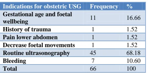
Table 4: Details of indications for antenatal ultrasonography.
Indications for obstetric USG Frequency %
Gestational age and foetal
wellbeing 11 16.66 History of trauma 1 1.52
Pain lower abdomen 1 1.52
Decrease foetal movements 1 1.52
Routine ultrasonography 45 68.18
Bleeding 7 10.60
Total 66 100
Table 5: Obstetric ultrasonography findings.
Obstetric ultrasonography
done Frequency %
Ist trimester pregnancy 9 13.63
IInd trimester pregnancy 22 33.33
IInd trimester pregnancy with
low lying placenta 6 9.10 IInd trimester pregnancy with
breech presentation 7 10.61 IIIrd trimester pregnancy 16 24.24
IIIrd trimester pregnancy
with breech presentation 2 3.03 IIIrd trimester with low lying
placenta 4 6.06
Total 66 100
DISCUSSION
The ultrasonography is an important tool in present day medical diagnosis. It helps in shaping clinical management. It is the first choice in most diagnostic algorithms used by a general practitioner. Ultrasound is relatively non-invasive, safe and well-tolerated by the patients; hence it is very frequently used in family practice. The primary health centre Hazratbal which is under the administrative control of Community Medicine, GMC, Srinagar allows trained primary care physician to perform ultrasound examinations in primary care. Upper abdominal pain was the most frequent complaint for which an ultrasound was requested. This is consistent with a previous study in which pain was the most common indication for an abdominal ultrasound.7 The ultrasound scan helped in diagnosis as well as management. The clinical value of ultrasound was notable. Of the 255 examinations performed, 80% (205) had ultrasound findings; which either added to clinical diagnosis or helped in clinching diagnosis influenced the outcome or decision regarding treatment for the patient. Among the patients referred for ultrasound, 80% had an abnormal report, which is inconsistent with the findings of a previous study.8 The large percentage of abnormal findings is striking and probably confirms that patients generally wait for extended periods before seeking medical care, until diseases have progressed, and they
tend not to seek medical care unless they think that a medical problem is serious. Our study showed ovarian cyst as the most common finding followed by fatty liver and nephrolithiasis. This could be because majority of our cases were female patients and so ovarian cyst was a common finding. In a study done by Alamri et al, fatty liver was also amongst the common findings on ultrasonography.9 The point of care evaluation by ultrasonography is not only a cost effective option in primary care settings, it also hugely helps in mitigating direct and indirect costs to health care facility as well as to the patient. In India rural poor already suffer from multiple other substantial burdens including large barriers to accessing care and (if and when care is accessed) a high risk of financial ruin.10,11 Primary care physician is able to avert large number of referrals and manage patients at the primary health centre by getting timely diagnosis at the primary health care setting. Furthermore, it helps in prioritization of referral of patients who need secondary or tertiary care.
CONCLUSION
Ultrasonography is proven diagnostic tool for evaluation and diagnosis. The utility of ultrasonography in the hands of primary care physician is of great value. It is cost effective option with great precision, especially in this part of the world, were skilled manpower scarcity is at galore. We need to give expertise to primary care physicians in order to provide better health care at the point of care in primary health care settings and, which will also lessen the burden of referrals. Community diagnostic ultrasound services should be made an integral part of primary health care.
Funding: No funding sources Conflict of interest: None declared
Ethical approval: The study was approved by the Institutional Ethics Committee
REFERENCES
1. Director General, World Health Organization.
World Health Report. Fighting Disease, Fostering Development, 1996.
2. Mindel S. Role of Imager in developing world. Lancet. 1997;350(9075):426-9.
3. Partners in Health Mobile Voluntary Counseling
and Testing Unit, Partners in Health: Kirehe District, Rwanda. Annual Report, 2007.
4. Filly RA. Ultrasound: the stethoscope of the future, alas. Radiol. 1988;167:400.
5. Relmann AS. Dealing with conflicts of interest. N Engl J Med. 1985;313:749-51.
6. Radecki SE, Steele JP. Effect of on-site facilities on use of diagnostic radiology by non-radiologists. Invest Radiol. 1990;25:190-3.
7. Aboud M, Mkony C, Wustner M. Elective
Dar-Es-Salaam, Tanzania. East Cent Afr J Surg. 2006;11:52–6.
8. Speets AM, Hoes AW, van der-Graaf Y, Kalmijn S,
de-Wit NJ, van-Swijndregt AD, et al. Upper
abdominal ultrasound in general practice:
Indications, diagnostic yield and consequences for patient management. Fam Pract. 2006;23:507–11.
9. Alamri AF, Khan I, Baig MIA, Iftikhar R. Trends in
ultrasound examination in family practice. J Family Community Med. 2014;21(2):107-11.
10. Katyal A, Singh PV, Bergkvist S, Samarth A, Rao M. Private sector participation in delivering tertiary health care: a dichotomy of access and affordability across two Indian states. Health Policy Plan. 2015;30(1):23-31.
11. Rao M, Ramachandra SS, Bandyopadhyay S,
Chandran A, Shidhaye R, Tamisettynarayana S, et al. Addressing healthcare needs of people living below the poverty line: a rapid assessment of the Andhra Pradesh Health Insurance Scheme. Natl Med J India. 2011;24(6):335-41.