Haplotypes and polymorphism in the CCR5
gene in sickle cell disease
A.F. Nascimento1, J.S. Oliveira2, J.C. Silva Junior2 and A.A.L. Barbosa2
1Programa de Pós-Graduação em Genética, Biodiversidade e Conservação,
Universidade Estadual do Sudoeste da Bahia, Jequié, BA, Brasil
2Departamento de Ciências Biológicas,
Universidade Estadual do Sudoeste da Bahia, Jequié, BA, Brasil
Corresponding author: A.F. Nascimento E-mail: alana20007@hotmail.com
Genet. Mol. Res. 16 (2): gmr16029675 Received March 20, 2017
Accepted May 3, 2017 Published June 29, 2017
DOI http://dx.doi.org/10.4238/gmr16029675
Copyright © 2017 The Authors. This is an open-access article distributed under the terms of the Creative Commons Attribution ShareAlike (CC BY-SA) 4.0 License.
ABSTRACT. Sickle cell disease shows several clinical manifestations in distinct levels of severity. This heterogeneity is due to the haplotype variability associated with the HbS gene, levels of fetal hemoglobin and environmental conditions, which modify the disease expression. Science community believes that the presence of a polymorphism in the CCR5 gene, which is related to chronic inflammatory state, could confer a higher survival rate and a lower number of inflammatory events to these patients since the deletion in CCR5Δ32 would knock out the
CCR5 gene. Therefore, this study aimed to identify the haplotypes in βS and βC genes, as well as to investigate the presence of the CCR5Δ32 deletion in patients with sickle cell disease. For this purpose, DNA was isolated with the QIAamp DNA Investigator Kit, and PCR was the method chosen to detect the mutant allele CCR5Δ32. The haplotypes in βS and βC genes were detected by RFLP with the restriction enzymes
(11.5%), Senegal (2.8%), and Cameroon (2.8%). No patients presented
CCR5Δ32 deletion. The increase in the frequency of atypical haplotypes suggests that these patients passed by variation in the genetic pattern from ancestral haplotypes throughout the years.
Key words: Haplotypes; Sickle cell disease; Hemoglobin S; Hemoglobin C; CCR5 receptor; CCR5Δ32
INTRODUCTION
Haplotypes in sickle cell disease can be used as markers for understanding patterns of population migration in anthropological studies, as well as, in the detection of genetic distances between main ethnic groups when studying human ethnicity origin (Nagel and Ranney, 1990).
Sickle cell disease haplotypes (βS) are classified into five different types according to their mainly common ethnic and geographical origins. Benin haplotype has been associated with West Africa; Bantu or the Central African Republic (CAR) to East Africa and Central South; Senegal to Atlantic West Africa; Arab-Indian to India and Arabian Peninsula; and Cameroon to the West African coast (Kan and Dozy, 1978; Gonçalves et al., 2003).
A remarkable characteristic of this disease is its clinical variability: while a few patients present a clinical condition with great severity and are related to numerous complications with possible hospitalization, other patients present a mild evolution that can be almost asymptomatic (Sebastiani et al., 2005).
These changes in hematological and clinical characteristics of the disease are attributed in part to the variability of linked and non-linked genes that modify the disease expression, as well as the diversity regarding climate, social, and economic conditions (Nagel et al., 1985). So, genetic, environmental, and social factors contribute to the clinical variability (ANVISA, 2002).
Different types of haplotypes are associated with varying levels of fetal hemoglobin (HbF) and consequent variation in the severity of the clinical condition of the patients. Therefore, the presence of HbF may change the sites in contact with HbS molecules, in such a way that the polymer generation becomes impaired, consequently, decreasing the sickling process (Adekile and Huisman, 1993). In other words, HbF acts as an inhibitor effect on the polymerization (Naoum, 1997).
It is known that the Senegal haplotype is associated with high levels of HbF (>15%) and less severe clinical course of the disease; Benin is associated with moderate levels of HbF (5to 15%) and intermediate clinical course; Bantu, to decreased levels of HbF (<5%) and more aggressive clinical condition; and Arab-Indian haplotype presents high levels of HbF and heterogeneous clinical course (Sebastiani et al., 2005; Silva et al., 2009).
clinical manifestations (Vargas et al., 2005); and polymorphism C677T in the MTHR gene, associated with an increase the risk of vascular disease (Couto et al., 2004).
Polymorphic associations of CCR5 with sickle cell disease are described in the literature. The presence of a variant harboring a deletion of 32 bp, called CCR5Δ32, would be associated with greater clinical advantages to the patients (Lopes et al., 2010). Since CCR5 is an important receptor of a pro-inflammatory chemokine, acting as an inflammatory mediator (Doodes et al., 2009; Schauren, 2010), the presence of CCR5Δ32 deletion makes it non-functional, conferring a lower inflammatory picture due to a less efficient response (Lopes et al., 2010; Doodes et al., 2009). This association was also observed in research performed by Chies and Hutz (2003), in which groups of individuals with sickle cell disease, a disease considered chronic inflammatory, showed a higher frequency of CCR5Δ32.
Thus, it is essential to investigate associations with polymorphisms, like haplotypes and CCR5Δ32 analysis and the variability of clinical severity of sickle cell disease. It is required a greater understanding of the mutation origin and the evolution of clinical aspects of the disease, to confer a proper monitoring of these patients (Galiza Neto and Pitombeira, 2003; Steinberg, 2005). Considering that, this study aimed to identify the haplotypes of βS and βC genes, and investigate the presence of theCCR5Δ32 deletion in individuals with sickle cell disease.
MATERIAL AND METHODS
Sample characterization
We searched all cases of sickle cell disease registered by the Health Department of Jequié, BA, from 2009 to 2012. We also performed an active search of new cases, which allowed us to make up a sample of 20 patients with sickle cell disease (15 SS and 5 SC) ranging from 4 to 50 years old. The project was approved by the Research Ethics Committee of UESB (No. 077/2011, CAAE: 0057.0.454.000-11) and all patients signed the Informed Consent after all clarifications related to procedures and aims of the study.
Genomic DNA isolation
Harvesting biological samples were performed in the Human Genetics Laboratory in UESB. From each, we harvested 10 mL whole blood through venous puncture with a Vacu-Tainer tube with EDTA. DNA was isolated with the DNA QIAamp DNA Investigator Kit.
Haplotype determination and analysis
The detection of haplotypes was performed with PCR and RFLP-PCR techniques in six polymorphic sites with the restriction enzymes XmnI, HindIII, HincII, and HinfI. PCR products were incubated with the restriction enzymes for 24 h at 37°C.
Each sample was marked by the presence (+) or absence (-) of the restriction sites in the analysis of six polymorphic sites located in cluster β, determining the most common haplotypes of the βs gene: Bantu or CAR [- + - - - -], Benin [- - - - + -], Senegal [+ + - + + +], Cameroon [- + + - + +], and Arab-Indian [+ + - + + -]. Taking these haplotypes as a pattern, any different combination of the presence and/or absence of these sites was classified as atypical haplotype; and the most common haplotypes of the βC gene: Type I [- + - - + +], Type II [- - - - + +], and Type III [- - - +].
Determination of the CCR5Δ32 polymorphism
Determination of the CCR5Δ32 polymorphism was performed through PCR tech -niques for CCR5 marker, using the following primers: 5'-ATCACTTGGGTGGTGGCTGTGT TTGCGTCTC-3' and 5'-AGTAGCAGATGACCATGACAAGCAGCGGCAG-3'.
PCRs with genomic DNA were analyzed in Applied Biosystems Veriti thermal cycler for amplification. The equipment was programmed for: 1) one cycle at 94°C for 4 min, 2) 30 cycles at 94°C for 1 min, 3) 70°C for 30 s (with the increment of 1 s per cycle), and 4) one cycle at 72°C for 10 min, added by 4°C indefinitely (Sousa, 2005).
The amplified product was separated by 3% agarose gel electrophoresis, stained with Gel RedTM and visualized by UV light transillumination. The amplification of the normal allele produces a fragment of 193 bp, while the mutant (CCR5Δ32) presents a fragment with 161 bp.
RESULTS
Through searching all cases of sickle cell disease registered by the Health Department of Jequié, BA, with the result of the newborn screening between 2009 and 2012, we could find 20 patients with the disease (15 HbSS and 5 HbSC), in which we could detect the haplotypes from the 40 chromosomes studied.
From 35 studied chromosomes for βs haplotype, we could observe Benin haplotypes in 10 patients (28.6%), Bantu in 4 patients (11.5%), Senegal in 1 patient (2.8%), Cameroon in 1 patient (2.8%), and atypical in 19 patients (54.3%), as observed in Table 1.
Table 1. Distribution of βS haplotypes in all 35 chromosomes evaluated.
βS haplotypes Benin Bantu Senegal Cameroon Atypical Total
Number of chromosomes 10 4 1 1 19 35
% 28.6 11.5 2.8 2.8 54.3 100
From five studied chromosome for the βC haplotype, we could observe in almost all of type II haplotype (four patients), just one type I and none from type III.
DISCUSSION
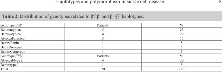
In this study, among common haplotypes, the one with the highest frequency was Benin (28.6%), followed by Bantu (11.5%), Senegal (2.8%), and Cameroon (2.8%). We could also find a great contribution of atypical haplotypes (54.3%), which scattered with all different common haplotypes, as shown in Table 2.
From 19 atypical haplotypes, eight had shown a more similar pattern to Benin haplotype, differing in a single or two restriction sites (? ? - - + -) for H01 and H23 markers. Through these results, we suggest that the atypical haplotypes were probably derived from Benin and that these differences were due to point mutations, through nearly 2000 years of existence (Zago et al., 2000).
The other atypical haplotypes did not present a common pattern with a specific type of common haplotypes. Possibly, they had been generated through a single or double crossing-over between two βs common haplotypes or, more frequently, between a common βs haplotype and a different chromosome βs, as observed by Zago et al. (2000).
The most frequent genotype in these patients was Benin/atypical, diverging from studies performed in Bahia, in which Bantu was the most frequent haplotype, followed by Benin and atypical. Furthermore, Figueiredo et al. (1994) performed a study in Salvador, BA, with 21 patients with sickle cell disease and observed the frequencies of 54.8% Bantu and 45.2% Benin. However, Gonçalves et al. (2003) when studying a sample of 80 patients with sickle cell disease, demonstrated slightly lower frequencies of Bantu (48.1%), a similar frequency of Benin (45.6%), and new haplotypes were observed as Senegal (0.6%) and atypical (5.6%).
After a few years, a study also performed in Salvador, by Adorno (2005), including 125 patients with sickle cell disease, reported a high frequency of Benin type, followed by Bantu haplotype, with a predominance of CAR/Ben genotype. The researcher verified the presence of the Senegal haplotype and for the first time in patients with sickle cell disease from Salvador, the presence of the Arab-Indian haplotype.
Accordingly, including the results of the present study, we realized a gradual change in the frequencies of the haplotypes observed, in which we could identify a persistent decrease in the prevalence of Bantu haplotype and an increase in the prevalence of Benin haplotype. Then, we suggest that these observations might be related to different mechanisms of distribution that goes from a likely miscegenation due to internal migration - which could explain the high frequency of the atypical haplotypes - to a decrease in Bantu individuals, due to the highest severity of the disease (Silva, 2008), corroborating with this study.
Table 2. Distribution of genotypes related to βs/ βs and βs /βC haplotypes.
Genotype βs/βs Patients %
Benin/atypical 5 25
Bantu/atypical 4 20
Atypical/atypical 3 15
Benin/Benin 1 5
Benin/Senegal 1 5
Benin/Cameroon 1 5
Genotype βS/βC Patients %
Atypical/type II 4 20
Benin/type I 1 5
The presence of the Senegal haplotype, also observed by Gonçalves et al. (Gonçalves et al., 2003), reinforces the hypothesis that the State of Bahia had gotten a gene flow from the Atlantic West Africa, as in other Brazilian states during the period of slave trade. We point out that the identification of a unique Senegal haplotype could be a reflection of the less severe clinical course of the disease in this population, which would decrease the requirement of special medical care, or even the recent origin of this mutation in Bahia’s population (Nagel and Steinberg, 2001; Adorno, 2005).
When related to CCR5 marker, 100% of the studied patients presented the normal genotype, which means that no patients presented the CCR5Δ32 mutation. It was proposed that the presence of the non-functional CCR5Δ32 allele would confer more significant clinical advantages to patients with sickle cell disease, a disease that is considered chronic inflammatory (Chies and Hutz, 2003). Additionally, the presence of this mutation would imply in a less efficient Th1 response, which is associated with inflammation, and consequently higher survival and lower number of inflammatory events (Lopes et al., 2010). However, this association could not be evaluated, since this marker was homomorphic in the sample.
Though the studies demonstrating that the CCR5Δ32 deletion is more prevalent among sickle cell disease patients (5.1%) than in a healthy control sample (1.3%) (Chies and Hutz, 2003), the absence of the deletion in the patients enrolled in this study, can possibly be related to the small proportion of the CCR5Δ32 allele (5.1%) and the number of individuals with sickle cell disease in the city. Even after an active search through the health units and agents, we faced limitations as A) the absence of a databank in the health department; B) few patients have registered in the health units and are attended in the region; C) the absence of a specialized center or a hematologist in the study location.
Thus, we could not observe CCR5Δ32 allele in the participants of the study. In respect to the identification of haplotypes, between common variants, we verified a higher frequency of Benin, followed by Bantu, Senegal, and Cameroon. We also evidenced a great contribution of the atypical haplotypes, which may indicate that these patients passed by variation in the genetic pattern of the ancestor haplotypes, throughout the years, which could make it associated with the disease prognosis. So, we suggest the implementation of other studies focused on the identification of an association between the genetic pattern of atypical haplotypes and the clinical course of sickle cell disease in these patients.
Conflicts of interest
The authors declare no conflict of interest.
ACKNOWLEDGMENTS
The authors thank all the patients and family members for their participation. Research supported by Universidade Estadual do Sudoeste da Bahia (UESB) and Fundo Nacional de Desenvolvimento da Educação (FNDE).
REFERENCES
Adorno EV (2005). Anemia falciforme em Salvador-Bahia: caracterização fenotípica, molecular e de sequências gênicas potencialmente importantes na expressão dos genes gama da hemoglobina fetal. Doctoral thesis, Universidade Federal da Bahia, Fundação Oswaldo Cruz, Salvador.
Alves PM (2007). Análise cromossômica dos linfócitos do sangue periférico e dos polimorfismos do gene de reparo do
DNA xrcc1 em indivíduos com anemia falciforme. Master’s thesis, Universidade Federal do Triângulo Mineiro, Uberaba.
ANVISA (Agência Nacional de Vigilância Sanitária) (2002). Manual de diagnóstico e tratamento de doenças falciformes. ANVISA, Brasília.
Chaar V, Tarer V, Etienne-Julan M, Diara JP, et al. (2006). ET-1 and ecNOS gene polymorphisms andsusceptibility to acute chest syndrome and painful vaso-occlusive crises in children with sickle cell anemia. Haematologica 91: 1277-1278. Chies JAB and Hutz MH (2003). High frequency of the CCR5delta32 variant among individuals from an admixed
Brazilian population with sickle cell anemia. Braz. J. Med. Biol. Res. 36: 71-75 https://doi.org/10.1590/S0100-879X2003000100010.
Costa RN, Conran N, Albuquerque DM, Soares PH, et al. (2005). Association of the G-463A myeloperoxidase polymorphism with infection in sickle cell anemia. Haematologica 90: 977-979.
Couto FD, Adorno EV, Menezes JF, Moura Neto JP, et al. (2004). C677T polymorphism of the MTHFR gene and variant hemoglobins: a study in newborns from Salvador, Bahia, Brazil. Cad. Saúde Publica 20: 529-533. http://10.1590/ S0102-311X2004000200021
Doodes PD, Cao Y, Hamel KM, Wang Y, et al. (2009). CCR5 is involved in resolution of inflammation in
proteoglycan-induced arthritis. Arthritis Rheum. 60: 2945-2953. http://10.1002/art.24842
Figueiredo MS, Silva MCBO, Guerreiro JF, Souza GP, et al. (1994). The heterogeneity of the β s cluster haplotypes in
Brazil. Gene Geogr. 8: 7-12.
Galiza Neto GC and Pitombeira MS (2003). Aspectos moleculares da anemia falciforme. J. Bras. Patol. Med. Lab. 39: 51-56. https://doi.org/10.1590/S1676-24442003000100011
Gonçalves MS, Bomfim GC, Maciel E, Cerqueira I, et al. (2003). BetaS-haplotypes in sickle cell anemia patients from
Salvador, Bahia, Northeastern Brazil. Braz. J. Med. Biol. Res. 36: 1283-1288. https://doi.org/10.1590/S0100-879X2003001000001
Kan YW and Dozy AM (1978). Polymorphism of DNA sequence adjacent to human β-globin structural gene: relationship
to sickle mutation. Proc. Natl. Acad. Sci. USA 75: 5631-5635. https://doi.org/10.1073/pnas.75.11.5631
Kutlar A (2005). Sickle cell disease: a multigenic perspective of a single gene disorder. Hematology 10 (Suppl 1): 92-99.
https://doi.org/10.1080/10245330512331390069
Lopes MP, Bezerra MAC, Albuquerque DM, Araújo AS, et al. (2010). Polimorfismo do gene CCR5 nas doenças falciformes. In: XVIII Congresso Interno de Iniciação Científica da UNICAMP, Campinas.
Nagel RL and Ranney HM (1990). Genetic epidemiology of structural mutations of the beta-globin gene. Semin. Hematol.
27: 342-359.
Nagel RL and Steinberg MH (2001). Genetics of the βS gene: origins, genetic epidemiology, and epistasis in sickle cell
anemia. In: Disorders of hemoglobin: genetics, pathophysiology, and clinical management (Steinberg MH, Forget BG, Higgs DR and Nagel RL, eds.). Cambridge University Press, New York, 711-755.
Nagel RL, Fabry ME, Pagnier J, Zohoun I, et al. (1985). Hematologically and genetically distinct forms of sickle cell anemia in Africa. The Senegal type and the Benin type. N. Engl. J. Med. 312: 880-884. https://doi.org/10.1056/ NEJM198504043121403
Naoum PC (1997). Hemoglobinopatias e talassemias. Sarvier, São Paulo.
Schauren JS (2010). Análise de uma variante gênica do receptor de quimiocinas CCR5 em pacientes do Rio Grande do Sul com lúpus eritematoso sistêmico. Undergraduation Final Paper, Universidade Federal do Rio Grande do Sul, Porto Alegre. Sebastiani P, Ramoni MF, Nolan V, Baldwin CT, et al. (2005). Genetic dissection and prognostic modeling of overt stroke
in sickle cell anemia. Nat. Genet. 37: 435-440. http://10.1038/ng1533
Silva LB, Gonçalves RP and Rabenhorst SHB (2009). Análise dos haplótipos da anemia falciforme em Fortaleza revela as origens étnicas da população cearense. J. Bras. Patol. Med. Lab. 45: 115-118. http://10.1590/S1676-24442009000200005
Silva AIM (2008). Importância dos haplótipos do gene da globina beta-S na hemoglobinopatia SC. Master’s thesis, Escola Paulista de Medicina, Unifesp, São Paulo.
Sousa SMB (2005). Diversidade genética de populações indígenas Pataxó da Bahia. Doctoral thesis. Faculdade de Medicina de Ribeirão Preto, USP, Ribeirão Preto.
Vargas AE, da Silva MA, Silla L and Chies JAB (2005). Polymorphisms of chemokine receptors and eNOS in Brazilian patients with sickle cell disease. Tissue Antigens 66: 683-690. http://10.1111/j.1399-0039.2005.00506.x