P R I M A R Y R E S E A R C H
Open Access
Whole exome sequencing of a single
osteosarcoma case
—
integrative analysis with
whole transcriptome RNA-seq data
Ene Reimann
1,2*, Sulev Kõks
1,2, Xuan Dung Ho
3,4, Katre Maasalu
3,5and Aare Märtson
3,5Abstract
Background:Osteosarcoma (OS) is a prevalent primary malignant bone tumour with unknown etiology. These highly metastasizing tumours are among the most frequent causes of cancer-related deaths. Thus, there is an urgent need for different markers, and with our study, we were aiming towards finding novel biomarkers for OS. Methods:For that, we analysed the whole exome of the tumorous and non-tumour bone tissue from the same patient with OS applying next-generation sequencing. For data analysis, we used several softwares and combined the exome data with RNA-seq data from our previous study.
Results:In the tumour exome, we found wide genomic rearrangements, which should qualify as chromotripsis— we detected almost 3,000 somatic single nucleotide variants (SNVs) and small indels and more than 2,000 copy number variants (CNVs) in different chromosomes. Furthermore, the somatic changes seem to be associated to bone tumours, whereas germline mutations to cancer in general. We confirmed the previous findings that the most significant pathway involved in OS pathogenesis is probably the WNT/β-catenin signalling pathway. Also, the IGF1/ IGF2 and IGF1R homodimer signalling and TP53 (including downstream tumour suppressor geneEI24) pathways may have a role. Additionally, the mucin family genes, especiallyMUC4and cell cycle controlling geneCDC27may be considered as potential biomarkers for OS.
Conclusions:The genes, in which the mutations were detected, may be considered as targets for finding biomarkers for OS. As the study is based on a single case and only DNA and RNA analysis, further confirmative studies are required.
Keywords:Osteosarcoma, Whole exome sequencing, Integrative analysis
Introduction
Osteosarcoma (OS) is a most prevalent primary malig-nant bone tumour and mostly occurs in children and adolescents—75% of patients with OS are 15 to 25 years old. The etiology is unknown; however, a genetic predis-position has been suggested [1,2]. Reviewed in [3], these tumours have high potential to metastasize and are one of the most frequent causes of cancer-related deaths. The survival rate increased up to 70% after chemotherapy became available [4]. However, no further improvements
have been made in the last decades in terms of survival. Thus, the survival plateau forces scientists to look for new biomarkers (diagnostic, disease monitoring, re-sponse, resistance markers, drug targets), which could lead to, i.e. applying new therapeutic agents. While OS is rare and very heterogeneous (patient, inter-tumour and intra-inter-tumour heterogeneity), the clinical study progress is slow; thus, the preclinical studies are vital. Furthermore, finding the biomarkers and detect-ing the potential targets for new drugs are essential to improve the present situation.
There are several next-generation sequencing (NGS) and genome-wide association studies (GWAS) about OS, which associate different genes and pathways with pathogenesis of OS [5-7]. With whole exome sequencing * Correspondence:[email protected]
1
Department of Pathophysiology, University of Tartu, 19 Ravila Street Tartu 50411, Estonia
2
Department of Reproductive Biology, Estonian University of Life Sciences, 64 Kreutzwaldi Street, Tartu, Estonia
Full list of author information is available at the end of the article
© 2014 Reimann et al.; licensee BioMed Central Ltd. This is an Open Access article distributed under the terms of the Creative Commons Attribution License (http://creativecommons.org/licenses/by/4.0), which permits unrestricted use, distribution, and reproduction in any medium, provided the original work is properly credited. The Creative Commons Public Domain Dedication waiver (http://creativecommons.org/publicdomain/zero/1.0/) applies to the data made available in this article, unless otherwise stated.
(WES) and whole genome sequencing (WGS) studies, TP53, PTEN and PRB2are found to be mutated in
sig-nificant frequency [5]. High mutation rate in TP53 has
also demonstrated in OS cell lines. Additionally, deletion
of CDKN2A/B locus and amplification of MDM2 were
detected [8]. With GWAS studies, a single nucleotide
variant (SNV) in GRM4 was detected as potential
bio-marker for OS [7]. Gene expression studies reveal that, i. e. WNT inhibitory factor (WIF1) has a loss of expression in OS cell lines [9]; however, we found in our previous work that the expression has increased significantly [10]. Thus, as demonstrated, the expression pattern of WNT pathway genes in different OS cases may not be similar. When correlating the expression patterns of miRNA/
mRNA pairs, miRNAs regulating TGFBR2, IRS1, PTEN
and PI3K have been detected [11]. In addition, several
serine/threonine kinases (mechanistic target of rapamy-cin (mTOR)) or tyrosine kinases (SRC, IGF1R, PDGFR, KIT) are considered as targets in OS treatment [3,12,13].
When observing the related pathways, the WNT/β
-ca-tenin pathway is one of the most thoroughly studied among bone malignancies. For example, the tumour growth is regulated through this pathway and the over-expression ofBMP9 suppresses its activity [14,15]. Fur-thermore, PI3K/AKT/mTOR signalling pathway was brought forward as a potential target for therapy, and also, pathways associated to TP53 may be altered [5].
Hypoxia-HIF-1α-CXCR4 pathway plays a crucial role
during the migration of human osteosarcoma cells [16].
These are just a few examples—the network of
associ-ated genes and pathways is complex.
OS has a very unstable genome—it may contain aberrant number of chromosomes, and in most cases, these chro-mosomes display major structural abnormalities including amplification, deletions and translocations. For example, several studies have demonstrated the gain of chromosomal arms 6p, 8q and 17p in the case of OS [17,18]. To be more precise, i.e.VEGFAamplification andLSAMPdeletion have been detected in OS [19,20]. Thus, it is suggested that gen-omic instability is linked to the development of this tumour [18,21-23]. Furthermore, the genomic aberrations are more frequent in metastases than in primary tumours [24]. The genes responsible for cell cycle regulation are suggested to be associated to DNA breakage and genomic instability, i.e. CDC5L overexpression and mutations in TP53 gene are correlated to the high genomic instability in OS [23,25]. Moreover, the chromothripsis event is characteristic to
OS—it generates new fusion products. This may explain
the sudden onset of OS and the complexity and heterogen-eity of OS genome [26]. All these changes make it difficult to find biomarkers suitable for targeting OS, as there are so many different subtypes.
In the present work, we analysed the whole exome of the tumorous and non-tumour bone tissue from the
same patient with osteosarcoma. We used next-generation sequencing to study how the coding region of the tumour genome has altered. Additionally, we analysed together the WES genotyping and RNA expression data (from our previous RNA-seq analysis).
Materials and methods Subject
The protocols and informed consent form used in this study were approved by the Ethical Review Committee on Human Research of the University of Tartu. The pa-tient signed a written informed consent, which also in-cludes the acceptance of the report to be published. A 16-year-old Caucasian male patient with an OS diagnosis was studied. In more detail, the patient became ill with complaints of pain in the left knee area. History of trauma was missing, and GP administered painkillers and vitamins. After 6 months, the patient returned to GP with complaints of pain, swelling and dysfunction in the left distal femur and knee area. The swelling line was observed in the left femoral distal region, and the area was thicker and painful to touch. No changes in skin colour were detected. The X-ray investigation showed additional shading and structural change in the distal part of the left femur. For detailed investigation, the MRI was performed and as a result, malignant process was suspected. Patient was hospitalized, and bone biopsy was taken for histological investigation. The diagnosis of osteosarcoma was confirmed. Chemotherapy for osteo-sarcoma started by Scandinavian Sarcoma Group (SSG) XIV treatment protocol. The patient responded well to the therapy—the histological analysis confirmed the nec-rotic tissue in tumour. After 3 month of chemotherapy, surgical removal of tumour (distal part of femoral bone with knee joint) and replacement of the knee and the lower part of the femur with megaprosthesis was per-formed. Pathologist confirmed that resection line was without tumour cells and OS was referred as NAS (Not Further Specified). After the patient had recovered from surgery, the SSG XIV chemotherapy treatment protocol was followed. Materials for this study were collected from the surgically removed tissue.
Exome sequencing
The genomic DNA (gDNA) was extracted from two
bone samples from different locations—one sample from
tumour area and another sample from the uninvolved normal bone tissue as a control. For gDNA extraction, the tissue was homogenized applying liquid nitrogen and a mortar, and after that, the PureLink Genomic DNA kit (Life Technologies Corp., Carlsbad, CA, USA) was used
according to manufacturer’s protocol. The Target Seq
Exome Enrichment System and SOLiD 5500 barcoded adaptors (Life Technologies Corp., Carlsbad, CA, USA)
Reimannet al. Human Genomics2014,8:20 Page 2 of 16
were used to prepare the libraries. The SOLiD 5500xl platform and paired-end DNA sequencing chemistry (75 bp forward and 35 bp reverse direction) were applied to sequence the samples.
The data analysis
Offline cluster was used for data processing and analysis. For bioinformatic analysis, LifeScope version 2.5 was ap-plied. LifeScope performed colour space mapping and pairing. Tertiary analysis consisted of SNV discovery (diBayes algorithm) and detection of small indels. Hg19 (GRCh37.p13) was used as a reference, and before map-ping, the multifasta file was verified in order to increase the mapping quality.
The SNVs and small indel .gff3 files were used as input in ANNOVAR software (AS; www.openbioinformatics. org/annovar/) [27] and Ingenuity Variant Analysis (IVA; http://www.ingenuity.com) QIAGEN, Redwood City, MD, USA) software. Applying refGene hg19, dbSNP135 and dbCOSMIC67 databases, AS annotated and pre-dicted the effects of SNVs and small indels we detected in our study samples. AS also provides other prediction tools in order to get prediction scores (PolyPhen-2, SIFT, ljb2 etc.) [28-30]. Comparative distribution of SNVs and small indels between different samples was performed with Galaxy software bundle [31,32]. IVA provided tools to annotate SNVs and small indels, which may be associ-ated to cancer. The tumour and control samples were compared, and the lists for diseases, processes and path-ways related to cancer were received as output.
The .bam and .bai files were used as input in CEQer soft-ware (CS) (www.ngsbicocca.org/html/ceqer.html), which is a tool for analysing copy number variants (CNVs) and loss of heterozygosity (LOH).
About the RNA-seq data analysis, please see our previ-ous article, where we used the bone samples from the same patient [10].
Results
For comparing the tumour tissue and non-tumour tissue (control tissue) from the same individual, different ap-proaches were applied. After mapping the data to a ref-erence genome, we used several tools to perform the tertiary analysis.
Sequencing statistics from LifeScope software
In the case of the tumour tissue, over 130 million (58%) mappable reads were in target and the enrichment fold was 48%. Eighty-five percent of the detected targets were cov-ered over 20 times, and the average coverage was 185.5. In the case of the control tissue, over 154 million (61%) map-pable reads were in target and the enrichment fold was 51%. Eighty-three percent of the detected targets were cov-ered over 20 times, and the average coverage was 157.
SNVs, small indels and CNVs
1) Results from ANNOVAR software
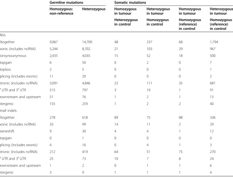
Using refGene hg19 database, AS was able to annotate 37,990 SNVs and 1,484 small indels. In the case of SNVs, we considered the data reliable, if the coverage was over 20; thus, 25,914 SNVs remained. In the case of SNVs, there were 23,767 germline mutations (9,067 in homozy-gous form and 14,700 in heterozyhomozy-gous form) and 2,147
somatic mutations (in the tumour tissue—116 in
homo-zygous form and 2,031 in heterohomo-zygous form) (Table 1, Additional file 1). Furthermore, there were 896 germline small indels (278 in homozygous form and 618 in het-erozygous form) and 588 somatic indels (in the tumour
tissue—177 in homozygous form and 411 in
heterozy-gous form).
Applying dbSNP135, we were able to annotate 5,281 SNVs and 239 small indels. With dbCOSMIC67, we
an-notated 2,569 SNVs and 59 small indels—none of these
were noted to be associated to bone cancer. Applying ljb2 database, we found 469 SNVs to potentially cause a disease (average ljb2 score over 0.918), including 31 germline mutations and 4 somatic mutations (ESX1: c.
A578G/p.K193R; CDC27: c.A17G/p.E6G; TMEM120B:
c.G274A/p.D92N; TMEM131: c.C3947T/p.P1316L) in
homozygous form in the tumour tissue.
2) Results from Ingenuity Variant Analysis software
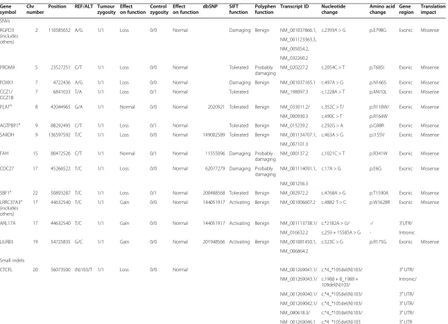
Altogether, 207 cancer driver variants (CD-SNVs) were found in 123 genes according to IVA (Additional file 2). Fourteen CD-SNVs potentially gain and 186 lose the gene function. Only seven SNVs may have no drastic effect on gene function in the tumour tissue. Further-more, according to IVA, none of these 207 SNVs affect the gene functionality in the control tissue. Thirteen of the CD-SNVs were homozygous in the tumour tissue (Table 2). There were no cancer-associated homozygous mutations present in the control tissue; thus, the homozygous CD-SNVs in the tumour tissue are all somatic.
According to IVA, six cancer-associated small indels were found (Table 2). Four of them are homozygous and two are heterozygous in the tumour tissue—the effect is most probably the loss of gene function. These indels are predicted to have no effect in the control tissue.
In most of the genes brought front by IVA, one CD-SNV was found in coding region in heterozygous form. However, some of the genes have more CD-SNVs in
cod-ing regions: MUC4 had even 22, ZNF717 had 8, CTBP2
had 7 and OR4C3had 5 CD-SNVs, whereas these were
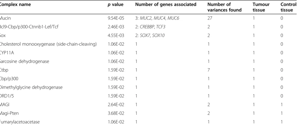
not present in the control tissue (data not shown). When observing from a slightly different angle—the gene com-plexes, we can see that the mucin complex has the highest
Reimannet al. Human Genomics2014,8:20 Page 3 of 16
significance—three genes and 27 CD-SNVs are considered (Table 3). There are also other gene complexes, which are potentially associated to cancer processes, and in different complexes, the CD-SNVs are either somatic or germline (Table 3).
In the case of cancer-associated small indels, the sta-tistically most significant results were with complexes
related to RELA gene—NFKB1-RELA and RELA-REL
complexes both hadpvalue 7.56E-4.
IVA provided the first 100 cancer-associated processes and diseases related to CD-SNVs and small indels. Seventy-three genes and 135 CD-SNVs were found
asso-ciated to process named as “disorder of genitourinary
system” (Table 4). These findings were present in both
the tumour and control tissues. There were also two processes associated to bone“myelopoiesis of bone
mar-row” (associated genes NPM1, RARA) and “quantity of
trabecular bone” (associated genes CREBBP, SMO)—
these findings were present only in the tumour tissue. In the case of small indels, all the findings were somatic
andALKandRELAgenes were associated to“outgrowth
of bone marrow cells” and “inflammatory response of
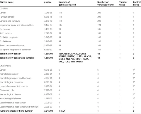
bone marrow-derived macrophages”, respectively. IVA found 111 genes with 202 germline CD-SNVs as-sociated to cancer (Table 5). Fifteen genes, which had 43
somatic CD-SNVs were associated to“bone marrow
can-cer and tumours”. In the case of small indel, all six
genes, with a finding, are associated to cancer and the found small indels are all somatic. The disease named as
“tumourigenesis of bone tumour” was associated to
small indel in ALK gene and was present only in the
tumour tissue.
With the osteosarcoma patient’s tumour and control
tissue, WES data IVA found six pathways associated to CD-SNVs and six to cancer driver small indels (Table 6). All the mutations considered here were somatic. In the case of CD-SNVs, the statistically most significant
asso-ciation was between tumour and WNT/β-catenin
signal-ling pathway. In the case of small indels, associations with different cytokine pathways were found. Also, a
pathway directly linked to the bone tissue—“RANK
sig-nalling in osteoclasts”was brought front.
Table 1 The numbers of SNV and small indel findings received from data analysis with ANNOVAR software
Germline mutations Somatic mutations Homozygous:
non-reference
Heterozygous Homozygous in tumour
Heterozygous in tumour
Homozygous in tumour
Heterozygous in tumour Heterozygous
in control
Homozygous in control
Homozygous (reference) in control
Homozygous (reference) in control
SNVs
Altogether 9,067 14,700 48 237 68 1,794
Exonic (includes ncRNA) 5,244 8,702 21 103 29 967
Nonsynounymous 2,435 4,035 15 52 18 500
Stopgain 6 50 0 2 0 7
Stoploss 2 5 0 0 0 1
Splicing (includes exonic) 11 20 0 0 0 2
Intronic (includes ncRNA) 3,091 4,846 22 111 35 681
5′UTR and 3′UTR 515 797 3 19 1 91
Downstream and upstream 51 76 1 2 1 13
Intergenic 155 259 1 2 2 40
Small indels
Altogether 278 618 89 75 88 336
Exonic (includes ncRNA) 33 99 14 11 2 29
Frameshift 9 30 4 4 1 12
Stopgain 0 1 0 0 0 0
Splicing (includes exonic) 4 16 0 4 1 3
Intronic (includes ncRNA) 212 419 64 51 75 270
5′UTR and 3′UTR 25 73 10 7 8 24
Downstream and upstream 1 2 0 1 1 6
Intergenic 3 9 1 1 1 4
Reimannet al. Human Genomics2014,8:20 Page 4 of 16
Table 2 The somatic cancer driver SNVs and small indels found in data analysis with Ingenuity Variant Analysis software
Gene symbol
Chr number
Position REF/ALT Tumour zygosity
Effect on function
Control zygosity
Effect on function
dbSNP SIFT function
Polyphen function
Transcript ID Nucleotide change
Amino acid change
Gene region
Translation impact
SNVs
RGPD3 (includes others)
2 110585652 A/G 1/1 Loss 0/0 Normal Damaging Benign NM_001037866.1, c.2393A > G p.E798G Exonic Missense
NM_001123363.3,
NM_005054.2,
NM_032260.2
PRDM9 5 23527251 C/T 1/1 Loss 0/0 Normal Tolerated Probably damaging
NM_020227.2 c.2054C > T p.T685I Exonic Missense
FOXK1 7 4722436 A/G 1/1 Loss 0/0 Normal Damaging Benign NM_001037165.1 c.497A > G p.N166S Exonic Missense
CCZ1/ CCZ1B
7 6841033 T/A 1/1 Loss 0/1 Normal Tolerated NM_198097.3 c.1228A > T p.M410L Exonic Missense
PLATa 8 42044965 G/A 1/1 Normal 0/0 Normal 2020921 Tolerated Benign NM_033011.2/ c.352C > T/ p.R118W/ Exonic Missense
NM_000930.3 c.490C > T p.R164W
AGTPBP1a 9 88292495 C/T 1/1 Loss 0/1 Normal Tolerated Benign NM_015239.2 c.292G > A p.G98R Exonic Missense
SARDH 9 136597592 T/C 1/1 Loss 0/0 Normal 149002589 Tolerated Benign NM_001134707.1, c.463A > G p.I155V Exonic Missense
NM_007101.3
FAH 15 80472526 C/T 1/1 Normal 0/1 Normal 11555096 Damaging Probably damaging
NM_000137.2 c.1021C > T p.R341W Exonic Missense
CDC27 17 45266522 T/C 1/1 Loss 0/0 Normal 62077279 Damaging Probably damaging
NM_001114091.1, c.17A > G p.E6G Exonic Missense
NM_001256.3
SBF1a 22 50893287 T/C 1/1 Loss 0/1 Normal 200488568 Tolerated Benign NM_002972.2 c.4768A > G p.T1590A Exonic Missense
LRRC37A3a (includes others)
17 44632540 T/C 1/1 Gain 0/0 Normal 144051917 Activating Benign NM_001006607.2 c.4882 T > C p.W1628R Exonic Missense
ARL17A 17 44632540 T/C 1/1 Gain 0/0 Normal 144051917 Activating Benign NM_001113738.1/ c.*2182A > G/ -/ 3'UTR/
NM_016632.2 c.259 + 15585A > G - Intronic
LILRB3 19 54725835 G/C 1/1 Gain 0/0 Normal 201948566 Activating Benign NM_001081450.1, c.523C > G p.R175G Exonic Missense
NM_006864.2
Small indels
CTCFL 20 56073500 (N)103/T 1/1 Loss 0/0 Normal NM_001269041.1/ c.*4_*105del(N)103/ 3′UTR/
NM_001269043.1/ c.1988 + 8_1988 + 109del(N)103/
Intronic/
NM_001269040.1/ c.*4_*105del(N)103/ 3′UTR/
NM_001269042.1/ c.*4_*105del(N)103/ 3′UTR/
NM_080618.3/ c.*4_*105del(N)103/ 3′UTR/
NM_001269046.1 c.*4_*105del(N)103 3′UTR
Reimann
et
al.
Human
Genomics
2014,
8
:20
Page
5
o
f
1
6
http://ww
w.humgenom
ics.com/co
Table 2 The somatic cancer driver SNVs and small indels found in data analysis with Ingenuity Variant Analysis software(Continued)
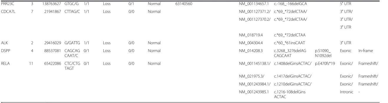
PRR23C 3 138763627 GTGC/G 1/1 Loss 0/1 Normal 63140560 NM_001134657.1 c.-168_-166delGCA 5′UTR
CDCA7L 7 21941867 CTTAG/C 1/1 Loss 0/0 Normal NM_001127371.2/ c.*69_*72delCTAA/ 3′UTR/
NM_001127370.2/ c.*69_*72delCTAA/ 3′UTR/
3′UTR
NM_018719.4 c.*69_*72delCTAA
ALK 2 29416029 G/GATTG 1/1 Loss 0/0 Normal NM_004304.4 c.*60_*61insCAAT 3′UTR
DSPP 4 88537081 CAGCAG CAAT/C
0/1 Loss 0/0 Normal NM_014208.3 c.3268_3276delAG
CAGCAAT
p.S1090_ N1092del
Exonic In-frame
RELA 11 65422086 CTC/CTG TAGT
0/1 Loss 0/0 Normal NM_001145138.1/ c.1408delGinsACTAC/ p.E470fs*19 Exonic/ Frameshift/
NM_021975.3/ c.1417delGinsACTAC/ Exonic/ Frameshift/
NM_001243984.1/ c.1210delGinsACTAC/ Exonic/ Frameshift/
NM_001243985.1 c.1216-108delGins ACTAC
Intronic
-a
The expression pattern of these genes has changed in the tumour tissue compared to that in the control tissue.
Reimann
et
al.
Human
Genomics
2014,
8
:20
Page
6
o
f
1
6
http://ww
w.humgenom
ics.com/co
3)Results from CEQer software
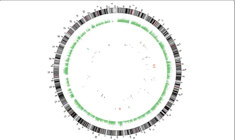
We applied CS to analyse CNVs in tumour and non-tumour tissue exomes. Compared to the control tissue, in the tumour tissue, the loss of coding sequences was found in 6 chromosomes and 183 genes and gain of cod-ing sequences in 4 chromosomes and 65 genes (Figure 1). The loss or gain of coding sequences was altogether in 8 chromosomes, and the most altered were chromosomes 2 and 19 (193,701 bp and 115,358 bp, respectively; Figure 2). The loss of heterozygosity was detected altogether in 68 regions in 37 genes, located in 15 differ-ent chromosomes (Additional file 3).
Integrative analysis
The integrative analysis narrows down the large list of findings from NGS data. When combining the results from WES data (AS, IVA, CS) and RNA-seq data [10], we found some interesting and rather logical associa-tions, which we would like to emphasize.
SNVs, small indels and RNA expression
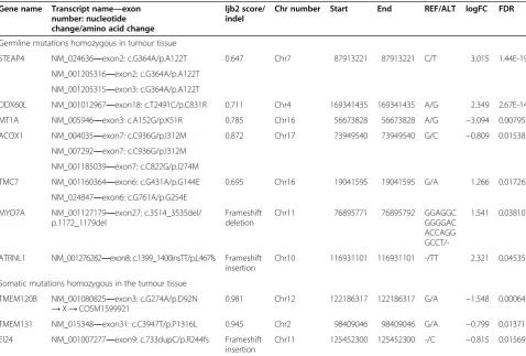
To reduce down the complexity of data we received from AS, we decided to perform as follows. In the case of SNV data, we observed both somatic and germline SNVs, which are homozygous in the tumour tissue and should have an effect on translation (nonsynonymous, stopgain, stoploss findings). Thus, we got 527 homozy-gous germline SNVs (in 392 genes) and 8 homozyhomozy-gous somatic SNVs (in 7 genes), which are located in genes with altered expression in the tumour tissue compared to that in the control tissue. If also considering the ljb2 database scores, seven homozygous SNVs with high disease-causing probability remained (Table 7).
In the case of small indels detected with AS, we ob-served the somatic and germline indels, which were homozygous in the tumour tissue. There was 52 germ-line and 26 somatic indels in introns of the genes, which expression pattern has also changed (data not shown). Furthermore, there was five germline and three somatic indels in exons of the genes with altered expression. Thus, we found altogether three frameshift small indels, which possibly have an effect on translation (frameshift insertions and deletion in exons) (Table 7).
In the case of homozygous cancer driver SNVs and small indels found with IVA (Table 2), only four genes have altered expression pattern in the tumour tissue compared to that in the control tissue. The mRNA
ex-pression was increased in the case of PLAT (log fold
change (logFC) = 3.65, false discovery rate (FDR;
cor-rected statistical significance) = 8.27E-27), AGTPBP1
(logFC = 0.91, FDR = 0.039) and LRRC37A3 (logFC =
1.14, FDR = 0.0072) and decreased in the case of SFB1
(logFC =−1.33, FDR = 0.0037).
CNVs, LOHs and RNA expression
When analysing the CNV results together with RNA ex-pression results, we found that with gained copy num-bers, there were altogether 22 genes, with altered expression profile—20 genes with increased and 2 genes with decreased mRNA expression. In the case of loss
copy of number, 74 genes’ expression profile had
chan-ged—11 genes with increased and 63 genes with
de-creased mRNA expression. In Table 8, the genes with the lowest FDR values for gene expression results are
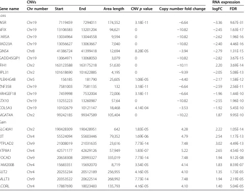
presented. Here, we would emphasize that the INSR,
which has copy number loss in area covering 174,552 bp has also a remarkable decrease in mRNA expression
Table 3 The gene complexes which are potentially associated to cancer processes
Complex name pvalue Number of genes associated Number of variances found
Tumour tissue
Control tissue
Mucin 9.54E-05 3:MUC2,MUC4,MUC6 27 1 0
Bcl9-Cbp/p300-Ctnnb1-Lef/Tcf 2.46E-03 2:CREBBP,TCF3 2 1 0
Sox 4.55E-03 2:SOX7,SOX10 2 1 0
Cholesterol monooxygenase (side-chain-cleaving) 1.06E-02 1 1 1 0
CYP11A 1.06E-02 1 1 1 0
Sarcosine dehydrogenase 1.06E-02 1 1 1 0
Ctbp 1.59E-02 1 7 1 0
Cbp/p300 1.59E-02 1 1 1 0
Dimethylglycine dehydrogenase 1.59E-02 1 1 1 0
DRD1/5 1.59E-02 1 1 1 0
MAGI 2.64E-02 1 2 1 1
Magi-Pten 3.68E-02 1 2 1 1
Fumarylacetoacetase 1.06E-02 1 1 1 1
There are both somatic and germline cancer driver SNVs found in the tumour and control tissues.
Reimannet al. Human Genomics2014,8:20 Page 7 of 16
(3.36 times; FDS = 9.67E-31). However, there are also several genes with CNVs, which could be associated to cancer.
Combining the LOH and mRNA expression data, we found that in the tumour tissue, the expression of four genes with LOH has increased significantly and expres-sion of five genes with LOH has decreased significantly (Table 9). The rest of the genes with LOHs had no sig-nificant changes in mRNA expression level, and two genes were not detected with RNA-seq (FLJ20518, MANSC4) [10].
For additional information, please see the supplemen-tary material as separate files for AS, IVA and CS com-bined with RNA-seq data.
Discussion
In this study, the exome profiles of the osteosarcoma
pa-tient’s tumour and normal bone tissue were compared.
Additionally, the RNA-seq data from our previous work was used [10]. For WES data analysis, several softwares were applied and possibly some of them are better in de-tecting some mutations and not so effective in dede-tecting others. Still, we think it is more beneficial to use differ-ent approaches and we believe it is easier to follow, if we discuss separately the results gained from each software.
The ANNOVAR software annotated a large amount of genes with SNVs and small indels, applying refGene hg19 database. Over 2,700 somatic SNVs and small indels were detected specifically in the tumour tissue, from which almost 300 are homozygous. These findings are located all over the exome. This demonstrates that the changes in OS genome are not concentrated into a single or few areas but are rather distributed.
When using ljb2 database, AS detected four homozy-gous somatic mutations in the tumour tissue, which could potentially cause a disease. These nonsynonymous
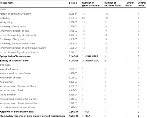
Table 4 The cancer-associated processes detected by IVA
Process name pvalue Number of
genes associated
Number of variances found
Tumour tissue
Control tissue
CD-SNVs
Disorder of genitourinary system 9.05E-14 73 135 1 1
Cell biology 4.08E-04 69 132 1 1
Cell signalling 3.83E-03 25 31 1 1
Morphology of body region 2.55E-03 23 24 1 1
Abnormal morphology of cells 1.73E-03 18 19 1 1
Abnormal morphology of body cavity 6.17E-04 17 18 1 1
Morphology of body cavity 1.36E-03 17 18 1 1
Morphology of cardiovascular system 5.80E-04 13 14 1 1
Abnormal morphology of cardiovascular system 7.22E-04 12 13 1 1
Abnormal morphology of thoracic cavity 1.22E-03 11 12 1 1
Myelopoiesis of bone marrow 3.64E-03 2: NPM1, RARA 2 1 0
Quantity of trabecular bone 4.08E-03 2: CREBBP, SMO 2 1 0
Small indels
Tissue development 1.19E-03 5 5 1 0
Developmental process of tissue 1.35E-03 5 5 1 0
Development of organ 5.05E-03 4 4 1 0
Organogenesis 5.32E-03 4 4 1 0
Colony formation of tumour cell lines 6.25E-05 3 3 1 0
Colony formation of cells 4.44E-04 3 3 1 0
Colony formation 5.46E-04 3 3 1 0
Developmental process of tumour cells 3.81E-03 3 3 1 0
Colony formation of carcinoma cell lines 5.94E-05 2 2 1 0
Apoptosis of nervous tissue cell lines 2.49E-04 2 2 1 0
Outgrowth of bone marrow cells 7.56E-04 1: ALK 1 1 0
Inflammatory response of bone marrow-derived macrophages 1.26E-03 1: RELA 1 1 0
The sorting is performed by number of genes.
The bold data reflects the processes directly associated to bone.
Reimannet al. Human Genomics2014,8:20 Page 8 of 16
mutations were located in ESX1, CDC27, TMEM120B
and TMEM131. Additionally, in the case ofTMEM120B
and TMEM131, the mRNA expression has decreased
substantially in the tumour tissue compared to that in the control tissue [10]; however, further studies are needed to confirm the possible associations between found mutations and gene expression level. Available data about the possible associations between OS and
these genes is very limited. InTMEM120B, a gene with
an unclear function, the mutation COSM1599921 has
been previously detected in glioma [33]. TheCDC27is a
gene possibly controlling the timing of mitosis and may have an important role in tumour cell division [34].
In addition to the somatic mutation, the CDC27 had
33 heterozygous germline disease-causing mutations (non-synonymous) (data not shown). In the case of breast
cancer, the CDC27 has been demonstrated to be a
promising biomarker in predicting the disease progression and prognostication [35]. Thus, these somatic mutations may have some effect on OS pathogenesis. Especially the
abundant changes in CDC27may be important in terms
of regulating OS tumour cell division.
In the tumour tissue, we detected homozygous som-atic small indels causing the frameshift in five genes—
EI24, ALG1L2, TIGD6, GPATCH4 and SSPO. None of
these genes have previously been associated to OS, and
according to our RNA-seq data, only EI24 of these five
genes has altered mRNA expression—it has decreased in
the tumour tissue [10], which could be due to the
inser-tion in exon 9. The EI24 encodes a tumour suppressor
and is an immediate-early induction target of
TP53-mediated apoptosis—it binds to antiapoptotic BLC2.
Furthermore, the EI24 has found to be highly mutated
in the case of aggressive breast cancer and is rather
Table 5 The diseases associated to CD-SNVs and small indels
Disease name pvalue Number of
genes associated
Number of variances found
Tumour tissue
Control tissue
CD-SNVs
Cancer 7.04E-23 111 202 1 1
Tumourigenesis 8.21E-16 111 202 1 1
Cancers and tumours 3.37E-15 111 202 1 1
Organismal injury and abnormalities 9.45E-17 105 194 1 1
Carcinoma 3.46E-25 99 186 1 1
Solid tumour 2.64E-24 99 186 1 1
Epithelial neoplasia 3.34E-23 99 186 1 1
Epithelioma 3.34E-23 99 186 1 1
Breast or colorectal cancer 5.45E-23 83 164 1 1
Malignant neoplasm of abdomen 6.93E-20 83 169 1 1
Bone marrow cancer 1.69E-03 15: CREBBP, EPHA2, FGFR2, KCNJ12, KMT2C, LILRB3, MUC17, MUC4, MYBPC3, NPM1, RARA, SMO, TCF3, TTN, TUBG1
43 1 0
Bone marrow cancer and tumours 1.69E-03 43 1 0
Small indels
Cancer 9.07E-03 6 6 1 1a
Hematologic cancer 2.36E-04 4 4 1 1a
Hematologic cancer and tumours 2.36E-04 4 4 1 1a
Hematological neoplasia 8.01E-04 4 4 1 1a
Lymphohematopoietic cancer 9.12E-04 4 4 1 1a
Disease of colon 7.88E-03 4 4 1 0
Hematological disease 8.15E-03 4 4 1 1a
Immunological disease 1.28E-02 4 4 1 1a
Gastrointestinal tract cancer 2.00E-02 4 4 1 0
Gastrointestinal tract cancer and tumours 2.02E-02 4 4 1 0
Tumourigenesis of bone tumour 7.04E-03 1: ALK 1 1 0
a
Here, only one gene PRR23C has a small indel in heterozygous form, which most likely does not affect the gene function. See Table2. The bold data reflects the diseases directly associated to bone.
Reimannet al. Human Genomics2014,8:20 Page 9 of 16
associated to tumour invasiveness than development of the primary tumour [36-38]. In the present case, we found no mutations inTP53nor was the expression altered [10]; thus, according to this data, we may suggest that the TP53 is functional in the tumour tissue. However, the TP53
pathway may still be suppressed due to mutated and
downregulated EI24. Moreover, the aggressive nature of
OS is correlated to this finding.
Appling Ingenuity Variant Analysis software, we found over 200 cancer driver variants and 93% of these possibly
Table 6 The pathways associated to cancer
Pathway name pvalue Number
of genes
Genes Number
of variants
Tumour tissue
Control tissue
CD-SNVs
Wnt/β-catenin signalling 7.07E-04 6 CREBBP, RARA, SMO, SOX10, SOX7, TCF3 6 1 0
Epithelial adherens junction Ssignalling 1.26E-02 4 IQGAP1, KEAP1, TCF3, TUBG1 4 1 0
Germ cell-sertoli cell junction signalling 2.10E-02 4 GSN, IQGAP1, KEAP1, TUBG1 5 1 0
Mouse embryonic stem cell pluripotency 2.59E-02 3 CREBBP, SMO, TCF3 3 1 0
Regulation of the epithelial-mesenchymal transition pathway
3.40E-02 4 FGFR2, SMO, TCF3, ZEB2 4 1 0
Hereditary breast cancer signalling 4.95E-02 3 CREBBP, NPM1, TUBG1 3 1 0
Small indels
IL-17A signalling in gastric cells 8.79E-03 1 RELA 1 1 0
Role of JAK1, JAK2 and TYK2 in interferon signalling 9.54E-03 1 RELA 1 1 0
Interferon signalling 9.79E-03 1 RELA 1 1 0
IL-15 production 1.00E-02 1 RELA 1 1 0
TNFR2 signalling 1.05E-02 1 RELA 1 1 0
RANK signalling in osteoclasts 2.86E-02 1 RELA 1 1 0
Figure 1Circos plot illustrating the CNVs and LOHs in the OS tissue compared to that in the control tissue.CNVs are marked as lines in the centre: red—gain and green—loss. LOHs are marked as dots in the centre: black—copy neutral, green—copy gain and red—copy loss.
Reimannet al. Human Genomics2014,8:20 Page 10 of 16
cause the loss of gene function. Thirteen homozygous
som-atic CD-SNVs were detected in different genes—RGPD3,
PRDM9, FOXK1, CCZ1, PLAT, AGTPBP1, SARDH, FAH,
CDC27, SBF1, LRRC37A3, ARL17A and LILRB3. The
mRNA expression ofPLAT,AGTPBP1andLRRC37A3has
increased and ofSFB1has decreased significantly [10]. We found no previous data about the associations between OS
and these genes, except SBF1. With previous OS studies,
another missense mutation (p.E1539K) has detected in
SBF1 [39]. SBF1 is a SET (a nuclear oncogene) binding
Figure 2The CNVs in chromosomes 2 and 19 in the osteosarcoma tissue compared to that in the control tissue.Data analysis performed with CEQer software.
Table 7 The integrative analysis—genes with altered expression pattern [10] and SNVs annotated with ANNOVAR software
Gene name Transcript name—exon number: nucleotide change/amino acid change
ljb2 score/ indel
Chr number Start End REF/ALT logFC FDR
Germline mutations homozygous in tumour tissue
STEAP4 NM_024636—exon2: c.G364A/p.A122T 0.647 Chr7 87913221 87913221 C/T 3.015 1.44E-19
NM_001205316—exon2: c.G364A/p.A122T NM_001205315—exon3: c.G364A/p.A122T
DDX60L NM_001012967—exon18: c.T2491C/p.C831R 0.711 Chr4 169341435 169341435 A/G 2.349 2.67E-14
MT1A NM_005946—exon3: c.A152G/p.K51R 0.785 Chr16 56673828 56673828 A/G −3.094 0.00795
ACOX1 NM_004035—exon7: c.C936G/p.I312M 0.872 Chr17 73949540 73949540 G/C −0.809 0.01538
NM_007292—exon7: c.C936G/p.I312M NM_001185039—exon7: c.C822G/p.I274M
TMC7 NM_001160364—exon6: c.G431A/p.G144E 0.695 Chr16 19041595 19041595 G/A 1.266 0.01726
NM_024847—exon6: c.G761A/p.G254E MYO7A NM_001127179—exon27: c.3514_3535del/
p.1172_1179del
Frameshift deletion
Chr11 76895771 76895792 GGAGGC
GGGGAC ACCAGG
GCCT/-1.541 0.03810
ATRNL1 NM_001276282—exon8: c.1399_1400insTT/p.L467fs Frameshift insertion
Chr10 116931101 116931101 -/TT 2.321 0.04535
Somatic mutations homozygous in the tumour tissue
TMEM120B NM_001080825—exon3: c.G274A/p.D92N →X→COSM1599921
0.981 Chr12 122186317 122186317 G/A −1.548 0.00064
TMEM131 NM_015348—exon31: c.C3947T/p.P1316L 0.945 Chr2 98409046 98409046 G/A −0.799 0.01371
EI24 NM_001007277—exon9: c.733dupC/p.R244fs Frameshift insertion
Chr11 125452300 125452300 -/C −0.815 0.01569
These germline or somatic SNVs are all nonsynonymous and homozygous in the tumour tissue and according to ljb2 database have a disease-causing effect.
Reimannet al. Human Genomics2014,8:20 Page 11 of 16
Table 8 The integrative analysis—CNVs and RNA expression data [10] is observed together
CNVs RNA expression
Gene name Chr number Start End Area length CNVpvalue Copy number fold change logFC FDR
Loss
INSR Chr19 7119459 7294011 174,552 3.18E-11 −6.64 −3.36 9.67E-31
NFIX Chr19 13106583 13201204 94,621 0 −10.82 −2.45 1.63E-17
FARSA Chr19 13034964 13044558 9,594 0 −10.82 −2.62 1.96E-16
RAD23A Chr19 13056627 13063667 7,040 0 −10.82 −2.40 4.46E-16
GINS4 Chr8 41386724 41399418 12,694 8.28E-05 −3.94 −2.79 1.31E-15
GADD45GIP1 Chr19 13064971 13068050 3,079 0 −10.82 −2.82 3.67E-15
IFIH1 Chr2 163123588 163175218 51,630 0 −10.11 2.20 3.69E-14
RPL31 Chr2 101618690 101622885 4,195 0 −9.39 −2.05 5.08E-13
PLEKHG4B Chr5 156185 181790 25,605 1.08E-05 −4.40 −2.17 1.58E-12
ZNF358 Chr19 7581003 7581135 132 3.18E-11 −6.64 −2.59 2.56E-11
ARHGEF18 Chr19 7459998 7532004 72,006 3.18E-11 −6.64 −1.96 1.44E-10
STX10 Chr19 13255223 13260987 57,64 0 −10.82 −2.55 1.96E-10
COL5A3 Chr19 10102679 10121147 18,468 4.14E-04 −3.53 −1.92 5.45E-10
MGAT4A Chr2 99242185 99347589 105,404 0 −10.22 1.87 9.95E-10
Gain
SLC40A1 Chr2 190428309 190428951 642 1.83E-05 4.28 2.22 1.05E-14
KIT Chr4 55524094 55603446 79,352 1.69E-06 4.79 2.54 1.17E-13
PTPLAD2 Chr9 21008019 21031635 23,616 7.73E-14 7.48 3.02 4.49E-13
ATP8A1 Chr4 42571177 42629126 57,949 1.83E-07 5.22 2.65 4.54E-10
FOCAD Chr9 20658308 20993327 335,019 7.73E-14 7.48 1.94 9.12E-08
FAM200B Chr4 15683351 15692070 8,719 3.54E-05 4.14 1.83 8.59E-07
SLIT2 Chr4 20255234 20512189 256,955 4.16E-05 4.10 1.35 1.73E-05
MLLT3 Chr9 20353522 20622514 268,992 7.73E-14 7.48 1.94 2.19E-05
LCORL Chr4 17887690 18023483 135,793 4.16E-05 4.10 1.40 5.04E-05
Only the genes with lowest FDR value are presented.
Table 9 The integrative analysis - loss of heterozygosity and RNA expression data observed together Gene name Chr
number
LOHs RNA expression
LOH position Alleles LOH LOHpvalue logFC FDR
MS4A14 Chr11 60165358–60165379 G/C CopyNeutralLOH 0.025 2.46 3.20E-08
DSC2 Chr18 28666554–28666556 A/C CopyNeutralLOH 0.025 1.87 3.82E-07
RPS4X ChrX 71495409–71495414 G/C CopyNeutralLOH 0.01 −1.44 7.25E-07
RPS23 Chr5 81571874 A/C CopyNeutralLOH 0.005 −1.43 1.04E06
IL7R Chr5 35874575 C/T 1AlleleGain 0.025 1.59 6.69E-06
PCNXL2 Chr1 233398713 C/T CopyNeutralLOH 0.01 1.20 0.00027
HILPDA (C7orf68) Chr7 128098270 T/G CopyNeutralLOH 0.0001 −1.12 0.00094
HRNR Chr1 152188041 C/T Allele(s)Loss 0.025 −3.09 0.00796
MUC4 Chr3 195515594, 195516630 C/G CopyNeutralLOH 0.025 −2.22 0.01230
Only the genes with significant mRNA expression changes in the tumour tissue compared to that in the control tissue are presented.
Reimannet al. Human Genomics2014,8:20 Page 12 of 16
factor 1 and may inhibit the cell division [40]. The de-creased expression in the tumour tissue may be responsible for the increased cell proliferation. Some other associations,
which might be interesting—PLATgene is important for
cell migration and tissue remodelling and the overexpres-sion might cause hyperfibrinolysis [41], which has not pre-viously described in the case of OS. Two mutations in ARL17Ahave detected in chondrosarcoma cells [42]. In the
case of CDC27, the same mutation (p.E6G) was also
brought front by AS as potentially disease causing, which is discussed above. Thus, it is highly likely that at least some of these genes participate in some level of OS pathogenesis.
Additionally, with IVA four homozygous somatic small indels were detected in the tumour tissue. These were in
noncoding regions of genes CTCFL, PRR23C, CDCA7L
and ALK; thus, the effect might be post-transcriptional. CTCFLis a genetic paralog ofCTCF; latter is an
import-ant methylation pattern regulator. In the case of CTCF,
it has previously demonstrated that in the OS tissue, the changes in its methylation pattern may also cause loss of
imprinting of IGF2 and H19 genes, which further alters
their expression pattern [43]. In our OS patient’s tumour
tissue, the mRNA expression of both IGF2 and H19 has
increased significantly (FDR = 3.46E-15 and FDR = 0.0015, respectively) [10]. Thus, the association may be valid here
also. In PRR23C, one missense mutation (p.R190W) has
detected previously in the OS tissue [42].ALK encodes a receptor tyrosine kinase and is rearranged, mutated or amplified in several tumours. However, in the case of OS, there are only few reports about ALK [44,45]. In addition, two heterozygous somatic small indels were detected in
DSPP and RELA exons; however, we found no previous
data about these findings and associations to OS. The small indels might have an effect on the expression of these genes both pre- and post-transcriptional level; how-ever, these suggestions need to be further studied.
According to IVA, there were several genes with more
than one mutation—inMUC4, there were even 22 somatic
mutations in exons and 44 in introns, although they all
were heterozygous. Thus, we foundMUC4locus to be the
most altered in the tumour tissue compared to that in the control tissue. This might explain why its mRNA expres-sion in the tumour tissue has decreased (FDR = 0.012) [10]. Mucin 4 is among major constituents of mucus, and it has demonstrated that primary bone tumours rarely ex-press MUC4 protein [46], which correlates to our finding. Furthermore, with IVA, we found mucin complex (MUC2,
MUC4, MUC6) to have a highest significance in OS
among others. However, there are also other mucin genes (MUC16, MUC17, MUC20) with somatic heterozygous CD-SNVs. The expression pattern of all other detected mucins has not changed significantly. Thus, mucins may have a role in OS pathogenesis, but we dear not to make any further conclusions.
With IVA, there was four bone-related processes brought front only in the case of the tumour tissue—“myelopoiesis
of bone marrow” (NPM1, RARA), “quantity of trabecular
bone” (CREBBP, SMO),“outgrowth of bone marrow cells”
(ALK) and “inflammatory response of bone
marrow-derived macrophages”(RELA). Furthermore, in disease list, 16 genes with over 40 somatic variations were associated to “bone marrow cancer” and“bone tumour”; however, there were also over 200 germline CD-SNVs associated to cancer. Thus, here, we would like to emphasize that in the case of both cancer-associated processes and diseases, the ones as-sociated with bone are somatic mutations; however, the findings possibly promoting cancer are germline mutations. This is one of the phenomena, which we would like to ob-serve in our future studies.
The most significant pathway found with IVA was
“WNT/β-catenin signalling pathway” (altered genes:
CREBBP, RARA, SMO, SOX10, SOX7, TCF3). Reviewed
in [15], the pathway is required for bone development and has demonstrated to be altered in pathogenesis of
OS—overexpression of numerous WNT pathway
com-ponents including WNT ligands, FZDs and LRP recep-tors and epigenetic silencing of the pathway inhibiting
genes, i.e. WIF1. However, in our previous study, we
found WNT7B and WNT11 to be downregulated and
WNT2Band WNT5Bupregulated;FZD4and FZD8
up-regulated and LRP8 and LRP12 downregulated and
DVL3 downregulated and WIF1and SOST upregulated.
Additionally, genes with CD-SNVs—RARA, SMO and
SOX7were upregulated [10]. Thus, our results are rather controversial to several previous studies demonstrating
the WNT/β-catenin pathway to be upregulated [47-49].
However, there are also studies correlating to our findings [50,51]. As our study is based on a single case, we dear
not to conclude, why the WNT/β-catenin pathway is
ra-ther downregulated here, but we suggest the controversial results may occur due to major heterogeneity of OS. Nevertheless, the present study demonstrates that in addition to altered expression patter, the genes involved in WNT/β-catenin signalling pathway carry the CD-SNVs.
In the case of small indels, the IVA brought front the pathways associated to RELA and these are mostly cyto-kine signalling pathways (Table 6). Previously, it has demonstrated that interaction of IL17A and IL17AR promotes metastasis in OS cells. Furthermore, IL17 stimulates osteoclast resorption [52]. In our previous study, we found IL17AR to be significantly upregulated
[10]. Osteoclasts are important in pathogenesis of OS—
the more active they are, the more aggressive the tumour is [53]. RELA is demonstrated to enhance the osteoclast differentiation [54]. As IVA predicts the loss of RELA functionality (at least partially, as the small indel is heterozygous), in the present case, the OS might not have been as aggressive as it usually would.
Reimannet al. Human Genomics2014,8:20 Page 13 of 16
Previously, it has demonstrated that chromothripsis
event is common to early stage of OS—hundreds of
gen-omic rearrangements will appear in a single instability event [26]. In the present case, the CEQer software de-tected nearly 2,400 gain and loss events in 8 chromosomes involved, which should qualify as the chromothripsis. However, the initiating cause of this massive rearrange-ment is unknown, as there were no traumas or other en-vironmental causes we are aware of.
In general, the gain of copy number should increase the mRNA expression and loss of copy number should decrease the expression [6]. In present work, this pattern was valid in the case of 86.5% of the genes with CNVs and altered expression. One of the strongest findings
here was the amount of CNVs in INSR, which
expres-sion has decreased remarkably (Table 8). The main physiological role of the insulin receptor appears to be metabolic regulation [55]. However, together with IGF1R it forms a hybrid receptor for IGF1, latter together with IGF2 is thought to have a key role in driving the prolifer-ation and survival of sarcoma cells [56]. Furthermore, the growth hormone and IGF1 axis controls the growth and bone modelling/remodelling [57]. Additionally, the IRS1, which is phosphorylated by the INSR, is important for both metabolic and mitogenic pathways [58]. In the
present case, the mRNA expression of both IGF1 and
IGF2 has increased (FDR = 4.65E-35 and FDR =
3.46E-15, respectively); however, the expression of IGF1R
remained the same in the tumour tissue compared to that in the control tissue [10]. Furthermore, inIGF1Rwe found a heterozygous germline nonsynonymous mutation (p.G1117R) with AS, which according to ljb2 database is a disease causing (data not shown). Similarly to INSR, the
mRNA expression of IRS1 is decreased in the tumour
tissue compared to that in the control tissue (FDR = 2.62E-10) [10]. Thus, in the present case it seems, the pro-liferation of tumour cells might be rather supported by increased effect of IGF1, IGF2 and IGF1R homodimer associations, than IGF1, IGF2 and INSR-IGF1R hetero-dimer associations or INSR effects on IRS1.
The loss of heterozygosity has been reported to be ex-tensive in OS exomes [39]. In the present case, we did not detect whole chromosome or gene region loss; how-ever, we did detect the loss of heterozygosity in smaller regions. The genes with LOH findings and increased
mRNA expression—MS4A14, DSC2, IL7R and PCNXL2
have not associated to OS previously. However, in the
case of DSC2, the overexpression has demonstrated to
be inversely correlated to bone metastasis-free survival
[59]. The mutations in IL7R exon 6 have been
demon-strated to be present in leukaemia patients’bone marrow samples but not associated to other solid tumours [60]. The five genes with LOHs and decreased mRNA
expres-sion—RPS4X, RPS23, HILPDA (C7orf68), HRNR and
MUC4 also do not have previous information associated
to OS. Nonetheless, also the LOH analysis brought for-ward different genes in mucin family. In addition to MUC4, there were also other genes with LOHs but with insignificant mRNA expression changes in the tumour
tissues—MUC2, MUC6andMUC17. Thus, these results
also support the idea that mucins might have a role in pathogenesis of osteosarcoma.
In summary, the present case has several characteris-tics previously demonstrated in OS. The wide genomic
arrangements have appeared—SNVs and small indels all
over the genome and CNVs in some chromosomes; and in several cases, these rearrangements may have an ef-fect on gene expression. Furthermore, the germline mu-tations seem to be associated to cancer in general and somatic mutations to bone tumours. The most signifi-cant pathway was the one probably most thoroughly
studied in the case of OS—the WNT/β-catenin
signal-ling pathway. We found several genes in this pathway carrying the cancer driver variances. Additionally, the IGF1/IGF2 and IGF1R homodimer signalling might have an essential effect on OS pathogenesis. Which also needs to be emphasized is that according to our data (based on DNA and RNA studies), there is no evidence of a
non-functional TP53; however, the TP53 pathway might be
suppressed in further levels—the downregulation of
EI24. In addition, with this study, we found associations between different genes and OS pathogenesis, which have not demonstrated before in earlier studies. We
found the MUC4 locus to be the most altered in the
tumour tissue compared to that in the control tissue; furthermore, several other mucin genes are also possibly
associated to OS. The somatic mutation in CDC27 was
brought front by two different data analysis softwares and might have a role in OS pathogenesis.
Conclusions
All genes, in which the mutations were detected, may be considered as potential targets for additional studies (i.e. functional, histopathological, clinical studies) for finding OS biomarkers. The present study brought front the WNT pathway genes, IGF1/IGF2 and IGF1R homodimer signalling pathway genes,TP53together withEI24,MUC4
together with other mucin genes andCDC27as potential
biomarkers for OS. Finally, as this study is based on a sin-gle case and only DNA and RNA analysis, these data may not be taken as conclusive evidence and further studies are needed to confirm the present findings.
Additional files
Additional file 1:ANNOVAR software.The file contains the list of SNVs (coverage at least 20 times) and small indels detected from WES study.
Reimannet al. Human Genomics2014,8:20 Page 14 of 16
Additionally, the dbSNP135, dbCOSMIC67, ljb2 scores and RNA-seq i nformation is added if available.
Additional file 2:Ingenuity Variant Analysis software.The file contains the list of SNVs, which according to IVA are associated to cancer. Additionally, the dbSNP135, SIFT and POLYPHEN functions and RNA-seq information is added if available.
Additional file 3:CEQer software.The file contains the list of genes where CNVs and LOHs were detected. Additionally, the RNA-seq information is added if available.
Competing interests
The authors declare that they have no competing interests.
Authors’contributions
ER participated in the research concept and design, data analysis and interpretation, writing of the article and critical revision of the article and performed the statistical analysis. SK involved in the research concept and design, data analysis and interpretation and critical revision of the article. XDH contributed in the research concept and design and carried out the collection and/or assembly of data. KM participated in the research concept and design, collection and/or assembly of data, data analysis and interpretation and critical revision of the article. AM involved in the research concept and design, collection and/or assembly of data, data analysis and interpretation and critical revision of the article. All authors read and approved the final manuscript.
Acknowledgements
This work was supported by the institutional research funding IUT (IUT20-46) of the Estonian Ministry of Education and Research, by the Centre of Translational Genomics of University of Tartu (SP1GVARENG) and by the European Regional Development Fund (Centre of Translational Medicine, University of Tartu).
Author details 1
Department of Pathophysiology, University of Tartu, 19 Ravila Street Tartu 50411, Estonia.2Department of Reproductive Biology, Estonian University of Life Sciences,
64 Kreutzwaldi Street, Tartu, Estonia.3Department of Traumatology and Orthopaedics, University of Tartu, 8 Puusepa Street, Tartu, Estonia.
4Department of Oncology, Hue University of Medicine and Pharmacy, 6 Ngo Quyen Street, Hue, Vietnam.5Traumatology and Orthopaedics Clinic, Tartu University Hospital, 8 Puusepa Street, Tartu, Estonia.
Received: 30 July 2014 Accepted: 10 November 2014
References
1. Picci P:Osteosarcoma (osteogenic sarcoma).Orphanet J Rare Dis2007,2:6. 2. Ottaviani G, Jaffe N:The etiology of osteosarcoma.Cancer Treat Res2009,
152:15–32.
3. Botter SM, Neri D, Fuchs B:Recent advances in osteosarcoma.Curr Opin Pharmacol2014,16C:15–23.
4. Allison DC, Carney SC, Ahlmann ER, Hendifar A, Chawla S, Fedenko A, Angeles C, Menendez LR:A meta-analysis of osteosarcoma outcomes in the modern medical era.Sarcoma2012,2012:704872. http://www.ashg. org/2013meeting/programguide/files/assets/basic-html/page258.html. 5. Kiezun A, Janeway K, Tonzi P, Mora J, Aguiar S, Mercado G, Melendez J,
Garraway L, Rodriguez-Galindo C, Orkin S, Golub T, Getz G, Yunes JA: Next generation sequencing of osteosarcoma identifies the PI3K/mTOR pathway as a unifying vulnerability to be exploited for targeted therapy. In.: ASHG meeting 2013; 2013.
6. Kuijjer ML, Hogendoorn PC, Cleton-Jansen AM:Genome-wide analyses on high-grade osteosarcoma: making sense of a genomically most unstable tumor.Int J Cancer2013,133(11):2512–2521.
7. Savage SA, Mirabello L, Wang Z, Gastier-Foster JM, Gorlick R, Khanna C, Flanagan AM, Tirabosco R, Andrulis IL, Wunder JS, Gokgoz N, Patiño-Garcia A, Sierrasesúmaga L, Lecanda F, Kurucu N, Ilhan IE, Sari N, Serra M, Hattinger C, Picci P, Spector LG, Barkauskas DA, Marina N, de Toledo SR, Petrilli AS, Amary MF, Halai D, Thomas DM, Douglass C, Meltzer PS,et al:Genome-wide
association study identifies two susceptibility loci for osteosarcoma.
Nat Genet2013,45(7):799–803.
8. Ottaviano L, Schaefer KL, Gajewski M, Huckenbeck W, Baldus S, Rogel U, Mackintosh C, de Alava E, Myklebost O, Kresse SH, Meza-Zepeda LA, Serra M, Cleton-Jansen AM, Hogendoorn PC, Buerger H, Aigner T, Gabbert HE, Poremba C:Molecular characterization of commonly used cell lines for bone tumor research: a trans-European EuroBoNet effort.
Genes Chromosomes Cancer2010,49(1):40–51.
9. Kansara M, Tsang M, Kodjabachian L, Sims NA, Trivett MK, Ehrich M, Dobrovic A, Slavin J, Choong PF, Simmons PJ, Dawid IB, Thomas DM: Wnt inhibitory factor 1 is epigenetically silenced in human osteosarcoma, and targeted disruption accelerates osteosarcomagenesis in mice.J Clin Invest2009,119(4):837–851.
10. Märtson A, Kõks S, Reimann E, Prans E, Erm T, Maasalu K:Transcriptome analysis of osteosarcoma identifies suppression of wnt pathway and up-regulation of adiponectin as potential biomarker.Genomic Discovery
2013,1:1–9.
11. Namlos HM, Meza-Zepeda LA, Baroy T, Ostensen IH, Kresse SH, Kuijjer ML, Serra M, Burger H, Cleton-Jansen AM, Myklebost O:Modulation of the osteosarcoma expression phenotype by microRNAs.PLoS One2012, 7(10):e48086.
12. Hingorani P, Zhang W, Gorlick R, Kolb EA:Inhibition of Src phosphorylation alters metastatic potential of osteosarcoma in vitro but not in vivo.
Clin Cancer Res2009,15(10):3416–3422.
13. Yap TA, Arkenau HT, Camidge DR, George S, Serkova NJ, Gwyther SJ, Spratlin JL, Lal R, Spicer J, Desouza NM, Leach MO, Chick J, Poondru S, Boinpally R, Gedrich R, Brock K, Stephens A, Eckhardt SG, Kaye SB, Demetri G, Scurr M:First-in-human phase I trial of two schedules of OSI-930, a novel multikinase inhibitor, incorporating translational proof-of-mechanism studies.Clin Cancer Res2013,19(4):909–919.
14. Lv Z, Wang C, Yuan T, Liu Y, Song T, Liu Y, Chen C, Yang M, Tang Z, Shi Q, Weng Y:Bone morphogenetic protein 9 regulates tumor growth of osteosarcoma cells through the Wnt/beta-catenin pathway.Oncol Rep
2014,31(2):989–994.
15. Cai Y, Cai T, Chen Y:Wnt pathway in osteosarcoma, from oncogenic to therapeutic.J Cell Biochem2014,115(4):625–631.
16. Guo M, Cai C, Zhao G, Qiu X, Zhao H, Ma Q, Tian L, Li X, Hu Y, Liao B, Ma B, Fan Q:Hypoxia promotes migration and induces CXCR4 expression via HIF-1alpha activation in human osteosarcoma.PLoS One2014,9(3):e90518.
17. Raymond A, Ayala A, Knuutila S:Conventional osteosarcoma, genetics.Lyon: IARC Press; 2002.
18. Lau CC, Harris CP, Lu XY, Perlaky L, Gogineni S, Chintagumpala M, Hicks J, Johnson ME, Davino NA, Huvos AG, Meyers PA, Healy JH, Gorlick R, Rao PH: Frequent amplification and rearrangement of chromosomal bands 6p12-p21 and 17p11.2 in osteosarcoma.Genes Chromosomes Cancer2004, 39(1):11–21.
19. Kresse SH, Ohnstad HO, Paulsen EB, Bjerkehagen B, Szuhai K, Serra M, Schaefer KL, Myklebost O, Meza-Zepeda LA:LSAMP, a novel candidate tumor suppressor gene in human osteosarcomas, identified by array comparative genomic hybridization.Genes Chromosomes Cancer2009,48(8):679–693.
20. Pasic I, Shlien A, Durbin AD, Stavropoulos DJ, Baskin B, Ray PN, Novokmet A, Malkin D:Recurrent focal copy-number changes and loss of heterozygosity implicate two noncoding RNAs and one tumor suppressor gene at chromosome 3q13.31 in osteosarcoma.Cancer Res2010,70(1):160–171. 21. Tarkkanen M, Karhu R, Kallioniemi A, Elomaa I, Kivioja AH, Nevalainen J,
Bohling T, Karaharju E, Hyytinen E, Knuutila S:Gains and losses of DNA sequences in osteosarcomas by comparative genomic hybridization.
Cancer Res1995,55(6):1334–1338.
22. Tarkkanen M, Elomaa I, Blomqvist C, Kivioja AH, Kellokumpu-Lehtinen P, Bohling T, Valle J, Knuutila S:DNA sequence copy number increase at 8q: a potential new prognostic marker in high-grade osteosarcoma.
Int J Cancer1999,84(2):114–121.
23. Overholtzer M, Rao PH, Favis R, Lu XY, Elowitz MB, Barany F, Ladanyi M, Gorlick R, Levine AJ:The presence of p53 mutations in human osteosarcomas correlates with high levels of genomic instability.Proc Natl Acad Sci U S A2003, 100(20):11547–11552.
24. Yen CC, Chen WM, Chen TH, Chen WY, Chen PC, Chiou HJ, Hung GY, Wu HT, Wei CJ, Shiau CY, Wu YC, Chao TC, Tzeng CH, Chen PM, Lin CH, Chen YJ, Fletcher JA:Identification of chromosomal aberrations associated with disease progression and a novel 3q13.31 deletion involving LSAMP gene in osteosarcoma.Int J Oncol2009,35(4):775–788.
Reimannet al. Human Genomics2014,8:20 Page 15 of 16
25. Lu XY, Lu Y, Zhao YJ, Jaeweon K, Kang J, Xiao-Nan L, Ge G, Meyer R, Perlaky L, Hicks J, Chintagumpala M, Cai WW, Ladanyi M, Gorlick R, Lau CC, Pati D, Sheldon M, Rao PH:Cell cycle regulator gene CDC5L, a potential target for 6p12-p21 amplicon in osteosarcoma.MCR2008,6(6):937–946. 26. Stephens PJ, Greenman CD, Fu B, Yang F, Bignell GR, Mudie LJ, Pleasance
ED, Lau KW, Beare D, Stebbings LA, McLaren S, Lin ML, McBride DJ, Varela I, Nik-Zainal S, Leroy C, Jia M, Menzies A, Butler AP, Teague JW, Quail MA, Burton J, Swerdlow H, Carter NP, Morsberger LA, Iacobuzio-Donahue C, Follows GA, Green AR, Flanagan AM, Stratton MR,et al:Massive genomic rearrangement acquired in a single catastrophic event during cancer development.Cell2011,144(1):27–40.
27. Wang K, Li M, Hakonarson H:ANNOVAR: functional annotation of genetic variants from high-throughput sequencing data.Nucleic Acids Res2010, 38(16):e164.
28. Adzhubei IA, Schmidt S, Peshkin L, Ramensky VE, Gerasimova A, Bork P, Kondrashov AS, Sunyaev SR:A method and server for predicting damaging missense mutations.Nat Methods2010,7(4):248–249. 29. Gonzalez-Perez A, Lopez-Bigas N:Improving the assessment of the outcome
of nonsynonymous SNVs with a consensus deleteriousness score.
Condel Am J Human Genetics2011,88(4):440–449.
30. Kumar P, Henikoff S, Ng PC:Predicting the effects of coding non-synonymous variants on protein function using the SIFT algorithm.Nat Protoc2009, 4(7):1073–1081.
31. Blankenberg D, Von Kuster G, Coraor N, Ananda G, Lazarus R, Mangan M, Nekrutenko A, Taylor J:Galaxy: a web-based genome analysis tool for experimentalists.Current protocols in molecular biology/edited by Frederick M Ausubel [et al.]2010,Chapter 19:Unit 19.10.1:1–21.
32. Giardine B, Riemer C, Hardison RC, Burhans R, Elnitski L, Shah P, Zhang Y, Blankenberg D, Albert I, Taylor J, Miller W, Kent WJ, Nekrutenko A:Galaxy: a platform for interactive large-scale genome analysis.Genome Res2005, 15(10):1451–1455.
33. Yost SE, Pastorino S, Rozenzhak S, Smith EN, Chao YS, Jiang P, Kesari S, Frazer KA, Harismendy O:High-resolution mutational profiling suggests the genetic validity of glioblastoma patient-derived pre-clinical models.
PLoS One2013,8(2):e56185.
34. Topper LM, Campbell MS, Tugendreich S, Daum JR, Burke DJ, Hieter P, Gorbsky GJ:The dephosphorylated form of the anaphase-promoting complex protein Cdc27/Apc3 concentrates on kinetochores and chromosome arms in mitosis.Cell Cycle2002,1(4):282–292.
35. Talvinen K, Karra H, Pitkanen R, Ahonen I, Nykanen M, Lintunen M, Soderstrom M, Kuopio T, Kronqvist P:Low cdc27 and high securin expression predict short survival for breast cancer patients.APMIS: acta pathologica, microbiologica, et immunologica Scandinavica2013,121(10):945–953. 36. Zhao YG, Zhao H, Miao L, Wang L, Sun F, Zhang H:The p53-induced gene
Ei24 is an essential component of the basal autophagy pathway.J Biol Chem2012,287(50):42053–42063.
37. Gu Z, Flemington C, Chittenden T, Zambetti GP:ei24, a p53 response gene involved in growth suppression and apoptosis.Mol Cell Biol2000,20 (1):233–241.
38. Zhao X, Ayer RE, Davis SL, Ames SJ, Florence B, Torchinsky C, Liou JS, Shen L, Spanjaard RA:Apoptosis factor EI24/PIG8 is a novel endoplasmic reticulum-localized Bcl-2-binding protein which is associated with suppression of breast cancer invasiveness.Cancer Res2005,65(6):2125–2129. 39. Joseph CG, Hwang H, Jiao Y, Wood LD, Kinde I, Wu J, Mandahl N, Luo J,
Hruban RH, Diaz LA Jr, He TC, Vogelstein B, Kinzler KW, Mertens F, Papadopoulos N:Exomic analysis of myxoid liposarcomas, synovial sarcomas, and osteosarcomas.Genes, chromosomes & cancer2014, 53(1):15–24.
40. Firestein R, Cleary ML:Pseudo-phosphatase Sbf1 contains an N-terminal GEF homology domain that modulates its growth regulatory properties.
J Cell Sci2001,114(Pt 16):2921–2927.
41. Booth NA, Bennett B, Wijngaards G, Grieve JH:A new life-long hemorrhagic disorder due to excess plasminogen activator.Blood1983, 61(2):267–275.
42. Tarpey PS, Behjati S, Cooke SL, Van Loo P, Wedge DC, Pillay N, Marshall J, O’Meara S, Davies H, Nik-Zainal S, Beare D, Butler A, Gamble J, Hardy C, Hinton J, Jia MM, Jayakumar A, Jones D, Latimer C, Maddison M, Martin S, McLaren S, Menzies A, Mudie L, Raine K, Teague JW, Tubio JM, Halai D, Tirabosco R, Amary F,et al:Frequent mutation of the major cartilage collagen gene COL2A1 in chondrosarcoma.Nat Genet2013,45(8):923–926.
43. Ulaner GA, Vu TH, Li T, Hu JF, Yao XM, Yang Y, Gorlick R, Meyers P, Healey J, Ladanyi M, Hoffman AR:Loss of imprinting of IGF2 and H19 in osteosarcoma is accompanied by reciprocal methylation changes of a CTCF-binding site.Hum Mol Genet2003,12(5):535–549.
44. Pant V, Jambhekar NA, Madur B, Shet TM, Agarwal M, Puri A, Gujral S, Banavali M, Arora B:Anaplastic large cell lymphoma (ALCL) presenting as primary bone and soft tissue sarcoma—a study of 12 cases.
Ind J Pathology & Microbiology2007,50(2):303–307.
45. Choy E, Hornicek F, MacConaill L, Harmon D, Tariq Z, Garraway L, Duan Z: High-throughput genotyping in osteosarcoma identifies multiple mutations in phosphoinositide-3-kinase and other oncogenes.
Cancer2012,118(11):2905–2914.
46. Tirabosco R, Berisha F, Ye H, Halai D, Amary MF, Flanagan AM:Assessment of MUC4 expression in primary bone tumours.Histopathology2013, 63(1):142–143.
47. Flores RJ, Li Y, Yu A, Shen J, Rao PH, Lau SS, Vannucci M, Lau CC, Man TK: A systems biology approach reveals common metastatic pathways in osteosarcoma.BMC Syst Biol2012,6:50.
48. Ma Y, Ren Y, Han EQ, Li H, Chen D, Jacobs JJ, Gitelis S, O’Keefe RJ, Konttinen YT, Yin G, Li TF:Inhibition of the Wnt-beta-catenin and Notch signalling pathways sensitizes osteosarcoma cells to chemotherapy.Biochem Biophys Res Commun2013,431(2):274–279.
49. Leow PC, Tian Q, Ong ZY, Yang Z, Ee PL:Antitumor activity of natural compounds, curcumin and p KF118–310, as Wnt/beta-catenin antagonists against human osteosarcoma cells.Investig New Drugs2010, 28(6):766–782.
50. Cleton-Jansen AM, Anninga JK, Briaire-de Bruijn IH, Romeo S, Oosting J, Egeler RM, Gelderblom H, Taminiau AH, Hogendoorn PC:Profiling of high-grade central osteosarcoma and its putative progenitor cells identifies tumourigenic pathways.Br J Cancer2009,101(12):2064. 51. Cai Y, Mohseny AB, Karperien M, Hogendoorn PC, Zhou G, Cleton-Jansen
AM:Inactive Wnt/beta-catenin pathway in conventional high-grade osteosarcoma.J Pathol2010,220(1):24–33.
52. Van Bezooijen RL, Papapoulos SE, Lowik CW:Effect of interleukin-17 on nitric oxide production and osteoclastic bone resorption: is there dependency on nuclear factor-kappaB and receptor activator of nuclear factor kappaB (RANK)/RANK ligand signalling?Bone2001,28(4):378–386.
53. Avnet S, Longhi A, Salerno M, Halleen JM, Perut F, Granchi D, Ferrari S, Bertoni F, Giunti A, Baldini N:Increased osteoclast activity is associated with aggressiveness of osteosarcoma.Int J Oncol2008,33(6):1231–1238. 54. Vaira S, Alhawagri M, Anwisye I, Kitaura H, Faccio R, Novack DV:RelA/p65
promotes osteoclast differentiation by blocking a RANKL-induced apoptotic JNK pathway in mice.J Clin Invest2008,118(6):2088–2097. 55. Lee J, Pilch PF:The insulin receptor: structure, function, and signalling.
Am J Physiol1994,266(2 Pt 1):C319–C334.
56. Kim SY, Toretsky JA, Scher D, Helman LJ:The role of IGF-1R in pediatric malignancies.Oncologist2009,14(1):83–91.
57. Canalis E, McCarthy T, Centrella M:Growth factors and the regulation of bone remodeling.J Clin Invest1988,81(2):277–281.
58. Dearth RK, Cui X, Kim HJ, Kuiatse I, Lawrence NA, Zhang X, Divisova J, Britton OL, Mohsin S, Allred DC, Hadsell DL, Lee AV:Mammary tumorigenesis and metastasis caused by overexpression of insulin receptor substrate 1 (IRS-1) or IRS-2.Mol Cell Biol2006,26(24):9302–9314. 59. Sanz-Pamplona R, Garcia-Garcia J, Franco S, Messeguer X, Driouch K, Oliva B,
Si-erra A:A taxonomy of organ-specific breast cancer metastases based on a protein-protein interaction network.Mol BioSyst2012,8(8):2085–2096. 60. Kim MS, Chung NG, Kim MS, Yoo NJ, Lee SH:Somatic mutation of IL7R exon
6 in acute leukemias and solid cancers.Hum Pathol2013,44(4):551–555.
doi:10.1186/s40246-014-0020-0
Cite this article as:Reimannet al.:Whole exome sequencing of a single osteosarcoma case—integrative analysis with whole transcriptome RNA-seq data.Human Genomics20148:20.
Reimannet al. Human Genomics2014,8:20 Page 16 of 16