C A S E R E P O R T
Open Access
A rare coincidence of different types of
driver mutations among uterine leiomyomas (UL)
Carsten Holzmann
1, Dominique Nadine Markowski
2, Sabine Bartnitzke
2, Dirk Koczan
3,
Burkhard Maria Helmke
4,5and Jörn Bullerdiek
1,2*Abstract
Mutations ofmediator subcomplex 12(MED12) and of high mobility group protein AT-hook 2 (HMGA2) are driver mutations in uterine leiomyomas (UL) that have not been observed to coexist in one tumor and even rarely coexist in different UL tumors of one patient. Here we describe a patient who underwent hysterectomy because of multiple leiomyomas which were studied by cytogenetics,MED12hotspot sequencing, and copy number variation arrays. Two of the UL tumors had differentHMGA2rearrangements not detected by G-banding. Two UL tumors had deletions of the long arm of chromosome 3, in one case associated with aMED12mutation. Both deletions lead to the loss of
MED12Lshowing strong similarity withMED12. It remains to be determined if this gene can play a role in leiomyomagenesis independent ofMED12. In summary, the patient presented exhibits an unusual coincidence of different driver mutations among her leiomyomas.
Keywords:Uterine leiomyoma, Genetic types, HMGA2, MED12, MED12L
Background
Uterine leiomyomas (UL) are likely to constitute the most frequent symptomatic human tumors at all. Des-pite a significant morbidity they can cause, the associ-ated health disparities as well as the enormous costs related to the disease surprisingly little is known about the roots of leiomyoma development. Statistical correlations and theories have been advanced to explain these frequent and clinically highly relevant neoplasms. Somatic alter-ations of the tumor genome still can be expected to give important novel insights thus representing a solid base for a better understanding.
As to their molecular pathogenesis UL present as a heterogeneous group of diseases due to different driver mutations part of which can be detected by cytogenetic investigations while other somatic mutations are re-stricted to single base exchanges, small deletions and small insertions [1, 2]. Of these, those affecting the genes encoding mediator subcomplex 12 (MED12) and high mobility group protein AT-hook 2 (HMGA2) apparently
characterize two independent types of UL [3–5]. In con-trast to other genetic abnormalities non-randomly seen in UL, both these genetic alterations have not been ob-served to coexist within one tumor and even their coex-istence in different UL tumors of one patient appears to be a very rare finding [6]. Recently, evidence has been presented that UL tumors with these mutations also dif-fer in their clinical behavior with e.g. HMGA2 -rear-ranged UL presenting with a larger average size than those with MED12 mutations [4, 6]. Also, they tend to occur as solitary nodules [6] whereas often multiple clonally independent leiomyomas with MED12 muta-tions have been described. Furthermore, the literature holds several examples of leiomyosarcomas and STUMP (smooth muscle tumors of uncertain malignant poten-tial) carrying MED12 mutations indistinguishable from those found in “ordinary” UL [7–13]. These latter cases suggest a rare but existing leiomyoma - STUMP - leio-myosarcoma sequence likely depending on the occur-rence of further genetic alterations in addition to the driver mutation of MED12. In contrast, neither STUMP nor uterine leiomyosarcomas with HMGA2 alterations akin to those seen in UL have been reported so far. However, considering the ongoing discussion of the risk of tumor spread due to power morcellation, any attempts * Correspondence:[email protected]
1
Institute of Medical Genetics, University Rostock Medical Center, Ernst-Heydemann-Strasse 8, D-18057 Rostock, Germany
2
Center of Human Genetics, University of Bremen, Leobener Strasse ZHG, D-28359 Bremen, Germany
Full list of author information is available at the end of the article
to gain further insights into the molecular pathogenesis of malignant transformation within UL are of high interest.
As to alterations of the two genes MED12 and
HMGA2there is ample evidence that we really deal with two pathogenetically and clinically distinct tumor en-tities. Here we describe the results of our genetic studies on UL of a woman apparently carrying both types of tumors along with those having other driver mutations. The implications of our findings with respect to the pathogenetic relevance of different driver mutations will be discussed.
Case presentation
A 48 year old patient underwent laparotomic hysterec-tomy because of symptomatic uterine leiomyomas. After hysterectomy, gross examination revealed the presence of four leiomyomas ranging in diameter between 3 cm and 8 cm. Histologic examination of all four UL tumors revealed typical benign smooth muscle tumors NOS (“not otherwise specified”) without evidence for UL vari-ants or STUMP lesions. Shortly after hysterectomy the
patient was diagnosed with an ER/PR-negative breast can-cer that showed overexpression of HER2/neu, but one year after hysterectomy no evidence for recurrence of uterine tumors, as e.g. peritoneal spreading, was obtained.
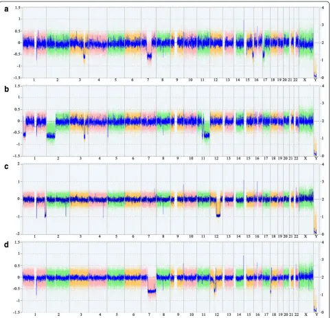
Pieces of four UL tumors were obtained from the hys-terectomy specimen. Molecular and cytogenetic analyses of these four UL tumors were carried out. Cytogenetic analyses failed to detect clonal karyotypic aberrations in two of the UL tumors (UL 709/3 and 709/4. Fig. 1a-d, Additional file 1: Table S1) and one of the UL tumors carried a MED12 mutation (709/2. c.131G > A, Fig. 1e). In contrast to the results obtained by G-banding, the CNV arrays revealed more or less gross genomic alter-ation in all four UL tumors (Fig. 2) that in neither of the UL tumors could have escaped detection by G-banding. E.g., in two UL tumors losses within the close surround-ing of or within the HMGA2 locus point to rearrange-ments of this gene (Fig. 3a, b). As other genomic alterations frequently seen in UL, two of the UL tumors were characterized by deletions of the long arm of chromosome 7 with different sizes.
Fig. 1Cytogenetic and molecular analysis of uterine leiomyomas.a-drepresentative G-banded karyotypes of four uterine leiomyomas.atumor 709/1, Karyotype: 45,XX,del(3)(q24q25.33),del(7)(q11.2),add(9)(q34),-15,der(17)t(15;17)(q?22;p?11.2)[3]/46,XX[3]. Of ten additional hypodiploid metaphases seven showed structural abnormalities and losses and three only losses of chromosomes. In contrast to the structural abnormalities, none of the losses observed were clonal.btumor 709/2, karyotype: 43 ~ 46,XX,del(1)(p34),der(2),-11 + mar.ctumor 709/3, karyotype: 46,XX[22].dtumor 709/4, karyotype: 46,XX[17].emutation ofMED12(c.131G > A) of tumor 709/2
Of note, UL 709/3, presented with two deletions located on either side of the HMGA2 locus. Of these, a short 5′proximal deletion mapped outside the putative transcription start of the gene removing a segment of only approximately 280 kbp (Fig. 3a). A much larger de-letion of roughly 42.79 Mb had removed part of the 3′ UTR of the gene. In terms of G-banding this latter dele-tion resulted in the partial monosomy for the bands 12q14.3-q24.11.HMGA2mRNA expression analysis car-ried out in this case revealed a slightly elevated expres-sion compared to other UL without clonal cytogenetic
abnormalities but was clearly lower than in cases with cytogenetically apparent rearrangement of the 12q14-15 region (Fig. 4). As not uncommon in UL with HMGA2
rearrangements this tumor in addition had a deletion of part of a terminal region of the long arm of chromosome 1 with a size of approximately 14.11 Mb affecting chromosomal bands 1q42.2-q44 including the fumarate hydratase gene (FH). Both the large deletion of 12q as well as those of 1q should be detectable by G-banding but neither clonal karyotypic abnormalities were seen in this case nor was even a single metaphase seen Fig. 2Results of CNV-array-analyses. Whole genome views of four uterine leiomyomas investigated. The weihghted log2 ratio of the probes are displayed as colored dots, the smooth signal (Gaussian smoothed calibrated copy number estimate) is displayed as blue line.atumor 709/1.b
presenting with these abnormalities. The second UL (709/4) with a putativeHMGA2rearrangement presented with deletions that both mapped proximal to the gene locus again including one large and one smaller deletion. The large deletion had a size of approximately 13.44 Mb and mapped within chromosomal region 12q12-q13.3 whereas the small deletions had a size of only approxi-mately 630 kbp and mapped in the direct vicinity of
HMGA2. From the array results it cannot even be ruled out that by this deletion part of the exons 1 and 2 of the gene had been removed (Fig. 3b). In addition, this UL car-ried three deletions on the long arm of chromosome 7 that were interspersed with short non-deleted fragments leading to a total loss of roughly 89,05 Mb from chromo-somal region 7q11.22-q36.3. In this case as well, neither evidence for the presence of chromosomal alterations in general nor for those involving the deleted regions as detected by the array analysis was obtained. Thus, as the most likely explanation, in both UL tumors affected by
HMGA2rearrangements the tumor cells did escape detec-tion by G-banding due to their reduced ability or even inability to proliferatein vitro.
Of the two UL tumors presenting no evidence for a
HMGA2rearrangements as detected by G-banding or array analysis (709/1, 709/2) one had a MED12 mutation but both showed large deletions of the short arm of chromo-some 3. Of note, the deleted segments in both UL tumors caused a removal of the gene encodingMED12L(Fig. 3c). In addition, both UL carried other large deletions that, in these both UL tumors akin to the deletions of 3p, did not escape detection by G-banding (Tab.1). However, of note in case of 709/2 with the MED12 mutation the cytogenetic preparation resulted in only two metaphases which could be investigated. Both shared cytogenetic abnormalities that thus can be considered being clonal (Fig. 1).
Conclusions
Among the few putative driver mutations observed in UL those affecting MED12 and these leading to rear-rangements of HMGA2 are particularly frequent and so far never have been reported to coincide within one in-dividual tumor. Also, patients harboring UL of both types seem to be very rare. The present case deals with one such coincidence. Being studied by G-banding as Fig. 3Rearrangements of chromosome 12 and deletions of the long arm of chromosome 3.alosses of parts of chromosome 12 in tumors 709/3 and 709/4. Black arrowheads indicate the region enlarged in (b).b. position of theHMGA2gene with intron/exon structure as indicated in a.c. Position of interstitial deletions of the long arm of chromosome 3 in tumors 709/1 and 709/2, respectively. The grey line and a black arrowhead indicate the position of theMED12Lgene
well as by molecular analyses i.e. MED12 sequencing and CNV arrays all four UL tumors revealed individual patterns of genomic alterations indicating their independ-ent clonal origin. Simultaneously, a rare coincidence of several putative driver mutations displayed by the single tumor was noted.
In two of them the profiles point to rearrangements of
HMGA2 that had escaped detection by G-banding. In general, two explanations are possible for this lack of de-tection. First, the underlying rearrangements may have occurred at a submicroscopic level. This explanation likely does not fit because the large deletions accom-panying both alterations are in a range easily detectable even when only applying a low resolution of chromo-somal bands. Alternatively, the findings can be explained by a reduced or even absent ability of the affected cells
to proliferatein vitro. Such a reduced ability recently has been observed for cell cultures of MED12 mutated UL [14]. Nevertheless, in that study cells of HMGA2 -mu-tated UL were able to proliferate for numerous in vitro
passages. At a first glance, this finding seems to contra-dict the latter explanation for the absence of metaphases with the said deletions. In the present study, however, both UL tumors with presumed HMGA2 rearrange-ments did also show other apparently independent ab-normalities i.e. a large deletion of the long arm of chromosome 1 in UL 709/3 and a large deletion of the long arm of chromosome 7 in UL 709/4. Both abnormal-ities have been described in UL before either in the pres-ence or in the abspres-ence of structural rearrangements of chromosome 12 [15–17]. Likewise, deletions of the long arm of chromosome 7 can by accompanied by MED12
Fig. 4Expression of HMGA2 mRNA in fibroid 709/3 as compared to ten other arbitrarily chosen fibroids without cytogenetically detectable rearrangements of chromosomal region 12q14-15 (white columns) as well as to 19 fibroids that showed a microscopically visible rearrangement of this chromosomal segment (grey columns), respectively
Table 1Karyotypes and culture conditions of the tumors
Tumor no Karyotype according to ISCN Days in culture until cytogenetic preparation
709/1 45,XX,del(3)(q2?),del(7)(q11.2),add(9)(q34),-15,der(17)t(15;17)?(q22;p11.2)[3]/46,XX[3]
14
Ten additional metaphases were hypodiploid. Of these, seven showed structural abnormalities and losses and three only losses of chromosomes. None of the losses observed were clonal.
709/2 43 ~ 46,XX,del(1)(p34)[[2],der(2)[2],-11[2],+mar[2][cp2] 35
709/3 46,XX[22] 35
mutations or occur independently [3]. In our recent study on the in vitro proliferation of UL cells the vitro “long term survivors” did not show one of these abnormalities [14]. It seems reasonable to assume that, independent of their coincidence with other driver mutations, both abnor-malities reduce in vitro proliferation. In turn, the actual frequency ofHMGA2 rearrangements may be underesti-mated when based solely on cytogenetics. In case of re-duced ability to proliferate, whole genome sequencing [4] as well as genomic array analysis seem to be more efficient to detect these rearrangements if they are accompanied by simultaneous losses of chromosomal material. Of note, even when showing cytogenetic abnormalities of 12q14-15 microscopic analyses may not reflect the complexity of underlying HMGA2 rearrangements sufficiently. Vice versa, the mere number of genetic imbalances as such does not allow diagnosing malignant growth [18].
Another interesting aspect in the UL tumors presented herein relates to the deletion of the long arm of chromo-some 3 found in two UL tumors including one without detectable rearrangements ofHMGA2 orMED12 muta-tions. Deletions of the long arm of chromosome 3 re-peatedly have been described before in UL. In their study on 52 uterine leiomyomas with clonal chromo-some abnormalities, Dal Cin P, Moerman P, Deprest J, Brosens I, Van den Berghe H. identified eight tumors with cytogenetic alterations that did not fit with any well-delineated cytogenetic subgroup [19]. In three of these cases the long arm of chromosome 3 was involved. Accordingly, it was considered that these changes characterize a new cytogenetic subgroup of uterine leio-myomas. Given that these deletions all point to the same target gene a loss-of-function for genes in that region as e.g. HLTF, SIAH2, RAP2B, MME, GMPS, MLF1 und
RARRES1 should be considered. On the other hand in both UL tumors presented herein MED12L mapped within the region of overlap. The strong similarity be-tween both genes and their proteins makes it reasonable to consider an independent role of alterations of that gene for the molecular pathogenesis in rare cases of UL.
In summary, the present case demonstrates the rare occurrence of a marked genetic heterogeneity among uterine leiomyomas with every single tumor displaying a unique CNV pattern. Loss of heterozygosity affecting the
MED12Llocus represents a candidate of a possible novel driver mutation in UL. Given the low but albeit existing probability of malignant transformation within UL the results point to the number of UL tumors as a risk factor associated with power morcellation of uterine tumors.
Methods
Tumor samples
From each of the UL tumors (lab code 709/1 - 709/4) one part was fixed for histologic examination, another
part was snap frozen in liquid nitrogen, and a third part was kept in Hank’s solution for subsequent cell cultures.
Histologic examination
The tumors were fixed in paraformaldehyde (4% in PBS) and processed for paraffin embedding. Tissue sections (1–2 μm thickness) were deparaffinized in xylene, rehy-drated through a series of ethanol, and stained with hematoxylin and eosin (H&E).
Cytogenetic studies
Chromosome analyses of cell cultures were performed following routine techniques as described earlier [20].
DNA isolation
For CNV analysis as well as MED12 mutation analysis DNA from the frozen tissue samples was isolated using the QIAamp DNA Mini Kit (Qiagen, Hilden, Germany) on a QIACube (Qiagen) according to the manufacturer’s instructions. The amount of double stranded DNA was measured using the Qubit dsDNA HS Assay Kit and a Qubit Fluorometer (Life Technologies, Carlsbad, CA).
PCR and sequencing
For PCR amplification 100 ng of genomic template DNA were used. Primers to amplify the desired human PCR fragment of the MED12 gene were those recently described [15]. Subsequently, PCR-products were sepa-rated by agarose gel-electrophoresis and the desired DNA-fragments/-bands were extracted by a QIAquick Gel Extraction Kit (Qiagen) using a QIACube (Qiagen) according to manufacturer’s instructions. DNA-sequencing of the purified PCR-products was performed using the CEQ DTCS Quick Start Sequencing Kit and a Beckman Coulter GenomeLab GeXP Genetic Analysis System (AB SCIEX, Framingham, MA, USA).
Arrays
CNV (copy number variation) analysis was performed using premade CytoScan HD Arrays (Affymetrix, Santa Clara, CA) consisting of more than 2.4 million markers for copy number and approximately 750,000 single nu-cleotide polymorphisms (SNPs). Enriched gene coverage for cancer and constitutional genes results in marker-base ratio coverages of 1/384 for ISCA, 1/553 for cancer genes, 1/486 for X-chromosomal genes and 1/659 for 12,000 OMIM genes. Labelling of 250 ng DNA and hybridization were done following the manufacturer’s instructions. After staining and washing using a Gene-Chip Fluidics Station 450 (Affymetrix) the arrays were scanned by an Affymetrix 3000 7G scanner. Arrays were analyzed through the Affymetrix Chromosome Analysis Suite (ChAS) software (ChAS analysis files for CytoScan®
HD Array version NA33). Numbering of map positions was based on hg19 (NCBI Build 37 reference sequence).
Quantification of HMGA2 mRNA
Expression of HMGA2mRNA was analysed as described earlier [21]. Briefly, RNA isolated from the samples had been was digested by DNAse and then used for cDNA synthesis. For quantification by real-time PCR (Applied Biosystems 7300, Applied Biosystems, Darmstadt, Germany), a commercially available gene expression assay (Hs00171569, Applied Biosystems) was used. HMGA2
mRNA expression was quantified relatively to HPRT
mRNA, which was used as an endogenous control.
Consent
The study was approved by the local ethics committee. Samples were obtained in accordance with the declar-ation of Helsinki and informed written consent was ob-tained from the patient prior to surgery.
Additional file
Additional file 1: Table S1.Results of array analysis. Overview of copy number variations with 200 kb minimal size. (DOCX 29 kb)
Competing interests
The authors declare that they have no competing interests.
Authors’contributions
CH: conception and design of the study; acquisition of data; analysis and interpretation of data; manuscript writing; final approval of the manuscript. DNM: conception and design of the study; acquisition of data; analysis and interpretation of data; manuscript writing; final approval of the manuscript. SB: acquisition of data; analysis and interpretation of data; manuscript writing; final approval of the manuscript. DK: acquisition of data; analysis and interpretation of data; final approval of the manuscript. BMH: analysis and interpretation of data; provision of study material; final approval of the manuscript. JB: conception and design of the study; analysis and interpretation of data; manuscript writing; revising the manuscript critically for important intellectual content; final approval of manuscript.
Acknowledgements
We thank Frauke Meyer for her excellent technical assistance.
Author details
1Institute of Medical Genetics, University Rostock Medical Center,
Ernst-Heydemann-Strasse 8, D-18057 Rostock, Germany.2Center of Human
Genetics, University of Bremen, Leobener Strasse ZHG, D-28359 Bremen, Germany.3Institute of Immunology, University of Rostock, University Rostock
Medical Center, Schillingallee 70, D-18057 Rostock, Germany.4Institute of
Pathology, University of Heidelberg, Heidelberg, Germany.5Present address:
Institute of Pathology, Elbe Kliniken, Klinikum Stade, Bremervörder Str. 111, D- 21682 Stade, Germany.
Received: 26 June 2015 Accepted: 22 September 2015
References
1. Schoenmakers EF, Wanschura S, Mols R, Bullerdiek J, Van den Berghe H, Van de Ven WJ. Recurrent rearrangements in the high mobility group protein gene, HMGI-C, in benign mesenchymal tumours. Nat Genet. 1995;10:436–44.
2. Mäkinen N, Mehine M, Tolvanen J, Kaasinen E, Li Y, Lehtonen HJ, et al. MED12, the mediator complex subunit 12 gene, is mutated at high frequency in uterine leiomyomas. Science. 2011;334:252–5.
3. Markowski DN, Bartnitzke S, Löning T, Drieschner N, Helmke BM, Bullerdiek J. MED12 mutations in uterine fibroids - their relationship to cytogenetic subgroups. Int J Cancer. 2012;131:1528–36.
4. Mehine M, Kaasinen E, Mäkinen N, Katainen R, Kämpjärvi K, Pitkänen E, et al. Characterization of uterine leiomyomas by whole-genome sequencing. N Engl J Med. 2013;369:43–53.
5. Bertsch E, Qiang W, Zhang Q, Espona-Fiedler M, Druschitz S, Liu Y, et al. MED12 and HMGA2 mutations: two independent genetic events in uterine leiomyoma and leiomyosarcoma. Mod Pathol. 2014;27:1144–53. 6. Markowski DN, Helmke BM, Bartnitzke S, Löning T, Bullerdiek J. Uterine
fibroids: do we deal with more than one disease? Int J Gynecol Pathol. 2014;33:568–72.
7. Pérot G, Croce S, Ribeiro A, Lagarde P, Velasco V, Neuville A, et al. MED12 alterations in both human benign and malignant uterine soft tissue tumors. PLoS One. 2012;7, e40015.
8. Kämpjärvi K, Mäkinen N, Kilpivaara O, Arola J, Heinonen H-R, Böhm J, et al. Somatic MED12 mutations in uterine leiomyosarcoma and colorectal cancer. Br J Cancer. 2012;107:1761–5.
9. Ravegnini G, Mariño-Enriquez A, Slater J, Eilers G, Wang Y, Zhu M, et al. MED12 mutations in leiomyosarcoma and extrauterine leiomyoma. Mod Pathol. 2013;26:743–9.
10. Markowski DN, Huhle S, Nimzyk R, Stenman G, Löning T, Bullerdiek J. MED12 mutations occurring in benign and malignant mammalian smooth muscle tumors. Genes Chromosomes Cancer. 2013;52:297–304.
11. Matsubara A, Sekine S, Yoshida M, Yoshida A, Taniguchi H, Kushima R, et al. Prevalence of MED12 mutations in uterine and extrauterine smooth muscle tumours. Histopathology. 2013;62:657–61.
12. De Graaff MA, Cleton-Jansen A-M, Szuhai K, Bovée JVMG. Mediator complex subunit 12 exon 2 mutation analysis in different subtypes of smooth muscle tumors confirms genetic heterogeneity. Hum Pathol. 2013;44:1597–604. 13. Schwetye KE, Pfeifer JD, Duncavage EJ. MED12 exon 2 mutations in uterine
and extrauterine smooth muscle tumors. Hum Pathol. 2014;45:65–70. 14. Markowski DN, Tadayyon M, Bartnitzke S, Belge G, Maria Helmke B,
Bullerdiek J. Cell cultures in uterine leiomyomas: rapid disappearance of cells carrying MED12 mutations. Genes Chromosomes Cancer. 2014;53:317–23.
15. Pandis N, Heim S, Bardi G, Flodérus UM, Willén H, Mandahl N, et al. Chromosome analysis of 96 uterine leiomyomas. Cancer Genet Cytogenet. 1991;55:11–8.
16. Sandberg AA. Updates on the cytogenetics and molecular genetics of bone and soft tissue tumors: leiomyoma. Cancer Genet Cytogenet. 2005;158:1–26. 17. Hennig Y, Deichert U, Bonk U, Thode B, Bartnitzke S, Bullerdiek J.
Chromosomal translocations affecting 12q14-15 but not deletions of the long arm of chromosome 7 associated with a growth advantage of uterine smooth muscle cells. Mol Hum Reprod. 1999;5:1150–4.
18. Hodge JC, Morton CC. Genetic heterogeneity among uterine leiomyomata: insights into malignant progression. Hum Mol Genet. 2007;16 Spec No:R7–13.
19. Cin PD, Moerman P, Deprest J, Brosens I, Van Den Berghe H. A new cytogenetic subgroup in uterine leiomyoma is characterized by a deletion of the long arm of chromosome 3. Genes Chromosomes Cancer. 1995;13:219–20.
20. Klemke M, Meyer A, Nezhad MH, Bartnitzke S, Drieschner N, Frantzen C, et al. Overexpression of HMGA2 in uterine leiomyomas points to its general role for the pathogenesis of the disease. Genes Chromosomes Cancer. 2009;48:171–8.
![Fig. 1 Cytogenetic and molecular analysis of uterine leiomyomas.Karyotype: 45,XX,del(3)(q24q25.33),del(7)(q11.2),add(9)(q34),-15,der(17)t(15;17)(q?22;p?11.2)[3]/46,XX[3]](https://thumb-us.123doks.com/thumbv2/123dok_us/276782.1520514/2.595.57.539.370.677/fig-cytogenetic-molecular-analysis-uterine-leiomyomas-karyotype-der.webp)