INTRODUCTION
The airways have constitutive host de-fense mechanisms including mucociliary clearance, alveolar macrophages sensing potential pathogens, and antimicrobial host defense proteins (HDPs) with a po-tency to eradicate bacteria (1). In addition to their antibacterial activity, HDPs may act as growth factors and regulate
leuko-cyte trafficking (2). Several HDPs are con-stitutively produced by airway epithelial cells to maintain host integrity, although many are induced by inflammation or stored preformed in innate immune cells (for example, LL-37 in neutrophils). Re-cently, we found that the growth factor midkine (MK) has potent bactericidal and fungicidal activities (in vitro) executed
through disruption of microbial mem-branes, similar to the activity exerted by other innate antibiotics such as the human β-defensins (hBDs), LL-37 and an-tibacterial chemokines (1,3,4). MK is stitutively expressed during healthy con-ditions in several tissues, for example, by epithelial cells in the large airways and by keratinocytes in the skin, where it may reach bactericidal concentrations in the microenvironment and thus contribute to host defense (5,6). Several factors present at sites of inflammation promote MK ex-pression including retinoic acid (RA), hy-poxia, reactive oxygen species and activa-tion of the transcripactiva-tion factor nuclear factor (NF)-κB (7–11).
Staphylococcus aureusis an important human pathogen. Although uncommonly
Chronic Obstructive Pulmonary Disease Exacerbations and
Ventilator-Associated Pneumonia Associated with
Staphylococcus aureus Infection
Helena M Linge,
1Cecilia Andersson,
1Sara L Nordin,
1Anders I Olin,
2Ann-Cathrine Petersson,
3Matthias Mörgelin,
2Amanda Welin,
4Johan Bylund,
4Leif Bjermer,
1Jonas Erjefält,
1and Arne Egesten
1*Sections for 1Respiratory Medicine and Allergology and 2Infection Medicine, Department of Clinical Sciences Lund, Lund University; 3
Clinical Microbiology, Regional Laboratories of Region Skåne, Lund, Sweden, University Hospital, Lund, Sweden; and 4Department of Rheumatology and Inflammation Research, Sahlgrenska Academy at University of Gothenburg, Göteborg, Sweden
Staphylococcus aureus is sometimes isolated from the airways during acute exacerbations of chronic obstructive pulmonary disease (COPD) but more commonly recognized as a cause of ventilator-associated pneumonia (VAP). Antimicrobial proteins, among them midkine (MK), are an important part of innate immunity in the airways. In this study, the levels and possible process-ing of MK in relation to S. aureus infection of the airways were investigated, comparprocess-ing COPD and VAP, thus comparprocess-ing a state of disease with preceding chronic inflammation and remodeling (COPD) with acute inflammation (that is, VAP). MK was detected in the small airways and alveoli of COPD lung tissue but less so in normal lung tissue. MK at below micromolar concentrations killed S. aureus in vitro. Proteolytic processing of MK by the staphylococcal metalloprotease aureolysin (AL), but not cysteine protease staphopain A (SA), resulted in impaired bactericidal activity. Degradation was seen foremost in the COOH-terminal portion of the molecule that harbors high bactericidal activity. In addition, MK was detected in sputum from patients suffering from VAP caused by S. aureus but less so in sputum from COPD exacerbations associated with the same bacterium. Recombinant MK was de-graded more rapidly in sputum from the COPD patients than from the VAP patients and a greater proteolytic activity in COPD sputum was confirmed by zymography. Taken together, proteases of both bacteria and the host contribute to degradation of the antibacterial protein MK, resulting in an impaired defense of the airways, in particular, in COPD where the state of chronic in-flammation could be of importance.
Online address: http://www.molmed.org doi: 10.2119/molmed.2013.00045
associated with respiratory tract infec-tions, it is sometimes isolated from spu-tum during acute exacerbations of chronic obstructive pulmonary disease (COPD), and it is a relatively common cause of hospital-acquired and, in partic-ular, ventilator-associated pneumonia (VAP). S. aureusis estimated to account for 10–20% of all nosocomial pneumonias (12). The reported mortality in
VAP patients ranges from 33% to 72%, with the upper range reflecting the in-creased risk for mortality among the elderly, patients with impaired cardio -pulmonary function, immune compro-mised patients and patients who require prolonged intubation of the airways. S. aureushas a plethora of countermeasures against host defense mechanisms, includ-ing surface proteins and secreted pro-teases (13). The continuing increase in an-tibiotics resistance among S. aureus strains, and in particular the spread of meticillin-resistant S. aureus(MRSA), con-stitute new challenges for health care and health care professionals (14).
The aim of this study was to investigate the levels and possible processing of MK in relation to S. aureusinfection of the air-ways, comparing COPD and VAP, with the former being characterized by a state of chronic inflammation and remodeling, whereas in VAP, the airways respond with an acute inflammation. We found expres-sion and location of MK within COPD air-ways. During infection with S. aureus, MK was undetectable in COPD sputum, but high levels were detected in aspirations from VAP patients. Staphylococcal pro-teases fragmented MK and thereby af-fected its bactericidal activity. COPD spu-tum showed a higher proteolytic activity than VAP aspirations. We believe this to be an important example of defense impair-ment in the airways during COPD.
MATERIALS AND METHODS
Patient Groups and Samples
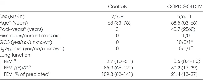
In the present study, lung tissue from 11 COPD (Global Initiative for Chronic Ob-structive Lung Disease [GOLD] stage IV) patients was used (Table 1). In addition,
healthy lung tissue from nine nonsmok-ing controls was obtained durnonsmok-ing lung lobectomy because of suspected lung can-cer in otherwise healthy nonatopic indi-viduals. Only patients with solid tumors with visible borders were included in the study, and tissues used were obtained as far from the tumor as possible. In patients with very severe COPD (GOLD IV), matching lung tissue was collected in as-sociation with lung transplantation. For all patient groups, care was taken to im-merse the tissue into fixative immediately after surgical excision, and multiple large tissue blocks were prepared for histologi-cal analysis. All subjects gave written in-formed consent to participate in the study, which was approved by the local ethics committee in Lund, Sweden (LU412-03).
Sputum from COPD patients and endo-tracheal aspirations from 20 consecutive patients diagnosed with VAP (both groups had positive cultures for S. aureus) were collected at Skåne University Hospi-tal between August 2011 and January 2012 and stored at –20°C until analysis. The study design was a cross- sectional study of patients suffering from acute and chronic airway inflammatory responses, respectively, as represented by patients with S. aureus–associated VAP or COPD exacerbation. Intervention was collection and culture of sputum or tracheal aspi-rates upon signs of airway infection/ exacerbation. Power analysis assuming a
difference between means of 2 ng/mL and 1 standard deviation (SD) indicated that six patients in each group were required to achieve a power of 0.8 at α= 0.05. Patients included in other trials (n = 3) or where sputum sample was too viscous (n = 3) were excluded from all parts of our study.
In patients suffering from VAP, endo-tracheal aspirates were collected by using a sterile suction catheter via an en-dotracheal tube or a tracheostoma. In pa-tients suffering from COPD, sputum was mobilized using the huffing technique, without inhalation of nebulized sodium chloride. Parts of the samples were used for culture and parts were frozen at –20°C until use in assays. After thawing, sputum was centrifuged at 5,000gfor 5 min and aliquots of the supernatant were used. Sputum was used without addition of dithiothreitol to make it pos-sible to use antibody-based measurement of MK (that is, enzymelinked immuno -sorbent assay [ELISA]). The protocol was approved by the ethics committee at Lund University (#492/2007).
Immunohistochemistry
Immediately after collection, tissues were placed in 4% buffered formaldehyde, dehydrated and embedded in paraffin, and thin sections (3 μm) were generated.
Single staining of MK.A single stain-ing protocol (EnVision™ Detection sys-tem, K5007; Dako, Glostrup, Denmark)
Table 1.Characteristics of patients with COPD and healthy controls whose tissue was used for histological analysis.
Controls COPD GOLD IV
Sex (M/F, n) 2/7, 9 5/6, 11
Agea(years) 63 (33–76) 58.5 (53–66)
Pack-yearsa(years) 0 40.7 (2560)
Exsmokers/current smokers 0 11/0
GCS (yes/no/unknown) 0 10/0/1b
β2Agonist (yes/no/unknown) 0 10/0/1
b
Lung function
FEV1a 2.7 (1.7–5.1) 0.6 (0.4–1.0)
FEV1/(F)VCa 85.9 (66–121) 30.2 (17–39)
FEV1% of predicteda 109.8 (82–141) 21.4 (13–27)
FEV1, forced expiratory volume; (F)VC, (forced) vital capacity; GCS, inhaled glucocorticosteroids.
aData are given as mean (range).
was used for visualization of MK. Briefly, MK was detected by a rabbit anti-MK an-tibody (Peprotech, London, UK), and sec-ondary antibodies were conjugated with peroxidase polymers. The immunohisto-chemistry protocols were performed by using an automated immunohistochem-istry robot (Autostainer; Dako). Sections were stained with Mayer’s hematoxylin for visualization of background tissue and were dehydrated in alcohol and xylene and mounted in Pertex (Histolab).
Double staining of MK and surfac-tant protein A (SP-A) or myeloperoxi-dase (MPO). After antigen retrieval and a blocking step (protein-blocking, ×0909; Dako), sections were incubated
overnight in 4°C with a rabbit anti-MK antibody, and immunoreactivity was vi-sualized after 1-h incubation at room temperature (RT) with a goat anti-rabbit Alexa Fluor 555– conjugated secondary antibody (1:200; Molecular Probes/Life Technologies, Carlsbad, CA, USA). SP-A was detected after 1-h incubation at RT with a mouse anti-SPA (1:50; Dako) and visualized after incubation at RT for 1 h with an Alexa Fluor 488–conjugated goat anti-mouse secondary antibody (1:200; Molecular Probes/Life Technologies). Consecutive sections were stained with a directly conjugated rabbit anti-MPO (1:12,000; Dako) by using a Rabbit Alexa Fluor 488 nm Zenon labeling kit accord-ing to the manufacturer’s instructions (Molecular Probes/Life Technologies). Nuclei were detected by Hoechts 33342, and sections were mounted in TBS/glyc-erin and frozen until quantification. For all immunohistochemical procedures, markers and tissues, staining was absent in sections using isotype-matched control antibodies (Dako) that were used instead of, and at the same concentration as, the primary antibody.
Bacterial Strains, Determination of Antibiotics Resistance Profile and Screening of Virulence-Associated Genes
The S. aureusstrains 5120, Newman, 8325-4 and 8325-4 sarA (sarA; deficient of the repressor sarA, resulting in
overex-pression of proteases released extracellu-larly [15]) were used for the in vitro stud-ies and routinely cultured in Todd-Hewitt medium (TH; BD, Franklin Lakes, NJ, USA) at 37°C with 5% CO2.
The antibiotics susceptibility of the S. aureusstrains isolated from COPD and VAP patients were tested by using the disc diffusion method according to the Swedish Reference Group for Antibiotics (SRGA) and its subcommittee on meth-odology (SRGA-M; www.srga.org) or by E-test (bioMeriéux, France). Discs with cefoxitin, clindamycin, fusidic acid, trimethoprim-sulfamethoxazole, nor-floxacin, tigecyclin, gentamicin, linezolid and rifampicin were obtained from Oxoid (Basingstoke, UK). The van-comycin susceptibility was tested using E-tests. All isolates were screened for the presence of virulence-associated genes encoding Panton Valentine leucocidine (lukS-PV and lukF-PV) and tssencoding the TSS-1 toxin using polymerase chain reaction and spatyped as previously described (16).
Bactericidal Assay
Bacteria was cultured to the midexpo-nential growth phase (OD620≈ 0.4),
washed in incubation buffer (10 mmol/L Tris/HCl, 5 mmol/L glucose; pH 7.5) and adjusted to a concentration of 2 ×10⁶ colony-forming units (CFUs)/mL. Fifty microliters bacterial solution (~105CFUs) was incubated in buffer or with various concentrations of the indicated protein or mixtures for 1 h at 37°C. To quantify the bactericidal activity, serial dilutions in in-cubation buffer were plated on TH-agar in duplicates and incubated overnight at 37°C. The percent bacterial killing was calculated by comparison with the num-ber of CFUs when incubated in buffer alone.
Electron Microscopy
Bacteria were incubated with MK (1μmol/L) in incubation buffer for 1 h at 37°C and then subjected to scanning elec-tron microscopy as described (17). Im-munogold labeling and transmission electron microscopy (TEM) of ultrathin
sections was performed as previously described (5). Specimens were examined in a JEOL (Tokyo, Japan) 1200EX trans-mission electron microscope operated at 60 kV accelerating voltage.
Protein Detection by ELISA, Sodium Dodecyl Sulfate–Polyacrylamide Gel Electrophoresis (SDS-PAGE) and Western Blotting
The MK ELISA was from Peprotech. For Western blotting, standard tech-niques were used. After separation on a Tris-tricine gel, samples were electroblot-ted onto a polyvinylidene fluoride mem-brane. After blocking, membranes were probed with antibodies toward MK (Peprotech), followed by a secondary goat–anti-rabbit antibody (Bio-Rad, Her-cules, CA). Blots were developed by using the Bio-Rad Chemidoc system (Bio-Rad).
MK Proteolytic Cleavage Assay MK (5 μg) was incubated for the indi-cated time points with 0.5 μg of either staphylococcal proteases aureolysin (AL) or staphopain A (SA; Biocentrum, Krakow, Poland). Sterile filtered bacterial super-natants (10–15 μL) from overnight cultures were incubated with recombinant MK pro-tein for indicated time points before either SDS-PAGE followed by Western blot anal-ysis or N-terminal sequencing. In a sepa-rate experiment, MK was incubated with COPD sputa or VAP aspirations (patient IDs: B6, A10, B9 and A2, A6, A7, respec-tively) for 1 and 18 h. Samples were then analyzed by SDS-PAGE and standard pro-tein staining methods.
Zymography
Zymography was performed by using precast gels containing collagen (Bio-Rad). The material used in zymography assays were from COPD patients A1, A9 and B5 and VAP patients A3, A4 and B3, respectively.
N-Terminal Sequencing
se-quencing by Edman degradation of eight sequential amino acids (HEJ Analyser; Karolinska Institutet, Stock-holm, Sweden).
Molecular Modeling
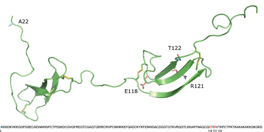
Molecular modeling of MK was per-formed as described (4).
RESULTS
Expression of MK in Lung Tissue from COPD
Immunohistochemistry followed by morphometric image analysis showed that COPD airways, including the airway wall (Figure 1A), epithelial cells (Figure 1B) and
the small airway parenchyma (Figure 1C) have a significantly higher expression of MK than the airways of controls. MK pro-tein, as detected by immunohistochem-istry in small airways (Figure 1D), as seen in a higher magnification (Figure 1E), is predominantly expressed in bronchial ep-ithelial cells. In alveoli, weaker presence
of MK was seen (Figure 1F). By immuno-fluorescent double staining, MK (red) was visualized in bronchial epithelium of small airways, showing weak colocaliza-tion with myeloperoxidase (green) of neutrophils. Neutrophils mainly showed submucosal positioning (Figure 1G). MK was also detected in the alveolar paren -chyma, showing to some degree colocal-ization with SP-A of type 2 pneumocytes (Figure 1H). In addition, alveolar macro -phages showed presence of both MK and SP-A (Figure 1I), capable of phagocytosing the latter (18).
To localize MK on an ultrastructural level, immunogold labeling followed by TEM analysis was performed (Figure 2). This process revealed the presence of MK
on the apical surface and association with cilia of bronchial epithelial cells in COPD airways (Figures 2A, B); for closer detail see Figures 2C, D.
Figure 2.Ultrastructural detection of MK by immunoelectron microscopy of lung tissue in COPD. (A) Immunogold-labeling of ultra-thin sections was performed on small airway lung tissue from a COPD patient. Colloidal gold particles indicate location of bound antibodies against MK. MK is present on the surface of cilia (B) and on the bronchial ep-ithelial cell surface (C and D). Scale bars: A = 10 μm, B = 1 μm and C and D = 100 nm.
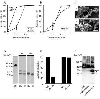
Bactericidal Activity of MK Against Different Strains of S. aureus
A previous study identified MK as an antibacterial against S. aureus(4). In the current study, we demonstrate bacterial killing of S. aureusstrains 5120, New-man and 8325-4 (Figures 3A, B). MK was the more potent antimicrobial agent, killing 5120 at lower concentra-tions than the classic HDP LL-37. Bacte-ria exposed to buffer alone remained in-tact, as imaged by standard error of the mean (Figure 3C, top panel i), whereas exposure to MK resulted in leakage of intracellular material from the bacteria, suggesting membrane disruption as a likely mode of action (Figure 3C, lower panel ii).
MK Fragments Generated by Staphylococcal Proteases
In the host–microbe encounter, bacte-rial proteases are likely to have access to constitutively expressed HDPs such as MK. Incubation of recombinant MK with the S. aureusproteases AL and SA for 3 h generated fragments, which re-mained present after 18 h of incubation (Figure 3D) as detected by protein staining. AL generated fragments of
MK of the approximate size of 5 and 6 kDa, as estimated from the stained gel. N-terminal sequencing combined with mass spectrometry identified the band of higher molecular weight to cor-respond to the true NH terminus of the holoprotein, whereas the other band constituted a mixture of peptides lo-cated in the COOH terminus of the holoprotein. SA generated an MK frag-ment of the apparent size of 4 kDa, and N-terminal sequencing of this band showed a mixture of two equally abun-dant peptides—one corresponding to the true NH terminus of the holopro-tein and one from the COOH terminus. These results are displayed in Figure 4. To investigate the effect of AL and SA cleavage on MK bactericidal activity, MK was preincubated with respective pro-tease and then used in the viable count assay. The results show that AL reduced MK killing to 25% (Figure 3E), whereas killing remained close to 100% after incu-bation with SA. MK was then incubated with supernatants from cultures of S. au-reuswild-type (wt) strain 8325-4 or its protease overexpressing derivative sarA (Figure 3F). Immunoblotting revealed that MK remained intact after incubation
with wtsupernatant, whereas it was cleaved by the sarA supernatant. Taken together, these data demonstrate that MK is bactericidal toward S. aureusand that AL, but not SA, by proteolytic cleavage acts as a bacterial countermeasure to limit MK-mediated eradication.
COPD Sputa and Endotracheal Aspirations from VAP Patients Associated with S. aureus Infection Show Differences in MK Levels and Proteolytic Activity
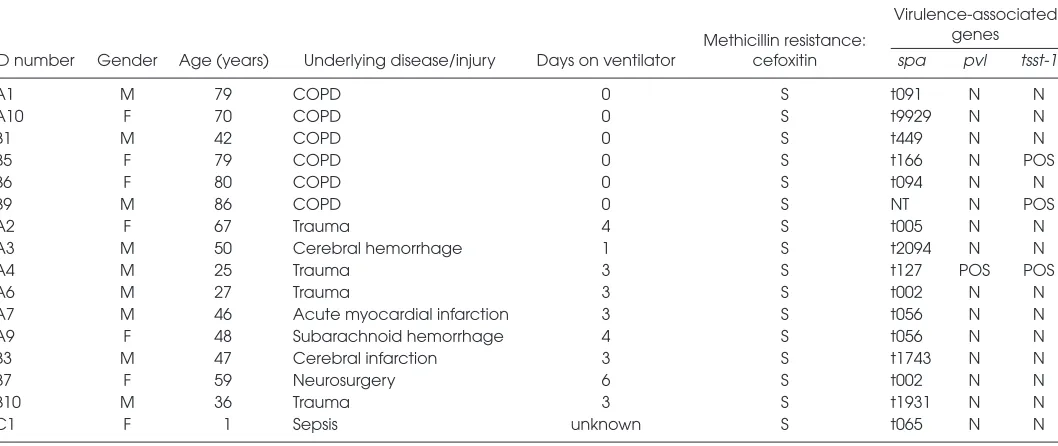
Patient characteristics and the results of the virulence-associated genes screening are shown in Table 2. The average age of the patient groups was 73 years for COPD and 41 years for VAP. The VAP group spent an average of 3.3 d on a ven-tilator before the introduction of antibi-otics or death occurred. None of the COPD patients required intubation. The results of the virulence gene screening re-vealed that only one bacterial isolate (A4) was positive for the pvlgene, and three (A4, B5, B9) were positive for the super-antigen tsst-1. The spa-types t002 and t056 were found in two isolates each (A6, B7 and A7, A9, respectively). All tested strains were sensitive to methicillin.
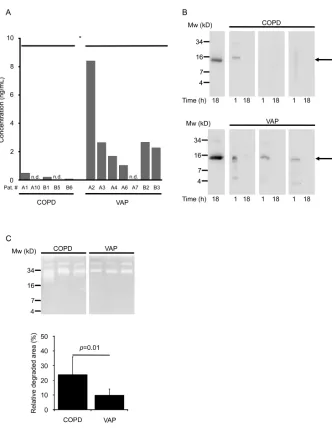
Significantly less MK was detected in sputum from COPD patients (mean: 0.15 ng/mL, range 0–0.49; n = 5, 2 of 5 undetectable) than in endotracheal aspi-rations from VAP patients (n = 8, mean: 2.62 ng/mL, range 0–8.41; 1 of 8 unde-tectable) (p = 0.030 as determined by the Mann-Whitney Utest; Figure 5A). Fig-ure 5B shows degradation patterns of re-combinant MK exposed for 1 or 18 h to either COPD sputa or VAP aspirations, respectively. In COPD sputum, MK deg-radation kinetics was more rapid than in aspirations from VAP patients. To eluci-date why MK was degraded more rap-idly in COPD sputa, zymography was performed. The substrate clearance area was quantified and shows that COPD sputa had a significantly higher prote-olytic activity than VAP aspirations (p= 0.01, Figure 5C).
DISCUSSION
According to the World Health Organ-ization, 65 million people are affected by COPD, and, based on data from 2005, the disease accounts for 5% of all deaths glob-ally (http://www.who.int/respiratory/ copd/burden/en). COPD exacerbations are linked to recurrent bacterial or viral
infections that influence the outcome of the disease negatively. The most com-monly isolated bacterial species are Streptococcus pneumoniae, Haemophilus in-fluenzae and Moraxella catarrhalis, but S. aureusspecies are also found (19,20), par-ticularly in elderly patients (21).
In healthy individuals, MK is constitu-tively expressed in bronchial epithelium of large airways and at least partially de-pends on retinoic acid (5). In the current study, we found COPD lung tissue to ex-press MK in small airways, in particular within the airway wall, epithelium and parenchyma. This result is in contrast to healthy lungs, where MK is expressed in large but not small airways (5). Addition-ally, we found the presence of MK in type 2 pneumocytes and alveolar macrophages in the present study. One important question is whether MK con-centrations reach bactericidal concentra-tions. In a previous study, using an air liquid system, we calculated the MK con-centration of the airway surface liquid to an amount of 0.7 μmol/L (that is, a bac-tericidal concentration) (5). In addition, MK was detectable in induced sputum of healthy individuals (5). Several factors are likely to contribute to increased MK
expression during S. aureus infection of the airways. The MKgene has a retinoic acid (RA)-responsive element in its pro-moter region, and several factors present during inflammation increase the genera-tion of RA from vitamin A, for example, activation of TLR2 by peptidoglycan of gram-positive bacteria (e.g., S. aureus) (22,23). In addition, MK expression is en-hanced by the proinflammatory tran-scription factor NF-κB, reactive oxygen species, tumor necrosis factor-α, and in-terleukin-1β(10,11). In severe infection, hypoxia of tissues may occur and, inter-estingly, hypoxia enhances MK expres-sion via the transcription factor hypoxia-inducible factor 1-α(HIF-1α) (9).
Others found significantly elevated MK in sera from hypoxemic patients compared with healthy controls (24), but this study did not include examination of lung tissue or sputa. The difference in MK detection in lung tissue versus sputa questions the reliability of sputum as a sampling method. In addition, process-ing of sputa, for example by addition of the reducing agent dithiothreitol to lower viscosity, may result in dramatic differ-ences with respect to detection limits of various readouts (25). The sputum and
Table 2.Characteristics of COPD and VAP patients, bacterial methicillin resistance and virulence-associated gene profile.
Virulence-associated
Methicillin resistance: genes ID number Gender Age (years) Underlying disease/injury Days on ventilator cefoxitin spa pvl tsst-1
A1 M 79 COPD 0 S t091 N N
A10 F 70 COPD 0 S t9929 N N
B1 M 42 COPD 0 S t449 N N
B5 F 79 COPD 0 S t166 N POS
B6 F 80 COPD 0 S t094 N N
B9 M 86 COPD 0 S NT N POS
A2 F 67 Trauma 4 S t005 N N
A3 M 50 Cerebral hemorrhage 1 S t2094 N N
A4 M 25 Trauma 3 S t127 POS POS
A6 M 27 Trauma 3 S t002 N N
A7 M 46 Acute myocardial infarction 3 S t056 N N
A9 F 48 Subarachnoid hemorrhage 4 S t056 N N
B3 M 47 Cerebral infarction 3 S t1743 N N
B7 F 59 Neurosurgery 6 S t002 N N
B10 M 36 Trauma 3 S t1931 N N
C1 F 1 Sepsis unknown S t065 N N
aspiration samples used in our study were without additions, increasing the reliability of the results.
Electron microscopy analysis located MK to the airway lumen and on the cilia of the epithelia of COPD airways (Figure
2). MK is believed to exert its antibacter-ial activity through bacterantibacter-ial membrane disruption and the extracellular location identified by electron microscopy, where it can encounter invading or colonizing pathogens, supporting the role of MK as an antibacterial factor (3). Earlier studies have localized the highest antibacterial activity of MK to the COOH-terminal half and identified its ability to kill gram-positive and gram-negative bacteria as well as fungi (3–5). S. aureusis named among the isolated pathogens found in acute exacerbations of COPD but, be-sides skin infections, it remains a more recognized cause of necrotizing pneumo-nia, HAP, VAP and upper airway infec-tions. For these reasons, we used S. au-reusas an experimental target for MK function and compared airway-expelled material from two different patient groups infected with this pathogen. As expected, MK readily killed all tested strains of S. aureus in vitro. AL but not SA destructed the antibacterial activity of MK. Both NH- and COOH-terminal re-gions of MK contain antibacterial activ-ity, but only the activity of the COOH domain including the tail region equates to that of the holoprotein. Our data sug-gest that AL degrades the terminal half where its most potent anti-bacterial activity is found, although further experiments are needed to con-firm this. Future studies will address what role, if any, the protease-generated fragments of MK have.
Not only do the proteases used in this study act on host proteins (26) such as MK, they also regulate the profile of the extracellular proteins on the bacterial surface (15). Thereby, these proteases have the ability to substantially influ-ence bacterial virulinflu-ence and host inter-play. The theme is known from other host–pathogen encounters (27), and re-cent work has shown that proteases from Streptococcus pyogenesand Fine-goldia magnaact proteolytically to medi-ate bacterial evasion of host defense functions in skin infections (6). Bacterial colonization has over the last decade has become more recognized as a driving
force behind exacerbations in COPD. The role of bacterial proteases from pathogens more commonly associated with COPD exacerbations remains an open area of research.
S. aureushas many ways of evading the immune system, with expression of surface protein A, an promoting surface protein encoded by the spagene, being one of them. The clin-ical isolates in our study showed genetic heterogeneity in terms of spatype; only two types were found in more than one patient. This result suggests a low risk of patient-to-patient or hospital- mediated spread. One of the strains in our study carried t127, which is also found in clones of US100. Methicillin resistance has historically been a larger problem in the U.S. than in Europe (14). Although staphylococcal virulence does not de-pend on methicillin resistance alone but relies on a combination of factors, it cre-ates major problems for hospitals and health care professionals. Frequency of MRSA isolation remains low in Sweden (http://www.smittskyddsinstitutet.se [continuously updated information]), and fortunately, we found all strains in this study sensitive to methicillin.
That we detected MK in COPD air-ways by histology analysis but not in sputa led us to investigate whether COPD sputa contained more proteolytic activ-ity than VAP aspirations. Degranulation of extravasated neutrophils in the air-ways together with a protease–protease inhibitor imbalance is a hallmark of ex-acerbations in COPD as described in the article by Celli and Barnes (28). That COPD samples showed larger clearing zones in the zymography assay is likely due to higher concentrations of pro-teases originating from the increased number of neutrophils, which is a hall-mark of the condition. The serine pro-teases not only degrade MK, as shown in this study, but also contribute to ep-ithelium injury, increased mucus pro-duction and chemokine propro-duction (29). Because both patient groups were in-fected with S. aureus, it appears that the contribution of host proteases to MK
degradation in COPD is greater than the contribution by bacterial proteases. The patient groups differ by age and ventila-tion therapy and the sample size is small. Also, the VAP group may not suf-fer from the protease inhibitor imbalance or other unknown COPD-related innate immunity deficits. The composition of sputum (COPD) compared with tracheal aspirates (VAP) could differ. One exam-ple is the levels of anionic molecules (for example, mucins, free DNA and osteo-pontin) that could interact with MK and thus affect measurements. In addition, we cannot rule out that contaminating saliva could have a diluting effect on the MK concentrations observed in COPD. However, in a previous study, sputum was induced through inhalation of nebu-lized sodium chloride in healthy indi-viduals. In these samples, MK was de-tectable using the same ELISA as used in the current study (5).
CONCLUSION
Our data demonstrate the possibility that S. aureusmodulates and corrupts host airway defense lines such as MK by frag-mentation in both immuno-competent and -suppressed patient groups.
ACKNOWLEDGMENTS
We thank Pia Andersson and Maria Baumgarten for excellent technical assis-tance and Staffan Arvidson for providing the S. aureusstrains 8325-4 and sarA.
This work was supported by the Swedish Research Council (projects A0615601 HML and 2010-4224AE); the Swedish Heart and Lung Foundation (20100164); the Medical Faculty of Lund University; Swedish Government Funds for Clinical Research (ALF); and the foundations of Bergh, Greta and Johan Kock, and Alfred Österlund.
DISCLOSURE
The authors declare that they have no competing interests as defined by Molec-ular Medicine, or other interests that might be perceived to influence the re-sults and discussion reported in this paper.
REFERENCES
1. Bartlett JA, Fischer AJ, McCray PB Jr. (2008) In-nate immune functions of the airway epithelium.
Contrib. Microbiol.15:147–63.
2. Gallo RL. (2008) Sounding the alarm: multiple functions of host defense peptides. J. Invest. Der-matol.128:5–6.
3. Svensson SL, et al.(2010) Midkine and pleiotrophin have bactericidal properties: preserved antibacterial activity in a family of heparin-binding growth fac-tors during evolution. J. Biol. Chem.285:16105–15. 4. Nordin SL, Sonesson A, Malmsten M, Morgelin M, Egesten A. (2012) The epithelium-produced growth factor midkine has fungicidal properties.
J. Antimicrob. Chemother.67:1927–36.
5. Nordin SL, et al.(2013) Midkine is part of the an-tibacterial activity released at the surface of dif-ferentiated bronchial epithelial cells. J. Innate Immun.5:519–30.
6. Frick IM, et al.(2011) Constitutive and inflammation-dependent antimicrobial peptides produced by epithelium are differentially processed and inactivated by the commensal Finegoldia magna and the pathogen Streptococ-cus pyogenes. J. Immunol.187:4300–9. 7. Kadomatsu K, Tomomura M, Muramatsu T.
(1988) cDNA cloning and sequencing of a new gene intensely expressed in early differentiation stages of embryonal carcinoma cells and in mid-gestation period of mouse embryogenesis.
Biochem. Biophys. Res. Comm.151:1312–8. 8. Tomomura M, Kadomatsu K, Matsubara S,
Muramatsu T. (1990) A retinoic acid-responsive gene, MK, found in the teratocarcinoma system: heterogeneity of the transcript and the nature of the translation product. J. Biol. Chem.
265:10765–70.
9. Reynolds PR, Mucenski ML, Le Cras TD, Nichols WC, Whitsett JA. (2004) Midkine is regulated by hypoxia and causes pulmonary vascular remod-eling. J. Biol. Chem.279:37124–32.
10. Hobo A, et al.(2009) The growth factor midkine regulates the renin-angiotensin system in mice.
J. Clin. Invest.119:1616–25.
11. You Z, et al.(2008) Midkine is a inducible gene that supports prostate cancer cell survival. BMC Med. Genomics. 1:6.
12. Lode H, Raffenberg M, Erbes R, Geerdes-Fenge H, Mauch H. (2000) Nosocomial pneumonia: epi-demiology, pathogenesis, diagnosis, treatment and prevention. Curr. Opin. Infect. Dis.13:377–84. 13. Veldkamp KE, van Strijp JA. (2009) Innate
im-mune evasion by staphylococci. Adv. Exp. Med. Biol.666:19–31.
14. Defres S, Marwick C, Nathwani D. (2009) MRSA as a cause of lung infection including airway in-fection, community-acquired pneumonia and hospital-acquired pneumonia. Eur. Resp. J.
34:1470–6.
mutants due to up-regulation of extracellular proteases. Infect. Immun.69:4742–8.
16. Petersson AC, Olsson-Liljequist B, Miorner H, Haeggman S. (2010) Evaluating the usefulness of spa typing, in comparison with pulsed-field gel electrophoresis, for epidemiological typing of methicillin-resistant Staphylococcus aureus in a low-prevalence region in Sweden 2000–2004.
Clin. Microbiol. Infect.16:456–62.
17. Oehmcke S, Morgelin M, Herwald H. (2009) Acti-vation of the human contact system on neutrophil extracellular traps. J. Innate. Immun.1:225–30. 18. Kingma PS, Whitsett JA. (2006) In defense of the
lung: surfactant protein A and surfactant protein D. Curr. Opin. Pharmacol.6:277–83.
19. Sethi S. (2010) Infection as a comorbidity of COPD. Eur. Resp. J.35:1209–15.
20. Sethi S, Evans N, Grant BJ, Murphy TF. (2002) New strains of bacteria and exacerbations of chronic obstructive pulmonary disease. N. Engl. J. Med.347:465–71.
21. Albertson TE, Louie S, Chan AL. (2010) The diag-nosis and treatment of elderly patients with acute exacerbation of chronic obstructive pul-monary disease and chronic bronchitis. J. Am. Geriatr. Soc.58:570–9.
22. Muramatsu T. (2002) Midkine and pleiotrophin: two related proteins involved in development, sur-vival, inflammation and tumorigenesis. J. Biochem.
132:359–71.
23. Manicassamy S, et al.(2009) Toll-like receptor 2-dependent induction of vitamin A-metabolizing enzymes in dendritic cells promotes T regulatory responses and inhibits autoimmunity. Nat. Med.
15:401–9.
24. Krzystek-Korpacka M, et al.(2008) Respiratory insufficiency related to COPD accelerates sys-temic inflammation, under-nutrition, and angio-genesis in esophageal malignancies. Exp. Oncol.
30:75–80.
25. Wang F, He B. (2009) The effect of dithiothreitol on chemotactic factors in induced sputum of chronic obstructive pulmonary disease patients.
Respiration.78:217–22.
26. Sieprawska-Lupa M, et al.(2004) Degradation of human antimicrobial peptide LL-37 by Staphylo-coccus aureus-derived proteinases. Antimicrob. Agents Chemother.48:4673–9.
27. Egesten A, et al.(2009) SpeB of Streptococcus pyogenes differentially modulates antibacterial and receptor activating properties of human chemokines. PLoS One.4:e4769.
28. Celli BR, Barnes PJ. (2007) Exacerbations of chronic obstructive pulmonary disease. Eur. Resp. J.29:1224–38.