INTRODUCTION
The G protein-coupled receptors (GPCRs) chemokine (C-X-C motif) recep-tor 4 (CXCR4) and atypical chemokine receptor 3 (ACKR3, formerly known as RDC1 and CXCR7 [1]) play important roles during the development of the car-diovascular system. CXCR4 deficiency results in cardiac and vascular defects,
whereas animals lacking ACKR3 show abnormal heart valve development (2–5). Both receptors share chemokine (C-X-C motif) ligand 12 (CXCL12, stromal cell-derived factor-1α) as a common cognate ligand (1,6,7). CXCR4 fulfills pleiotropic functions in the immune system and contributes to various pathophysiologi-cal processes, such as tissue repair,
can-cer metastases or human immunodefi-ciency virus infection (2,8,9). Functions of ACKR3 after birth are less well under-stood. Whereas ACKR3 was described initially as a scavenger receptor for CXCL12, recent evidence suggests that it is an active cell surface receptor, which induces G protein independent signaling (10–12).
CXCR4 and ACKR3 are expressed in many tissues after birth, including the heart and vasculature (10,13,14). Infor-mation on their possible roles in the reg-ulation of cardiovascular function, how-ever, is sparse. While interactions between CXCR4 and β2-adrenergic recep-tors (ARs) have been described in car-diomyocytes, the physiological relevance of this observation remains unclear (15–17).
Chemokine Receptor 3 Regulate Vascular
α
1
-Adrenergic
Receptor Function
Harold H Bach IV,
1,2Yee M Wong,
1Abhishek Tripathi,
1Amanda M Nevins,
3Richard L Gamelli,
1Brian F Volkman,
3Kenneth L Byron,
2and Matthias Majetschak
1,21Department of Surgery, 2Department of Molecular Pharmacology and Therapeutics, Loyola University Chicago, Maywood, Illinois,
United States of America; 3Department of Biochemistry, Medical College of Wisconsin, Milwaukee, Wisconsin, United States of America
Chemokine (C-X-C motif) receptor (CXCR) 4 and atypical chemokine receptor (ACKR) 3 ligands have been reported to mod-ulate cardiovascular function in various disease models. The underlying mechanisms, however, remain unknown. Thus, it was the aim of the present study to determine how pharmacological modulation of CXCR4 and ACKR3 regulate cardiovascular function. In vivo administration of TC14012, a CXCR4 antagonist and ACKR3 agonist, caused cardiovascular collapse in normal animals. Dur-ing the cardiovascular stress response to hemorrhagic shock, ubiquitin, a CXCR4 agonist, stabilized blood pressure, whereas coac-tivation of CXCR4 and ACKR3 with CXC chemokine ligand 12 (CXCL12), or blockade of CXCR4 with AMD3100 showed opposite effects. While CXCR4 and ACKR3 ligands did not affect myocardial function, they selectively altered vascular reactivity upon
α1-adrenergic receptor (AR) activation in pressure myography experiments. CXCR4 activation with ubiquitin enhanced α1 mediated vasoconstriction, whereas ACKR3 activation with various natural and synthetic ligands antagonized α1-AR-mediated vasoconstriction. The opposing effects of CXCR4 and ACKR3 activation by CXCL12 could be dissected pharmacologically. CXCR4 and ACKR3 ligands did not affect vasoconstriction upon activation of voltage-operated Ca2+channels or endothelin receptors. Effects of CXCR4 and ACKR3 agonists on vascular α1-AR responsiveness were independent of the endothelium. These findings sug-gest that CXCR4 and ACKR3 modulate α1-AR reactivity in vascular smooth muscle and regulate hemodynamics in normal and pathological conditions. Our observations point toward CXCR4 and ACKR3 as new pharmacological targets to control vasore-activity and blood pressure.
Online address: http://www.molmed.org doi: 10.2119/molmed.2014.00101
Address correspondence toMatthias Majetschak, Loyola University Chicago, 2160 South First Avenue, Maywood, IL 60153. Phone: (708) 327-2472; Fax: (708) 327-2813; E-mail: mmajetschak@luc.edu.
Several lines of evidence, however, suggest that CXCR4 and ACKR3 may contribute to the regulation of cardiovas-cular function in pathological conditions. Ubiquitin, a noncognate CXCR4 agonist (1,18), improved hemodynamic stability in large animal models of endotoxic and traumatic-hemorrhagic shock, while blockade of CXCR4 with the selective antagonist AMD3100 (1,1′ -[1,4- phenylenebis(methylene)]bis-1,4,8,11-tetraazacyclotetra-decane octahydrochlo-ride, generic name: plerixafor) impaired hemodynamic stability (19–21). Further-more, AMD3100 decreased chronic hy-poxia-induced pulmonary hypertension and the selective CXCR4 antagonist AMD3465 (N-[(4-[1,4,8,11-tetraazacy- clotetradec-1-ylmethyl]phenyl)methyl]-2-pyridinemethanamine hexahydrobro-mide), which has a several-fold higher affinity for CXCR4 than AMD3100 (22), attenuated mineralocorticoid excess-in-duced hypertension in mice and rats (23,24). CCX771, an ACKR3 ligand (25), also reduced chronic hypoxia-induced pulmonary hypertension in a mouse model (26). The mechanisms underlying these cardiovascular effects of the CXCR4 and ACKR3 modulators, how-ever, remain to be determined. Because a better understanding of the roles of CXCR4 and ACKR3 in cardiovascular physiology and pathology may identify new approaches to control hemodynam-ics and blood pressure, it was the aim the present study to assess how pharmaco-logical modulation of CXCR4 and ACKR3 regulate cardiovascular function. Thus, we studied the effects of a panel of natural and synthetic CXCR4 and ACKR3 ligands on cardiovascular func-tion in rats in vivoand on vascular reac-tivity of isolated mesenteric rat vessels in pressure myography experiments.
MATERIALS AND METHODS
Proteins and Reagents
Ubiquitin, anti-ACKR3 (CXCR7) 11G8 and IgG1isotype control were purchased from R&D Systems, Minneapolis, MN, USA. TC14012 was from Tocris
Bio-science, Minneapolis, MN, USA. Phenyle-phrine, endothelin-1 and AMD3100 were from Sigma-Aldrich, St. Louis, MO, USA. CCX771 and CCX704 (inactive analogue of CCX771) were kindly provided by Mark Penfold, ChemoCentryx, Mountain View, CA, USA. CXCL12 was produced as described previously (27).
Human CXCL11 was cloned into pre-viously described pQE30 vectors that in-corporate an N-terminal His6 and Saccha-romyces cervisiaesmall ubiquitin-related modifier (SUMO) protein (Smt3) fusion tag (28–30) to be used for purification. The final, purified CXCL11 construct has a native N-terminal sequence vital for proper function. All expression vector in-serts have been verified by DNA sequenc-ing. His6Smt-CXCL11 vectors were trans-formed into E. colistrain BL21 (pREP4). Cells were then grown at 37°C in Terrific Broth medium. His6Smt-CXCL11 produc-tion was induced with 1 mmol/L iso-propyl β-D-1-thiogalactopyranoside per liter upon reaching OD600nmof 0.6. After 5 h of incubation at 37°C cells were pel-leted at 5,000gand then stored at –80°C until further processing. Cell pellets were resuspended in 20 mL of 50 mmol/L Na2PO4, 300 mmol/L NaCl, 10 mmol/L imidazole, 0.2% sodium azide, 1 mmol/L phenylmethylsulfonyl fluoride and 0.1% β-mercaptoethanol. Resuspended pellets were then lysed via three passages through a French press. Cell lysates were clarified by centrifugation at 15,000g. The supernatant was loaded onto 5 mL of Ni-NTA resin. After 30 min, the column was washed with 2 × 10 mL of buffer AD (6 mol/L guanidinium chloride, 50 mmol/L Na2PO4[pH 8.0], 300 mmol/L NaCl, 10 mmol/L imidazole, 0.2% sodium azide). The insoluble inclusion body pellet was dissolved in 20 mL buffer AD and loaded onto the equili-brated resin. After 1 h, the column was washed with 4 × 10 mL of buffer AD plus 0.1% β-mercaptoethanol followed by elution with 6 mol/L guanidinium chloride, 50 mmol/L Na2PO4[pH 7.4], 300 mmol/L NaCl, 500 mmol/L imida-zole, 0.2% sodium azide, and 0.1% β-mercaptoethanol. The eluate was
pooled and refolded via infinite dilution into a 100-mmol/L Tris (pH 8.0), 10 mmol/L cysteine and 0.5 mmol/L cystine solution. After refolding
overnight, the solution was concentrated by ultrafiltration (molecular weight cut-off [MWCO] 10 kDa), and the tag was cleaved through incubation with 500 μg of Ulp1 protease supplemented with 10 mmol/L cysteine at 30°C for 12 h. The His6Smt was separated from the protein through cation- exchange chromatogra-phy. Samples were loaded onto SP Sepharose Fast Flow resin (GE Health-care, Chalfont St Giles, Buckinghamshire, UK) and washed with 100 mmol/L Tris (pH 8.0), 50 mmol/L NaCl to remove the His6Smt-tag. Protein was then eluted with a buffer containing 100 mmol/L Tris (pH 8.0) and 2 mol/L NaCl. Finally, samples were purified using reverse-phase high-performance liquid chro-matography with a 30 min gradient from 30% to 60% acetonitrile in aqueous 0.1% trifluoroacetic acid. CXCL11 was frozen, lyophilized and stored at –20°C. Purification of CXCL11 was verified by sodium dodecyl sulfate–polyacrylamide gel electrophoresis (SDS-PAGE), matrix-assisted laser desorption/ionization time-of-flight (MALDI-TOF) spec-troscopy, and nuclear magnetic reso-nance (NMR) spectroscopy.
All reagents used in pressure myogra-phy experiments and in in vivostudies were tested for endotoxin contamination utilizing the ToxinSensor Chromogenic LAL Endotoxin Assay Kit (GenScript, Pis-cataway Township, NJ, USA), as de-scribed (21). The endotoxin concentra-tions in all soluconcentra-tions were less than 0.05 EU/mL, which would be suitable for parenteral use in humans (31 [Bacterial Endotoxins/Pyrogens]).
Animals
Materiel Command Animal Care and Use Review Office. Male Lewis rats (300 to 350 g) were purchased from Harlan (Indianapolis, IN, USA).
Pressure Myography
Pressure myography was performed as described in detail previously (33–36). In brief, animals were euthanized by car-diac extirpation under 4% isoflurane anesthesia. The mesentery was immedi-ately removed, placed in 145 mmol/L NaCl, 4.7 mmol/L KCl, 1.2 mmol/L NaH2PO4, 1.2 mmol/L MgSO4, 2 mmol/L CaCl2, 2 mmol/L pyruvic acid, 0.02 mmol/L EDTA, 3 mmol/L 3-(N-morpholino)propanesulfonic acid (MOPS), and 5 mmol/L D-glucose with 1% BSA, pH 7.4 at 0°C, with the osmolar-ity adjusted to 300 ± 1 mOsm/L with
D-glucose. Third or fourth order mesen-teric vessels were then dissected free from the mesentery, mounted onto two glass cannulae with 11-0 sutures and pressurized as required in a DMT 110P Pressure Myograph System (DMT-USA Inc., Ann Arbor, MI, USA). The intralumi-nal solution contained 145 mmol/L NaCl, 4.7 mmol/L KCl, 1.2 mmol/L NaH2PO4, 1.2 mmol/L MgSO4, 2 mmol/L CaCl2, 2 mmol/L pyruvic acid, 0.02 mmol/L EDTA, 3 mmol/L MOPS, 5 mmol/L
D-glucose and 1% BSA, pH 7.4, osmolar-ity adjusted to 300 ± 1 mOsm/L with
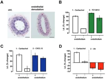
D-glucose. The vessel bath solution con-tained the same solution without BSA. The inner diameter (i.d.) of the pressur-ized vessel was then continuously mea-sured and recorded via digital video-edge detection. For each condition or drug treatment, the vessel was observed for at least 15 min or until the inner di-ameter remained stable. Mechanical en-dothelial denudation was performed in mesenteric arteries by rubbing the arte-rial lumen with a human hair, followed by air insufflation and washing the lumen with normal saline to remove the endothelium, as described (34).
Measurement of Sarcomere Length
Adult rat (Sprague Dawley) left ven-tricular cardiomyocytes, isolated using a
standardized enzymatic dispersion tech-nique (37), were kindly provided by X Ji and S Sadayappan, Loyola University Chicago. Left ventricular cardiomyocytes were superfused in 141.4 mmol/L NaCl, 4 mmol/L KCl, 0.33 mmol/L NaH2PO4, 1 mmol/L MgCl2, 10 mmol/L HEPES, 5.5 mmol/L glucose, 1.8 mmol/L CaCl2and 14.5 mmol/L mannitol, pH 7.4, field-stimulated (20 V, 2 Hz, 6 ms pulse dura-tion) and changes in sarcomere length during contractions after addition of ve-hicle or various concentrations of CXCR4/ACKR3 ligands (1 nmol/L to 10μmol/L) measured using a sarcomere detection system (IonOptix Corp), as described (38,39). Measure-ments of 60 steady state contractions were averaged for each condition. All ex-periments were carried out at 36 ± 1°C.
Light Microscopy
Arteries with and without mechanical endothelial denudation were fixed in 10% formalin and embedded in paraffin. Sections were stained with hematoxylin and eosin (H&E). Slides were examined under light microscopy (magnification 40×, 100×, 200×, 400×), as described (40).
In Vivo Experiments
Animals were anesthetized with 100 mg/kg ketamine and 10 mg/kg xy-lazine intraperitoneally (IP). This dose al-lowed the animals to be deeply sedated but able to breathe spontaneously throughout the experiment. After a mid-line laparotomy, the aorta was instru-mented with a 22-gauge angiocatheter for monitoring of arterial blood pressure, blood withdrawal and administration of the CXCR4/7 modulators. The abdomen was then closed with monofilament su-ture. Core body temperature was main-tained using warming lamps. For left ventricular pressure-volume (PV) loop analyses, animals were instrumented with a 1.9 F PV catheter (FTS-1912B-8018; Scisense, London, ON, Canada) that was placed into the right carotid artery and advanced through the aortic valve into the left ventricle. Left ventricular function was then monitored using the Scisense
ADVantage PV System with the iWorx data acquisition and analysis software. Left ventricular function indices were de-termined from 25 to 40 consecutive PV loops. CXCR4/7 modulators were in-jected in a total volume of 0.5 mL in 0.9% NaCl under normal conditions, in a hem-orrhagic shock model with fluid resusci-tation and in a lethal hemorrhagic shock model without fluid resuscitation, as de-scribed previously (41). In brief, in the hemorrhage and resuscitation model, ani-mals were hemorrhaged to a mean arte-rial blood pressure (MAP) of 30 mmHg for 30 min. The hemorrhage blood vol-ume to achieve MAP of 30 mmHg was similar in all groups (p> 0.05). At t= 30 min, CXCR4/7 modulators or vehicle (0.9% NaCl) were administered and the animals were then resuscitated with nor-mal saline until MAP returned to 70 mmHg. MAP was then maintained at 70 mmHg by continuous fluid adminis-tration for a total of 45 min, as required. At the end of the resuscitation period, the animals were euthanized by rapid exsan-guination, followed by thoracotomy re-sulting in bilateral pneumothorax. In the lethal hemorrhagic shock model, animals underwent withdrawal of 40% of the blood volume within 10 min. Blood was withdrawn at a rate of approximately 1 mL/min. At t= 15 min, CXCR4/7 mod-ulators or vehicle were administered. Thereafter, 2% of the blood volume were withdrawn every 15 min until death, as defined by asystole or disappearance of a pulse pressure. Animals after administra-tion of CXCR4/7 modulators under nor-mal conditions were euthanized by exsanguination and bilateral pneumotho-rax at the end of the observation period while being under deep anesthesia.
Data Analyses
sum-of-squares F test. Survival was plotted using the Kaplan-Meier method and survival be-tween groups was compared with the log-rank test. All data were analyzed using the GraphPad Prism 5 software (GraphPad Software Inc., La Jolla, CA, USA). A two-tailed p< 0.05 was considered significant.
All supplementary materials are available online at www.molmed.org.
RESULTS
CXCR4 and ACKR3 Ligands Modulate Hemodynamics and Blood Pressure
To assess whether modulation of CXCR4 and ACKR3 alters blood pressure
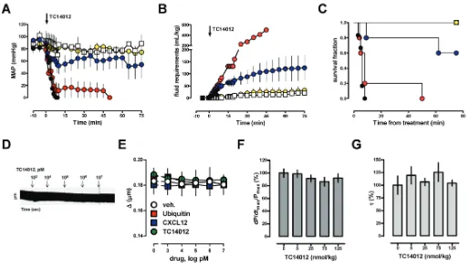
in normal animals, we first studied the effects of CXCL12, the cognate CXCR4 and ACKR3 agonist (1), ubiquitin, a noncognate CXCR4 agonist without affinity for ACKR3 (1,18,42), AMD3100, a selective CXCR4 antagonist (43), and TC14012, a CXCR4 antagonist and ACKR3 agonist (44). As expected (19,21,45–47), CXCL12, AMD3100 and ubiquitin did not affect normal blood pressure or fluid requirements in rats when administered systemically in a dose of 350 nmol/kg (Supplementary Figure 1). Systemic administration of TC14012, however, resulted in cardiovas-cular collapse despite fluid resuscitation (Figures 1A–C). The deleterious effects of
TC14012 on hemodynamics occurred dose dependently. TC14012 did not affect sarcomere length of isolated cardiomy-ocytes (Figures 1D, E) or left ventricular function in vivo(Figures 1F, G). The he-modynamic effects, however, severely compromised survival of normal animals and therefore precluded further in vivo testing of TC14012.
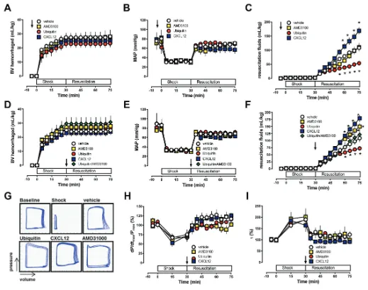
Next, we studied the effects of the CXCR4 and ACKR3 ligands that did not influence normal blood pressure in a hemorrhagic shock model. This model consisted of 30 min hemorrhage to a mean arterial blood pressure (MAP) of 30 mmHg followed by crystalloid fluid resuscitation to a MAP of 70 mmHg.
When the same doses were administered 5 min before blood withdrawal, CXCR4 activation with ubiquitin reduced whereas coactivation of CXCR4 and ACKR3 with CXCL12 increased systemic fluid requirements to maintain MAP at the resuscitation target of 70 mmHg. AMD3100 pretreatment did not affect
re-suscitation fluid requirements in this model (Figures 2A–C). As pretreatment protocols lack translational relevance, we then tested the effects of the CXCR4 and ACKR3 ligands when administered at the end of the shock period, prior to fluid resuscitation (Figure 2D–F). Whereas CXCR4 activation with
ubiqui-tin stabilized hemodynamics, coactiva-tion of CXCR4 and ACKR3 with CXCL12 and blockade of CXCR4 with AMD3100 reduced hemodynamic stability, as esti-mated based on the systemic fluid re-quirements to maintain MAP. The effects of ubiquitin and AMD3100 were abol-ished when both CXCR4 ligands were
coadministered. As observed for TC14012, none of the CXCR4/ACKR3 ligands affected sarcomere length of iso-lated cardiomyocytes (Figure 1E) or left-ventricular function in vivo(Figures 2G–I).
Because the resuscitation target of a MAP of 70 mmHg could be achieved in
all animals, we then tested the effects of the CXCR4/ACKR3 ligands in a shock model without fluid resuscitation to be able to assess their direct effects on blood pressure regulation during the cardiovas-cular stress response to hemorrhage. In this fixed volume hemorrhage model,
40% of the blood volume were with-drawn within 10 min followed by an ad-ditional 2% blood volume hemorrhage every 15 min without intervention. The CXCR4/ACKR3 ligands or vehicle were administered at t= 15 min. As shown in Figures 3A–C, median survival time was 41 min with vehicle administration. CXCR4 activation with ubiquitin stabi-lized blood pressure and prolonged me-dian survival time to 129 min (p< 0.001 versus vehicle, hazard ratio [95% confi-dence interval] 7.5 [2.9–18.8]). AMD3100 and CXCL12, however, reduced blood pressure and median survival time to 22 min (p= 0.0045 versus vehicle, hazard ratio [95% confidence interval] 0.12 [0.03–0.5]) and 18 min (p< 0.001 versus vehicle, hazard ratio [95% confidence in-terval] 0.11 [0.03–0.4]), respectively. To further assess whether the prolonged survival time after ubiquitin treatment could provide a therapeutic benefit, we then evaluated if a resuscitation target of MAP of 70 mmHg can be achieved when resuscitation is initiated at a time point when mortality was 100% in vehicle-treated animals. As shown in Figures 3D–G, in five out of seven animals with ubiquitin treatment, blood pressure could be restored to the resuscitation endpoint when crystalloid infusion was started at t= 85 min.
CXCR4 and ACKR3 Agonists Have Opposing Effects on α1-AR-Mediated Vasoconstriction
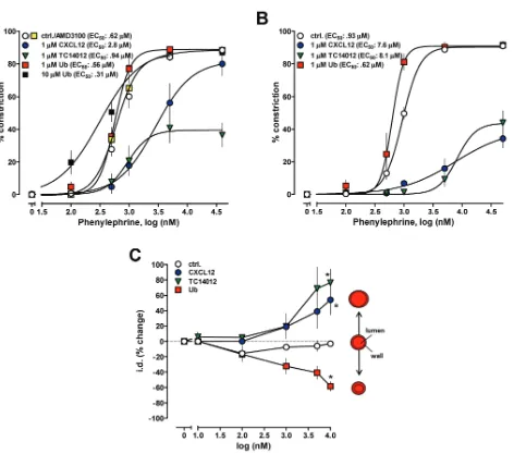
The observation that none of the CXCR4/ACKR3 ligands affected contrac-tility of isolated cardiomyocytes or left ventricular function in vivosuggested that their effects on blood pressure are mediated through modulation of vascular function. Thus, we then utilized pressure myography to study the effects of various natural and synthetic CXCR4 and ACKR3 ligands on isolated mesenteric arteries and veins. The CXCR4/ ACKR3 ligands did not affect the diameter of arteries in the absence of vasoconstrictors. Ubiquitin enhanced vascular reactivity to the selec-tive α1-AR agonist phenylephrine, whereas CXCL12 and TC14012 reduced
Figure 3.CXCR4/ACKR3 ligands regulate blood pressure during the cardiovascular stress response to hemorrhage. Rats underwent 40% blood volume (BV) hemorrhage followed by 2% BV hemorrhage every 15 min without intervention. CXCR4/ACKR3 ligands (350 nmol/kg) were injected at t = 15 min (arrow). Open circles: vehicle (n = 19). Yellow squares:
phenylephrine responsiveness (Figure 4A; p< 0.01 versus groups). AMD3100 did not affect phenylephrine responsive-ness (see Figure 4A). In these experi-ments, CXCR4/ ACKR3 ligands were ad-ministered to the organ bath; more robust effects of CXCL12 and TC14012 were ob-served when the CXCR4/ACKR3 modu-lators were administered intraluminally (Figure 4B; p< 0.01 versus groups).
When arteries were constricted with the EC50concentration of phenylephrine, ad-dition of ubiquitin enhanced, whereas CXCL12 and TC14012 antagonized vaso-constriction in a dose-dependent manner (Figure 4C).
We next tested whether CXCR4- and ACKR3-mediated effects of CXCL12 can be differentiated by blockade of CXCR4 with AMD3100. Consistent with the
op-posing effects of CXCR4 and ACKR3, blockade of CXCR4 by AMD3100 en-hanced the antagonistic effects of CXCL12 (Figures 5A, B) on phenyle-phrine-induced vasoconstriction. In con-trast, AMD3100 antagonized the effects of ubiquitin on phenylephrine-induced vasoconstriction (Figures 5 A, C).
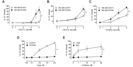
To further confirm the role of ACKR3, we then tested chemokine (C-X-C motif)
ligand 11 (CXCL11, interferon-inducible T cell αchemoattractant), an alternative natural agonist of ACKR3 that does not bind or activate CXCR4 (48), and CCX771, a small molecule ACKR3 lig-and, which has often been described as an antagonist at ACKR3 (25). As shown in Figure 6A, CXCL11 also antagonized phenylephrine-induced vasoconstriction and its effects could be antagonized with 100 nmol/L CCX771. CCX771 (100 nmol/L) also antagonized the ef-fects CXCL12 (see Figure 4B) and TC14012 (Figure 4C) on phenylephrine-induced vasoconstriction. CCX704, an in-active analogue of CCX771, did not affect the effects of the ACKR3 agonists (Fig-ures 6A–C). At higher concentrations (1 to 2 μmol/L), however, CCX771 alone antagonized vasoconstriction in response to phenylephrine (Figure 6D), whereas CCX704 was inactive. The specific ACKR3 antibody 11G8 (49) also antago-nized phenylephrine-induced
vasocon-Figure 5.Blockade of CXCR4 with AMD3100 antagonizes effects of ubiquitin (Ub) and en-hances effects of CXCL12 on phenylephrine-induced vasoconstriction. Mesenteric arter-ies were constricted with the EC50dose of phenylephrine (1 μmol/L) and the effects of the CXCR4/7 modulators tested. The effects of the CXCR4/7 modulators are expressed as % change of i.d. *: p < 0.05 versus control (ctrl). (A) Dose response of ubiquitin and CXCL12 in the presence and absence of 50 μmol/L AMD3100; n = 4. (B,C) Addition of AMD3100 (50 μmol/L) (B) enhances CXCL12-mediated effects and (C) antagonizes mediated effects; n = 4.
striction, whereas a nonspecific IgG1 iso-type control antibody was inactive (Fig-ure 6E). The receptor selectivity of the various CXCR4 and ACKR3 ligands and their effects on phenylephrine-induced vasoconstriction are summarized in Table 1.
Effects of CXCR4 and ACKR3 Ligands Are Specific and Independent of the Vascular Endothelium
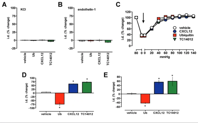
To assess the specificity of the ob-served effects of the CXCR4 and ACKR3 ligands on vascular reactivity, we next tested their effects on vasoconstriction induced via activation of operated Ca2+channels by KCl or vaso-constriction mediated through the GPCR endothelin receptor. None of the CXCR4 and ACKR3 agonists, however, influenced vasoconstriction induced by KCl (Figure 7A) or endothelin-1 (Fig-ure 7B).
To further characterize the vascular ef-fects of the CXCR4 and ACKR3 ligands, we then tested the influence of the trans-mural vascular pressure in pressure myography experiments. The CXCR4 and ACKR3 ligands did not affect the di-ameter of arteries in the absence of vaso-constrictors at various transmural pres-sures (Figure 7C). As observed for arteries pressurized to 80 mmHg, the CXCR4/ACKR3 ligands also modulated phenylephrine-induced vasoconstriction in arteries maintained at a low trans-mural pressure (30 mmHg, Figure 7D)
the CXCR4/ACKR3 agonists on phenyle-phrine-mediated vasoconstriction in mesenteric arteries after mechanical en-dothelial denudation (Figure 8A). En-dothelial denudation abolished the ef-fects of the endothelium-dependent vasodilator carbachol on phenylephrine-induced vasoconstriction, but did not significantly influence the modulatory actions of TC14012 (Figure 8B), CXCL12 (Figure 8C) and ubiquitin (Figure 8D).
DISCUSSION
In the present study, we demonstrate that pharmacological modulation of CXCR4 and ACKR3 in vivoinfluences blood pressure in normal and pathologi-cal conditions. Furthermore, our findings suggest that activation of CXCR4 and ACKR3 have opposing effects on α1 -AR-mediated vasoconstriction of isolated
Table 1.Receptor selectivity of CXCR4 and ACKR3 ligands and their effects on
α1-AR-mediated vasoconstriction of isolated mesenteric arteries.
Receptor selectivity Effect on vascular
Ligand CXCR4 ACKR3 References α1-AR reactivity
CXCL11 NB + (1,48) ↓
CXCL12 + + (1,6,7) ↓
TC14012 – + (44) ↓
11G8 NB + (49) ↓
CCX771 NB + (25) ↓
AMD3100 – (+) (1,67,68) ~
Ubiquitin + NB (1,18,42) ↑
NB, not binding; +, agonist; (+),weak allosteric agonist; –, antagonist; ↓, antagonizes phenylephrine-induced vasoconstriction; ↑, enhances phenylephrine-induced vasoconstriction; ~, no effect on phenylephrine-induced vasoconstriction.
Figure 7.Effects of CXCR4/ACKR3 ligands on vascular α1-AR reactivity are specific. Vaso-constriction was measured as change of the i.d. in pressure myography experiments. *: p < 0.05 versus control. (A,B) Arteries were constricted to 40% to 60% of i.d. with (A) 60 mmol/L KCl (n = 3 to 4), (B) 800 pmol/L endothelin-1 (n = 3 to 6) and the effects of CXCR4/ACKR3 ligands (10 μmol/L) tested. The effects of the CXCR4/ACKR3 modulators are expressed as % change of i.d. (C) CXCR4/ACKR3 modulators do not affect the i.d. of mesenteric arteries at various transmural pressures; n = 4 to 8. Arrow: Addition of CXCR4/ACKR3 modulators, 10 μmol/L . (D) Mesenteric arteries pressurized to 30 mmHg were constricted with the EC50 dose of phenylephrine (1 μmol/L) and the effects of the CXCR4/ACKR3 modulators (10 μmol/L) tested; n = 3. (E) Mesenteric veins pressurized to 12 mmHg were constricted with the EC50 dose of phenylephrine (1 μmol/L) and the effects of the CXCR4/ACKR3 modulators (10 μmol/L) tested; n = 3 to 6.
and in mesenteric veins pressurized to 12 mmHg (Figure 7E).
vessels, which are independent of the dothelium. While CXCR4 activation en-hances vascular α1-AR responsiveness, ACKR3 activation results in opposite ef-fects. In combination with the observa-tion that the CXCR4 and ACKR3 ligands did not directly affect cardiac function, our findings imply that CXCR4 and ACKR3 regulate cardiovascular function through their modulatory actions on vas-cular smooth muscle reactivity.
The observed in vivoactions of TC14012 indicate that simultaneous blockade of CXCR4 and activation of ACKR3 causes cardiovascular collapse in normal animals. As the natural CXCR4 and ACKR3 ligands CXCL11, CXCL12 and ubiquitin are constitutively ex-pressed in normal plasma (50–52), these data imply that CXCR4 and ACKR3 may act as an intricate control system that
regulates vascular function and blood pressure under normal conditions.
The findings that blockade of CXCR4 with AMD3100 and coactivation of CXCR4 and ACKR3 with CXCL12 im-paired blood pressure during the cardio-vascular stress response to hemorrhage, whereas selective CXCR4 activation with ubiquitin-stabilized blood pressure is consistent with the effects of CXCR4 and ACKR3 activation on vascular phenyle-phrine responsiveness ex vivo. In combi-nation with previously described effects of ubiquitin in various shock models (19–21,45), these data indicate that selec-tive CXCR4 activation stabilizes blood pressure during the cardiovascular stress response to hemorrhagic shock, provid-ing an extended therapeutic window during which blood pressure can be re-stored with fluid resuscitation.
Ubiquitin is known to be cosecreted with catecholamines from chromaffin cells of the adrenal gland (53). In connec-tion with the findings from the present study, these data point toward a physio-logical linkage between α1-AR and CXCR4 activation during the fight-or-flight response.
The observation that AMD3100 did not affect blood pressure and fluid require-ments when administered as a pretreat-ment in the hemorrhagic shock model can be explained by its pharmacological properties. After systemic administra-tion, the volume of distribution of AMD3100 is small, its half-life in rats is approximately 60 min and binding of AMD3100 to CXCR4, unlike binding of CXCL12 and ubiquitin to CXCR4, does not result in ligand-induced receptor in-ternalization (14,43,54). Thus, in our ex-periments, withdrawal of blood removed large proportions of AMD3100 from the systemic circulation and subsequent fluid resuscitation further reduced the re-maining drug concentrations. In addi-tion, the finding that AMD3100 did not affect phenylephrine-induced vasocon-striction of isolated arteries is consistent with the absence of natural agonists in this model system.
The biological properties that we ob-served for CXCL12 are in agreement with the much higher affinity of CXCL12 for ACKR3, as compared with its affinity for CXCR4 (7). Furthermore, pharmacologi-cal blockade of CXCR4 with AMD3100 and partial blockade of ACKR3 with CCX771 permitted differentiation of the opposing effects of CXCR4 and ACKR3 upon activation with CXCL12. The find-ing that AMD3100 was able to block the effects of ubiquitin is in agreement with the known receptor selectivity of ubiqui-tin (42) and the observed effects after in vivocoadministration of both CXCR4 lig-ands in the present study.
CXCL11 is known to be highly in-duced following endotoxin and inter-feron exposure (55). Therefore, it appears likely that CXCL11 may contribute to the development of vasodilatory shock and catecholamine refractoriness, which is
characteristic for endotoxic and septic shock.
All natural, synthetic and antibody ACKR3 ligands that we tested attenu-ated phenylephrine responsiveness of arteries, which indicates that these lig-ands function as receptor agonists with varying efficacy. Nevertheless, the syn-thetic ACKR3 ligand CCX771 acted as a low efficacy agonist that partially antag-onized the effects of the endogenous lig-ands CXCL11 and CXCL12. A more de-tailed pharmacological exploration of the role of ACKR3 in the regulation of cardiovascular function is currently not possible because ACKR3 ligands with-out intrinsic activity are not available. While genetic models may be useful to dissect the mechanistic basis for our ob-servations, such studies, however, would lack translational relevance and would not provide preclinical evidence for possible new therapeutic opportuni-ties. Thus, the development of ACKR3 antagonists without intrinsic activity will be important to provide a more de-tailed evaluation of the role of ACKR3 as a therapeutic target.
The finding that the CXCR4 and ACKR3 ligands did not affect the diame-ter of ardiame-teries in the absence of vasocon-strictors at various transmural pressures suggests that they do not affect myo-genic tone. The observation that endothelin-1 or KCl-induced vasocon-striction was not affected by CXCR4/ ACKR3 ligands documents specificity for α1-AR-mediated functions. As CXCR4 and ACKR3 ligands also modulated vas-cular reactivity at low transmural pres-sures and in mesenteric veins, these data indicate that CXCR4 and ACKR3 ligands modulate effects of α1-AR on vascular re-sistance and capacitance over a wide range of vascular pressures.
CONCLUSION
Our findings identify CXCR4 and ACKR3 as regulators of vascular α1-AR function and provide evidence for a new physiological role of chemokines beyond their known biological properties, the regulation of vascular function. We
cur-rently cannot exclude that CXCR4 or ACKR3 activation affects α1-AR receptor expression. Such effects could explain our observations on phenylephrine reac-tivity of isolated arteries when arteries were pretreated with CXCR4 and ACKR3 ligands, but are unlikely to account for the enhancing and antagonizing effects of the CXCR4 and ACKR3 agonists on vasoconstriction in response to the EC50 dose of phenylephrine, respectively. Reg-ulation of GPCR function is complex and cross-talk between GPCR-signaling path-ways, whereby activation of one receptor system positively or negatively affects the signaling of another signaling recep-tor system in the same cell, is a recognized principle that permits fine tuning of GPCR functions (56–61). Re-ceptor cross-talk has been attributed to various molecular mechanisms, such as interactions between intracellular signal transduction pathways, receptor transac-tivation or receptor hetero-oligomeriza-tion (56–63). The formahetero-oligomeriza-tion of homo-and/or heteromers between GPCRs is known to be important for many aspects of GPCR function (64). α1a-AR and α1b-AR have been reported to exist as homo- and hetero-oligomers, which is thought to provide a mechanism that regulates their physiological responses (65). Interestingly, α1a-AR have recently been described to colocalize with CXCR2 in the smooth muscle layer of prostate tissue and to form heteromeric com-plexes with CXCR2 in a recombinant sys-tem, resulting in signaling events distinct from the mono-/homomers (66). Thus, it appears possible that heteromeric com-plexes between α1a-AR and CXCR2, CXCR4, ACKR3 or other chemokine re-ceptors also exist in vascular smooth muscle, which may modulate vascular α1-AR function.
Although the exact molecular events underlying the regulatory functions of CXCR4 and ACKR3 on vascular α1-AR reactivity remain to be determined, our findings provide initial mechanistic in-sights, which explain pharmacological effects of CXCR4 and ACKR3 ligands on cardiovascular function and point
to-ward CXCR4 and ACKR3 as new thera-peutic targets to regulate vascular func-tion and blood pressure. This may enable development of new classes of drugs for the treatment of a broad variety of dis-eases that are associated with impaired vascular function, such as hypertension, pulmonary arterial hypertension, va-sodilatory shock or catecholamine refrac-toriness in critical illness.
ACKNOWLEDGMENTS
The authors thank Heather M La Porte for technical help and P de Tombe, X Ji, S Sadayappan, and R. Tiniakov, Loyola University Chicago, for help with myo-cardial function analyses. This research was made possible, in part, by a grant that was awarded and administered by the U.S. Army Medical Research & Ma-teriel Command (USAMRMC) and the Telemedicine and Advanced Technology Research Center (TATRC), at Fort Det-rick, MD, USA, under contract number W81XWH1020122. The views, opinions and/or findings contained in this re-search are those of the author(s) and do not necessarily reflect the views of the Department of Defense and should not be construed as an official DoD/Army position, policy or decision unless so designated by other documentation. No official endorsement should be made. This research was also supported, in part, by grants from the American Heart Association (13GRNT17230072), the NIH (T32GM008750) and the Dr. Ralph and Marian Falk Medical Research Trust.
DISCLOSURES
The therapeutic use of ubiquitin has been patented (US patent #7,262,162). M Majetschak is an inventor and has not received any income related to the patent.
REFERENCES
1. Bachelerie F, et al. (2013) International Union of Basic and Clinical Pharmacology. [corrected]. LXXXIX. Update on the extended family of chemokine receptors and introducing a new nomenclature for atypical chemokine receptors.
CXCR4 is essential for vascularization of the gas-trointestinal tract. Nature. 393:591–4.
3. Gerrits H, et al. (2008) Early postnatal lethality and cardiovascular defects in CXCR7-deficient mice. Genesis. 46:235–45.
4. Nagasawa T, et al. (1996) Defects of B-cell lym-phopoiesis and bone-marrow myelopoiesis in mice lacking the CXC chemokine PBSF/SDF-1.
Nature. 382:635–8.
5. Sierro F, et al. (2007) Disrupted cardiac develop-ment but normal hematopoiesis in mice deficient in the second CXCL12/SDF-1 receptor, CXCR7.
Proc. Natl. Acad. Sci. U. S. A. 104:14759–64. 6. Bleul CC, et al. (1996) The lymphocyte
chemoat-tractant SDF-1 is a ligand for LESTR/fusin and blocks HIV-1 entry. Nature. 382:829–33.
7. Balabanian K, et al. (2005) The chemokine SDF-1/ CXCL12 binds to and signals through the orphan receptor RDC1 in T lymphocytes. J. Biol. Chem. 280:35760–6.
8. Teicher BA, Fricker SP. (2010) CXCL12 (SDF-1)/ CXCR4 pathway in cancer. Clin. Cancer Res. 16:2927–31.
9. Nagasawa T, Tachibana K, Kishimoto T. (1998) A novel CXC chemokine PBSF/SDF-1 and its re-ceptor CXCR4: their functions in development, hematopoiesis and HIV infection. Semin. Im-munol. 10:179–85.
10. Rajagopal S, et al. (2010) Beta-arrestin- but not G protein-mediated signaling by the “decoy” receptor CXCR7. Proc. Natl. Acad. Sci. U. S. A. 107:628–32. 11. Boldajipour B, et al. (2008) Control of
chemokine-guided cell migration by ligand sequestration.
Cell. 132:463–73.
12. Kumar R, et al. (2012) CXCR7 mediated Giα inde-pendent activation of ERK and Akt promotes cell survival and chemotaxis in T cells. Cell. Immunol. 272:230–41.
13. Regard JB, Sato IT, Coughlin SR. (2008) Anatomi-cal profiling of G protein-coupled receptor ex-pression. Cell. 135:561–71.
14. Tripathi A, Davis JD, Staren DM, Volkman BF, Majetschak M. (2013) CXC chemokine receptor 4 signaling upon co-activation with stromal cell-derived factor-1alpha and ubiquitin. Cytokine. 65:121–5.
15. LaRocca TJ, et al. (2010) β2-Adrenergic receptor signaling in the cardiac myocyte is modulated by interactions with CXCR4. J. Cardiovasc. Pharmacol. 56:548–59.
16. Agarwal U, et al. (2010) Role of cardiac myocyte CXCR4 expression in development and left ven-tricular remodeling after acute myocardial infarc-tion. Circ. Res. 107:667–76.
17. Levoye A, Balabanian K, Baleux F, Bachelerie F, Lagane B. (2009) CXCR7 heterodimerizes with CXCR4 and regulates CXCL12-mediated G pro-tein signaling. Blood. 113:6085–93.
18. Saini V, Marchese A, Majetschak M. (2010) CXC chemokine receptor 4 is a cell surface receptor for extracellular ubiquitin. J. Biol. Chem. 285:15566–76. 19. Majetschak M, Cohn SM, Nelson JA, Burton EH,
Obertacke U, Proctor KG. (2004) Effects of exoge-nous ubiquitin in lethal endotoxemia. Surgery. 135:536–43.
20. Baker TA, Romero J, Bach HHt, Strom JA, Gamelli RL, Majetschak M. (2012) Effects of ex-ogenous ubiquitin in a polytrauma model with blunt chest trauma. Crit. Care Med. 40:2376–84. 21. Bach Iv HH, Saini V, Baker TA, Tripathi A,
Gamelli RL, Majetschak M. (2012) Initial assess-ment of the role of CXC chemokine receptor 4 after polytrauma. Mol. Med. 18:1056–66. 22. Bodart V, et al. (2009) Pharmacology of AMD3465:
a small molecule antagonist of the chemokine re-ceptor CXCR4. Biochem. Pharmacol. 78:993–1000. 23. Chu PY, et al. (2011) CXCR4 antagonism attenu-ates the cardiorenal consequences of mineralo-corticoid excess. Circ. Heart Fail. 4:651–8. 24. Yu L, Hales CA. (2011) Effect of chemokine
re-ceptor CXCR4 on hypoxia-induced pulmonary hypertension and vascular remodeling in rats.
Respir. Res. 12:21.
25. Zabel BA, et al. (2009) Elucidation of CXCR7-me-diated signaling events and inhibition of CXCR4-mediated tumor cell transendothelial migration by CXCR7 ligands. J. Immunol. 183:3204–11. 26. Sartina E, et al. (2012) Antagonism of CXCR7
at-tenuates chronic hypoxia-induced pulmonary hypertension. Pediatr. Res. 71:682–8.
27. Veldkamp CT, Seibert C, Peterson FC, Sakmar TP, Volkman BF. (2006) Recognition of a CXCR4 sul-fotyrosine by the chemokine stromal cell-derived factor-1alpha (SDF-1alpha/CXCL12). J. Mol. Biol. 359:1400–9.
28. Veldkamp CT, et al. (2008) Structural basis of CXCR4 sulfotyrosine recognition by the chemokine SDF-1/CXCL12. Sci. Signal. 1:ra4.
29. Veldkamp CT, et al. (2009) Monomeric structure of the cardioprotective chemokine SDF-1/CXCL12.
Protein Sci. 18:1359–69.
30. Takekoshi T, Ziarek JJ, Volkman BF, Hwang ST. (2012) A locked, dimeric CXCL12 variant effec-tively inhibits pulmonary metastasis of CXCR4-expressing melanoma cells due to enhanced serum stability. Mol. Cancer Ther. 11:2516–25. 31. (1985) Bacterial endotoxins/pyrogens [Internet].
Silver Spring (MD): FDA; [cited 2014 Aug 29]. (Inspection technical guide). Available from: http://www.fda.gov/ICECI/Inspections/ InspectionGuides/InspectionTechnicalGuides/ ucm072918.htm (web page last updated 2009 Feb 17).
32. Committee for the Update of the Guide for the Care and Use of Laboratory Animals, Institute for Laboratory Animal Research, Division on Earth and Life Studies, National Research Coun-cil of the National Academies. (2011) Guide for the Care and Use of Laboratory Animals. 8th edition. Washington (DC): National Academies Press. 33. Henderson KK, Byron KL. (2007)
Vasopressin-induced vasoconstriction: two dependent signaling pathways. J. Appl. Physiol. 102:1402–9.
34. Brueggemann LI, Mackie AR, Mani BK, Cribbs LL, Byron KL. (2009) Differential effects of selec-tive cyclooxygenase-2 inhibitors on vascular smooth muscle ion channels may account for dif-ferences in cardiovascular risk profiles. Mol.
Pharmacol. 76:1053–61.
35. Brueggemann LI, Mani BK, Haick J, Byron KL. (2012) Exploring arterial smooth muscle Kv7 potas-sium channel function using patch clamp electro-physiology and pressure myography. J. Vis. Exp. 14:e4263.
36. Mani BK, Brueggemann LI, Cribbs LL, Byron KL. (2011) Activation of vascular KCNQ (Kv7) potas-sium channels reverses spasmogen-induced con-strictor responses in rat basilar artery. Br. J. Phar-macol. 164:237–49.
37. Dow JW, Harding NG, Powell T. (1981) Isolated cardiac myocytes. I. Preparation of adult my-ocytes and their homology with the intact tissue.
Cardiovasc. Res. 15:483–514.
38. Govindan S, et al. (2012) Pathogenic properties of the N-terminal region of cardiac myosin binding protein-C in vitro. J. Muscle Res. Cell Motil. 33:17–30.
39. Jin CZ, et al. (2013) Myofilament Ca desensitiza-tion mediates positive lusitropic effect of neu-ronal nitric oxide synthase in left ventricular my-ocytes from murine hypertensive heart. J. Mol.
Cell. Cardiol. 60:107–15.
40. Geng Q, et al. (2009) A subset of 26S proteasomes is activated at critically low ATP concentrations and contributes to myocardial injury during cold ischemia. Biochem. Biophys. Res. Commun. 390:1136–41.
41. Bach HHt, Laporte HM, Wong YM, Gamelli RL, Majetschak M. (2013) Proteasome inhibition pro-longs survival during lethal hemorrhagic shock in rats. J. Trauma Acute Care Surg. 74:499–507. 42. Saini V, et al. (2011) The CXC chemokine receptor
4 ligands ubiquitin and stromal cell-derived fac-tor-1αfunction through distinct receptor interac-tions. J. Biol. Chem. 286:33466–77.
43. Hatse S, Princen K, Bridger G, De Clercq E, Schols D. (2002) Chemokine receptor inhibition by AMD3100 is strictly confined to CXCR4. FEBS Lett. 527:255–62.
44. Gravel S, et al. (2010) The peptidomimetic CXCR4 antagonist TC14012 recruits beta-arrestin to CXCR7: roles of receptor domains. J. Biol. Chem. 285:37939–43.
45. Majetschak M, Cohn SM, Obertacke U, Proctor KG. (2004) Therapeutic potential of exogenous ubiquitin during resuscitation from severe trauma. J. Trauma. 56:991–9.
46. O’Boyle G, Mellor P, Kirby JA, Ali S. (2009) Anti-inflammatory therapy by intravenous delivery of non-heparan sulfate-binding CXCL12. FASEB J. 23:3906–16.
47. Misra P, et al. (2008) Quantitation of CXCR4 expres-sion in myocardial infarction using 99mTc-labeled SDF-1alpha. J. Nucl. Med. 49:963–9.
recep-tor for SDF-1 and I-TAC involved in cell survival, cell adhesion, and tumor development. J. Exp.
Med. 203:2201–13.
49. Berahovich RD, Penfold ME, Schall TJ. (2010) Nonspecific CXCR7 antibodies. Immunol. Lett. 133:112–4.
50. Majetschak M. (2011) Extracellular ubiquitin: im-mune modulator and endogenous opponent of damage-associated molecular pattern molecules.
J. Leukoc. Biol. 89:205–19.
51. Butera D, et al. (2005) Plasma chemokine levels correlate with the outcome of antiviral therapy in patients with hepatitis C. Blood. 106:1175–82. 52. Derdeyn CA, et al. (1999) Correlation between
circulating stromal cell-derived factor 1 levels and CD4+ cell count in human immunodefi-ciency virus type 1-infected individuals. AIDS Res. Hum. Retroviruses. 15:1063–71.
53. Kieffer AE, et al. (2003) The N- and C-terminal fragments of ubiquitin are important for the an-timicrobial activities. Faseb J. 17:776–8. 54. Hendrix CW, et al. (2000) Pharmacokinetics and
safety of AMD-3100, a novel antagonist of the CXCR-4 chemokine receptor, in human volun-teers. Antimicrob. Agents Chemother. 44:1667–73. 55. Widney DP, Xia YR, Lusis AJ, Smith JB. (2000)
The murine chemokine CXCL11 (IFN-inducible T cell alpha chemoattractant) is an IFN-gamma-and lipopolysaccharide-inducible glucocorti-coid-attenuated response gene expressed in lung and other tissues during endotoxemia. J. Im-munol. 164:6322–31.
56. Moore CA, Milano SK, Benovic JL. (2007) Regu-lation of receptor trafficking by GRKs and ar-restins. Annu. Rev. Physiol. 69:451–82.
57. Busillo JM, Benovic JL. (2007) Regulation of CXCR4 signaling. Biochim. Biophys. Acta. 1768:952–63. 58. Marchese A, Chen C, Kim YM, Benovic JL. (2003)
The ins and outs of G protein-coupled receptor trafficking. Trends Biochem. Sci. 28:369–76. 59. Rockman HA, Koch WJ, Lefkowitz RJ. (2002)
Seven-transmembrane-spanning receptors and heart function. Nature. 415:206–12.
60. Diviani D, et al. (1996) Effect of different G pro-tein-coupled receptor kinases on phosphoryla-tion and desensitizaphosphoryla-tion of the alpha1B-adrener-gic receptor. J. Biol. Chem. 271:5049–58. 61. Collins S, Bouvier M, Lohse MJ, Benovic JL,
Caron MG, Lefkowitz RJ. (1990) Mechanisms in-volved in adrenergic receptor desensitization.
Biochem. Soc. Trans. 18:541–4.
62. McGraw DW, et al. (2006) Airway smooth muscle prostaglandin-EP1 receptors directly modulate beta2-adrenergic receptors within a unique het-erodimeric complex. J. Clin. Invest. 116:1400–9. 63. Dzimiri N. (2002) Receptor crosstalk. Implica-tions for cardiovascular function, disease and therapy. Eur. J. Biochem. 269:4713–30.
64. Milligan G, Canals M, Pediani JD, Ellis J, Lopez-Gimenez JF. (2006) The role of GPCR dimerisa-tion/oligomerisation in receptor signalling. Ernst Schering Found. Symp. Proc. 2:145–61.
65. Stanasila L, Perez JB, Vogel H, Cotecchia S. (2003) Oligomerization of the alpha 1a- and alpha 1b-adrenergic receptor subtypes. Potential implica-tions in receptor internalization. J. Biol. Chem. 278:40239–51.
66. Mustafa S, et al. (2012) Identification and profil-ing of novel alpha1A-adrenoceptor-CXC chemokine receptor 2 heteromer. J. Biol. Chem. 287:12952–65.
67. Schols D, Struyf S, Van Damme J, Este JA, Hen-son G, De Clercq E. (1997) Inhibition of T-tropic HIV strains by selective antagonization of the chemokine receptor CXCR4. J. Exp. Med. 186:1383–8.
68. Kalatskaya I, Berchiche YA, Gravel S, Limberg BJ, Rosenbaum JS, Heveker N. (2009) AMD3100 is a CXCR7 ligand with allosteric agonist properties.