R E V I E W
Open Access
Genetic mouse models to study blood
–
brain
barrier development and function
Fabien Sohet
*and Richard Daneman
*Abstract
The blood–brain barrier (BBB) is a complex physiological structure formed by the blood vessels of the central nervous system (CNS) that tightly regulates the movement of substances between the blood and the neural tissue. Recently, the generation and analysis of different genetic mouse models has allowed for greater understanding of BBB development, how the barrier is regulated during health, and its response to disease. Here we discuss: 1) Genetic mouse models that have been used to study the BBB, 2) Available mouse genetic tools that can aid in the study of the BBB, and 3) Potential tools that if generated could greatly aid in our understanding of the BBB.
Keywords:Blood–brain barrier, Mouse models, Endothelial cells, Neuro-vascular unit, Astrocytes, Pericytes, Central Nervous system
Review Introduction
The blood–brain barrier (BBB) is a functional physio-logical structure formed by the blood vessels of the cen-tral nervous system (CNS) that tightly regulates the exchange of molecules, ions and cells between the blood and the CNS, and is critical for maintenance of homeo-stasis within the nervous tissue. Many of the properties of the BBB are possessed by the endothelial cells (ECs) that form the walls of the blood vessels, and these prop-erties are tightly regulated by both neural and immune cells. Important BBB properties include: 1) CNS ECs are joined together by tight junctions (TJs) which create a paracellular barrier, 2) CNS ECs undergo extremely low rates of transcytosis creating a transcellular barrier to hydrophilic molecules, 3) CNS ECs express transporters to efflux potential toxins from the CNS, 4) CNS ECs express selective transporters to deliver specific nutrients to the CNS, 5) CNS ECs express very low levels of leukocyte ad-hesion molecules limiting the entry of immune cells into the CNS. ECs interact with immune cells in the blood, as well as different cells within the CNS parenchyma, inc-luding pericytes, astrocytes, macrophages, microglia and neurons, and these interactions are important to regulate the formation of the BBB during development, the
function of the BBB during health, and the response of the BBB to injury and disease.
In this review we will discuss mouse genetic models that can be utilized to study the BBB during health and disease. First we will discuss selected genetic models that have been used to identify novel aspects of BBB function in-cluding endothelial barrier function, CNS angiogenesis and BBB development, and interactions of different cell types within the neuro-vascular unit (see Additional file 1: Supplementary Table 1. Genetic mouse models to study the BBB). In the second section we will discuss current genetic tools available for analysis of BBB function. In the final section we will suggest several potential genetic tools that if generated could greatly increase our ability to study and understand the BBB.
Types of genetic mouse models
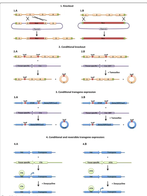
In general, mouse genetic models fall under two categories: gene silencing or ectopic gene expression (Figure 1). Pub-lished mouse lines can be found in the Mouse Genomic Informatics (MGI) data base (http://www.informatics.jax. org/).
For gene silencing, the most common tools include mouse knockout or conditional knockout technologies. Mouse knockout models use homologous recombination to delete a specific gene, or a section of a specific gene, from its endogenous chromosomal locus. This technique * Correspondence:[email protected];[email protected]
UCSF Department of Anatomy, 513 Parnassus Ave HSW1301, San Francisco, 94143, California, USA
leads to mice that lack the specific gene in all cells, and has widely been utilized to study the role of genes in mouse development and function. For genes on the som-atic chromosomes, each mouse receives a chromosome from each parent, and thus one can create homozygous knockout mice in which alleles on both parental chromo-somes are mutated or heterozygous mice in which only one allele is mutated. For genes on the sex chromosomes the details are more complicated. For instance, males only receive one X-chromosome from their mother, and thus for genes on the X-chromosome males can be either mu-tant or wild type, but not heterozygous. Females receive an X chromosome from each parent, and thus can be wild type, heterozygous or homozygous for mutant alleles, however because of X inactivation, heterozygous mutation can lead to mosaicism as a different X chromosome may be inactivated in different cells.
More recently, generation of conditional mutant mouse lines has allowed for spatial and temporal control over gene silencing. Specifically, homologous recombination is used to flank a critical exon (or exons) within a specific gene with lox-p sites. The lox-p sites do not alter gene function, but upon expression of a Cre recombinase gene, the recombinase deletes the section of the gene flanked by the lox-p sites. Therefore, expression of Cre-recombinase by transgenics, viral infection or other methods can trol the cell specificity of the gene deletion. Further con-trol of the timing of gene deletion can be achieved by using a CreERT or CreERT2 recombinase, in which the recombinase is fused to a modified estrogen receptor and thus is only targeted to the nucleus upon injection of tam-oxifen [1]. Therefore use of the CreERT allows for spatial control (where the CreERT is expressed) and temporal control (when tamoxifen is injected) of gene deletion. The Cre/lox systems irreversibly delete sequences flanked by lox-p sites, and thus several different methodologies have been used to deliver double-stranded RNA, either shRNA or siRNA, to reversibly silence specific genes.
For ectopic expression, several techniques can be used to introduce novel genetic sequences into the mouse
genome, including homologous recombination into a specific locus in the mouse genome, or random integra-tion of transgenes through injecintegra-tion into an embryo. These techniques have been used to express mutant forms of genes, over-express genes, mis-express genes in different cell types, express exogenous genes such as GFP or LacZ reporters, or express toxins to kill specific cell types [2]. Several methods have been used to control the specificity of the expression of the transgenes (Figure 1). The transgene can be generated downstream from a defined promoter, and thus the expression will be con-trolled by the specificity of the promoter. The transgene can be generated downstream from a strong promoter and a stop cassette that is flanked by lox-p sites (lox-stop-lox). In this case, the stop cassette will inhibit the expression of the gene, unless the cassette is excised by Cre recombinase, and thus the onset of the expression is controlled by the Cre recombinase, but expression is controlled by the up-stream promoter once the lox-p sites have been removed. Often the lox-stop-lox transgene cassette is inserted in the ROSA locus by homologous recombination. The ROSA locus has been shown to ubiquitously express genes, and thus inserting a lox-stop-lox reporter cassette into this locus marks all cells downstream of the cell in which the cre-recombinase excision has occurred. Zambrowiczet al. showed that insertion of the β-galactosidase gene at the ROSA locus in mice induced a broad β-gal activity throughout the body [3].
Another common technique utilized is to generate the transgene downstream of the tetracycline response element (TRE) (Figure 1). The TRE element promotes the expression of genes when the reverse tetracycline transactivator (rtTA) and doxycycline are both present. Therefore, spatial con-trol of gene expression can be achieved by the expression of rtTA in response to cell specific promoters, and tem-poral expression can be reversibly achieved by altering the levels of doxycycline in the diet. This method can also be used with a tetracycline transactivator (tTA) that induces expression from the TRE reporter when doxycycline is removed from the diet. Additionally different methods of (See figure on previous page.)
viral infection, electroporation, liposomal transfer and other techniques have been utilized to deliver genetic ma-terial to specific cells in mice.
Mouse models used to study the BBB Targeting endothelial cell function
Tight Junctions
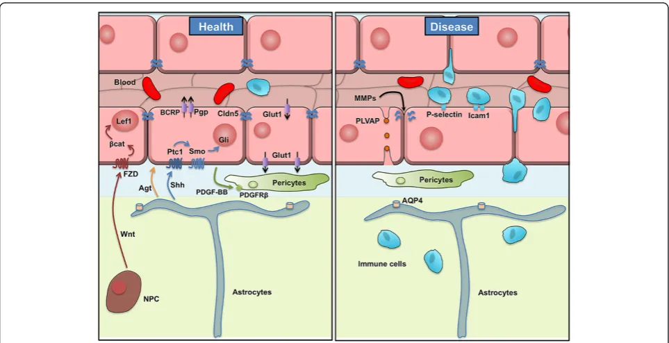
CNS ECs differ from ECs in non-neural tissues in that they are held together by TJs which greatly restrict the paracellular movement of molecules and ions between the blood and brain. Most of the TJ proteins have been identified by work on epithelial cells, which has demon-strated that TJs are formed by a series of transmembrane proteins, including the claudins [4,5], occludin [6] and junctional adhesion molecules (JAMS) [7] which are linked to the cytoskeleton and adherens junctions by adaptor molecules including ZO-1, ZO-2, Cingulin and others. In particular, claudins are a family of >20 tetra-spanin genes in mammals and expression of specific claudin family members in different cellular barriers is thought to be important for the specific paracellular physiology of the barrier [8]. Claudin 5 has been identi-fied as a major constituent of the TJs of CNS ECs (Figure 2). Nitta and colleagues have generated Cldn5 knockout mice [9]. These mice die at birth, and embryos have been shown to have a size selective leakiness of the BBB, with leaks to small molecules (up to 800 Da) but
not large molecules (serum albumin, 68 kDa and micro-peroxidase, 1.9 kDa). The BBB TJs look ultrastructurally normal in the absence of claudin 5 suggesting that other TJ proteins are sufficient to form the structural junc-tions. In fact, claudin 3 and 12 have been identified as being expressed by CNS ECs [10,11]. TheCldn5 knock-out mouse strain is a complete knockknock-out and thus this mouse model cannot be utilized to study the cell au-tonomous action of claudin 5 in CNS ECs.
Pfeiffer and colleagues have developed tools utilizing the tTA/TRE system to ectopically express claudin 1 in ECs [12]. This group used a double transgenic model in which the tTA was expressed from the Tie2 pan-endothelial promoter, and claudin 1 was expressed from the TRE promoter, therefore claudin 1 would be ectopi-cally expressed in ECs if the mouse diet lacks doxycyc-line. They used this model to express claudin 1 in ECs during neuroinflammation in experimental autoimmune encephalomyelitis (EAE), a mouse model of multiple sclerosis (MS) [13]. During EAE there is a breakdown of the BBB which allows the entry of immune cells and molecules into the CNS which attack the CNS myelin causing damage to the CNS. This group showed that ec-topic expression of claudin 1 seals the BBB during this disease and lessens the symptoms of EAE.
Occludin is a tetraspanin found at TJs in all epithe-lial cells, and has been identified as being expressed
PLVAP1 by CNS ECs [6,14]. Saitou and colleagues have generated
Ocln knockout mice, which are viable but the males are infertile [15]. The TJs in the epithelial cells and CNS ECs appear ultrastructurally normal in the Ocln knockout mice, and the measurements of the electrical resistance of the intestinal epithelial cells is also unperturbed, suggest-ing that TJs form a functional barrier in the absence of occludin. Interestingly, theOclnknockout mice have calci-fication of the brain suggesting that there could be specific defects in the regulation of the paracellular movement of calcium.
Transcytosis
Transcytosis is the process by which a vesicle is trafficked through the cell from one surface to the other and it can be accomplished through: a receptor-mediated mechanism by specific binding from a ligand to its receptor, by a non specific uptake called pinocytosis, or an adsorptive-mediated mechanism initiated by electrostatic forces between the negatively charged ECs membrane and posi-tively charged proteins. CNS ECs undergo extremely low rates of transcytosis compared with ECs in non-neural tis-sues, which greatly limits the transcellular movements of hydrophilic molecules between the blood and the brain. An increase in the number of transcytotic vesicles in CNS ECs has been observed in several diseases in which there is breakdown of the BBB [16-18]. The vesicle-mediated transport is primarily mediated through caveolin based vesicles ([19] for review). Several groups have madeCav1 knockout mice, including a caveolin-1 conditional lox-p flanked allele, however the complex phenotype in the mice throughout the vascular network makes it very difficult to study the role of caveolin-1 specifically at the BBB [20-25]. Plasmalemmal vesicle-associated protein-1 (PLVAP) is a transmembrane protein associated with the caveolae of fenestrated microvascular ECs [16]. In rodents, PLVAP ex-pression is enriched in non-CNS ECs compared to CNS ECs [26]. Interestingly, during diseases like ischemia/
stroke, acute ischemia, tumors or diabetic retinopathy, PLVAP1 is upregulated in CNS ECs (Figure 2) [18,27,28].
both knockout and conditional alleles, however mutant mice have not yet been described.
Efflux transport
CNS ECs express efflux transporters to eliminate poten-tial toxins from the CNS. These include members of the ATP-binding cassette (ABC) transporters, which utilize the hydrolysis of ATP to transport a wide variety of sub-strate molecules against their concentration gradient. In particular, CNS ECs express P-glycoprotein (Pgp/Mdr1/ Abcb1) and the breast cancer resistance protein (Bcrp/ Abcg2) (Figure 2), each of which has diverse but poten-tially overlapping substrate specificity [29-31]. The mouse genome contains two Pgp genes: Abcb1a and Abcb1b. Several mouse lines are available for studying Abcb1a, including targeted gene disruption (Abcb1atm1bor), a Cre/ lox regulated luciferase targeted into the Abcb1a locus (Abcb1atm1Kane) and a spontaneous mutation (Abcb1amds) that has a long terminal repeat of the ecotropic murine leukemia virus inserted into an intron [32-34]. Mice homozygous for Abcb1atm1bor allele have a BBB that is more permeable to specific molecules including different xenobiotics and drugs [32]. Because Pgp has 2 isoforms, Doran and colleagues generated a double knockout mouse ofAbcb1aandAbcb1bto study drug delivery [35]. Finally, as Pgp and Bcrp can transport some of the same substrates, theAbcb1a/Abcb1b/Bcrptriple knockout mouse was genet-ically engineered [36] and found to have a leaky BBB for many lipophilic xenobiotics, including rhodamine123, com-pared to their wild type (WT) littermates (Figure 3). These mice are very useful to study brain neuroprotection and neurotoxicity [37]. However one has to keep in mind that in these widely-used knockout strains, the efflux transpor-ters are deleted in every cell throughout the body, and not specifically in CNS ECs.
Figure 3Abcb1a/Abcb1b/Bcrptriple knockout mice have a leaky BBB to Rhodamine123.Adult wild type (C) orAbcb1a/Abcb1b/Bcrptriple knockout mice (3KO) were given an intravenous injection of Rhodamine123 (10 ng). After 1 hour the blood compartment was removed by transcardiac perfusion with PBS, the brains were removed, hemisected down the midline and imaged for Rhodamine123 fluorescence with a Fujifilm imager LAS 4000. Figure 3 represents an image of the hemisected brains with the cerebellum at the top of the image and frontal cortex towards the bottom of the image. More Rhodamine123 (dark color) was observed inAbcb1a/Abcb1b/Bcrptriple knockout mice compared with littermate controls. To demonstrate the consistency of the phenotype between different animals, 3 wild-type and 3Abcb1a/Abcb1b/Bcrptriple knockout mice brains were utilized.
Influx transport
CNS ECs express a series of solute transporters that transport specific nutrients into the brain including: glu-cose (GLUT1/Slc2a1), lactate (MCT1/Slc16a1), amino acids (Slc7a1, Slc7a5) and others (for review see [38]). Mouse null knockout models have been generated for a number of different transporters, however these often have phenotypes throughout the organism, as diverse cell types often require transport of these nutrients. For instance, Slc7a1 knockout mice die in the neonatal period with severe anemia [39], whereas Slc7a5 knock-out mice display embryonic lethality [40]. Therefore, for many of these solute carriers it may be critical to de-velop conditional alleles to specifically study their func-tion at the BBB.
GLUT1 has been largely studied for its role in deliv-ery of glucose to the CNS (Figure 2). Glucose is the primary source of energy for the brain, and human
GLUT1 deficiency results in an epileptic syndrome. A
Glut1 knockout allele has been generated with targeted disruption of the promoter and exon1 of the gene [41]. Mice homozygous for this allele die during embryogen-esis with pleiotropic phenotypes, whereas mice hetero-zygous for this Glut1 mutation display a 66% decrease of GLUT1 protein in the brain, and have similar symp-toms as the GLUT1 deficiency syndrome found in humans including epileptic events and impaired motor activity. A second group used a gene-trap method to disrupt the Glut1 locus [42]. For this gene-targeting model, the trapping vector contains a splice acceptor site along with the neomycin coding sequence and a polyade-nylation sequence, and thus when inserted into theGlut1 locus, the upstream Glut1 sequence is spliced to the trapped sequence forming a truncated mRNA. Following the polyadenylation sequence, the vector also contains a Bruton’s tyrosine kinase (btk) gene with a splice donor site, and thus a fusion mRNA is expressed with the Btk mRNA fused to the downstream Glut1 sequence. Mice homozy-gous for this gene trap demonstrated embryonic lethality, whereas heterozygous mice displayed no phenotype. The difference in phenotype observed between the Glut1 he-terozygotes generated from these two studies may be due to the differing affects of the targeting on gene expression, or compensation from other transporters such as the monocarboxylic acid transporters MCT1 and MCT2. A lox-p flanked conditionalGlut1allele has now been gener-ated that can be utilized to study GLUT1 in specific cell types, such as CNS endothelial cells [43]. In addition, Hei-lig and colleagues developed a transgenic mouse line in which the expression of antisense-GLUT1 sequence was driven from theb-actinpromoter in order to knockdown the glucose transporter during development [44]. Hemizy-gous or homozyHemizy-gous embryos for this transgene display reduced glucose uptake.
Leukocyte adhesion
The healthy CNS has an extremely low level of immune surveillance with the almost complete absence of subsets of leukocytes including neutrophils, T-cells and B-cells, however changes to the BBB during diseases including multiple sclerosis (MS), stroke, and neurodegenerative disorders can allow entry of immune cells into the CNS which is an important component of the pathogenesis of these diseases. Entry of immune cells into a tissue is a multi-step process that involves the binding of a series of adhesion molecules expressed on the immune cells to adhesion molecules on the post-capillary venule ECs [45]. This process involves tethering of the leukocyte to the endothelium, rolling along the endothelium, activation of the leukocyte, firm adhesion to the endothelium, and transmigration between or through the ECs. Several adhe-sion molecules on the endothelium have been identified, including P-Selectin and E-Selectin for rolling adhesion, and Icam1 and Vcam1 for firm adhesion (Figure 2). The expression of these adhesion molecules is low in healthy CNS ECs, but can be dramatically upregulated during in-jury and disease.
Several genetic mouse models have been developed to study leukocyte adhesion and transmigration in different models for CNS diseases. This review focuses on the dif-ferent genetic manipulations of the BBB, however here we briefly introduce a few of the disease models in which BBB dysfunction is commonly analyzed. MS is an inflammatory demyelinating disease of the CNS with numerous neuro-logical symptoms that can lead to physical and/or cogni-tive disability. The most common animal model of MS used to study its immune and inflammatory components is called experimental autoimmune encephalomyelitis (EAE). The disease is induced by immunization of a mye-lin peptide (such as MOG) emulsified in an adjuvant that will induce the inflammatory response, with pertussis toxin often used to facilitate the induction of EAE. A stroke is a loss of function of brain cells caused by an al-teration of blood flow, most of the time in a cerebral ar-tery, that limits the supply of oxygen and glucose to specific regions of the CNS (ischemia). Many studies fo-cussing on ischemic stroke use the middle cerebral artery occlusion (MCAO) model. In this model the middle cere-bral artery is ligated for minutes or hours which then may or may not be followed by a release of the ligation leading to reperfusion of the injured tissue. In addition, mouse models for different neurodegenerative diseases with BBB dysfunction are often utilized including amyotrophic lat-eral sclerosis, Alzheimer’s disease and Parkinson’s disease.
In an epilepsy model, PSGL-1 knockout mice displayed attenuated seizures suggesting that leukocyte CNS infiltra-tion was an important component of the disease [47]. On the other hand P-selectin knockout or PSGL-1 knockout did not affect the disease severity in several different EAE models [48-50]. E-selectin knockout mice have also been generated and have defects in neutrophil infiltration in dif-ferent tissues that are exacerbated when combined withP -selectin knockout mice [51], but in an EAE model defi-ciency in E-selectindoes not affect the progression of the disease [50]. However E-selectinknockout mice have not been extensively utilized to study neurological disease.
Firm adhesion is mediated through binding of CD11/ CD18 andαVβ1 integrins on leukocytes to EC Icam1 and Vcam1, respectively. Icam1 is an adhesion molecule com-posed of repeating immunoglobulin-like domains, and several different gene disruption strategies have been engi-neered to develop Icam1 knockout mice including the Icam1tm1Jcgr allele which disrupts exon 4 (Ig domain 3) [52], theIcam1tm1Bayallele which disrupts exon 5 (Ig do-main 4) [53], orIcam1tm1Alballele which deletes the entire coding sequence [54]. Whereas the Icam1tm1Alb allele lacks all Icam1 isoforms, theIcam1tm1JcgrandIcam1tm1Bay alleles each produce specific alternatively spliced Icam1 isoforms. Therefore comparing the phenotype of each knockout mouse can identify roles for different Icam1 iso-forms. Interestingly, during EAE,Icam1null mice (harbor-ingIcam1tm1Alballeles) have attenuated disease symptoms [55], whereas mice with Icam1tm1Bay alleles had worse EAE symptoms [55,56]. These data suggest that specific isoforms of Icam1 may have differing functions in regulat-ing neuroinflammation. Furthermore, Huet al. used adop-tive transfer of encephalitogenic T-cells from wild type to mutant strain or vice versa to determine the cell autono-mous function of these isoforms [55]. Vcam1 knockout mice have also been generated and die during embryogen-esis [57], however several lox-p flanked alleles have been generated in order to study the postnatal and cell specific function of Vcam1 [58-61].
Several adhesion molecules have been identified that regulate the adhesion of specific subsets of leukocytes to the endothelium. For instance, Th17 cells express MCAM which binds to laminin 411 on the EC basement membrane [62], CD4+ lymphocytes express CD6 which binds to EC ALCAM [63], and ninjurin-1 on myeloid cells homotypi-cally interacts with ninjurin-1 on inflamed ECs [64]. Mouse knockouts for Alcam [65] and Mcam (Mcamtm1Lex) have been generated, however these knockout models have not been extensively utilized to study neurological disease.
Matrix metalloproteinases
Matrix metalloproteinases (MMPs) are secreted zinc-dependent endopeptidases that can degrade components of the extracellular matrix. Twenty-eight MMP family
members have been reported so far, but in particular MMP2, MMP9 and MMP12 have been suggested to play a key role during CNS disease by disrupting the BBB. By deleting Mmp2 or Mmp9, researchers have found that the mice were protected after ischemia/reperfusion with attenuated inflammation of the brain [66-68]. Recently
Mmp12 deficient mice have been studied during
Thei-ler’s murine encephalomyelitis (TME), a virus-induced model of MS [69], and lack of MMP12 produced a re-duction in macrophage infiltration and demyelination with an intact BBB (Figure 2). It will be interesting to define the role played by each MMP during different neurological disorders.
CNS angiogenesis and BBB development
BBB development involves the complex interaction of CNS cells with different neural and immune cells. The process of BBB regulation begins with induction signals as ECs invade the CNS during development and continues with maintenance signals throughout life and ageing. Here we discuss selected genetic models that have been used to dissect this process including manipulating pathways that affect angiogenesis (VEGF, Notch), CNS-specific angio-genesis (Wnt/β-catenin, Gpr124), BBB maintenance (Shh, Agt) and BBB ageing (ApoE) (Figure 2).
Notch: Notch signaling is an evolutionarily conserved mechanism that is best known for its function in cell-fate decision in various tissues [80]. In mammals four Notch receptors and five ligands have been identified with diverse expression patterns [81]. In mouse embryos Notch1 and Notch4 are predominantly expressed on the arterial endothelium. When the Notch1 gene is inacti-vated specifically in the endothelium, mutant embryos die at embryonic day E10.5 with normal vasculogenesis but important defects of angiogenesis [82]. Notch4 -deficient mice exhibit normal development without any vascular abnormality [83]. However, the double Notch1/
Notch4 mutant mice have vascular defects more severe
than the single Notch1 mutant suggesting overlapping functions of both receptors during development [83-85]. By using the Tie2-tTa system coupled with the TRE-caNotch4, a constitutively active Notch4 mutant was specifically expressed in the endothelium of postnatal mice [27]. These mice show abnormal connections be-tween arteries and veins associated with ectopic expres-sion of the arterial marker ephrin B2 in veins. Activation of constitutively active Notch4 in the blood vessels of the developing mouse brain induces vessel enlargement followed by hemorrhages in the cerebellum and the neo-cortex, neurological damage and death [86].
Four of the 5 known Notch ligands (Delta-like 4 (Dll4), Dll1, Jagged1 and Jagged2) are specifically localized in ar-terial but not in the venous endothelium [81,83,87]. During early vascular development, Dll4 share the same expression pattern as Notch1 and Notch4 [83]. Homozygous and het-erozygous gene inactivation of Dll4 leads to embryonic lethality in several mouse strains between embryonic days E9.5 and E10.5 due to severe vascular deffects [88,89]. However, in the outbred ICR strain, the heterozygous mu-tation leads to limited embryonic lethality [88,89]. There-fore, by usingDll4+/−mice retinas in the ICR strain, it was shown that absence of oneDll4allele leads to an increase of endothelial tip cells that sense and respond to guidance cues during angiogenesis [90-92]. Moreover, Hellstromet al. demonstrated a similar phenotype whenNotch1 gene was inactivated specifically in ECs, suggesting that during angiogenesis, signaling through Dll4/Notch1 is responsible for the regulation of endothelial tip cell formation [90] in response to VEGF [90-92]. Although Dll1 is not involved in arterial cell fate, it has recently been shown to be required for maintaining arterial identity by using a trans-genic mouse line that inducibly deletes Dll1 in endothelial cells [93].
Wnt/ β-catenin: Several groups have demonstrated that Wnt/β-catenin signaling is specifically activated in CNS ECs during development and is required for angiogen-esis into the CNS as well as development of the BBB [94-96]. Wnts are secreted ligands that bind to Frizzled
receptors at the cell surface, which leads to inactivation of a protein complex that degrades β-catenin. Stabilized
β-catenin is then able to translocate to the nucleus and activate transcription along with Lef1/Tcf complexes [97]. A number of different genetic mouse models have been used to analyze different aspects of Wnt/beta-catenin signaling at the BBB [94,96]. Several transgenic Wnt re-porter mouse lines have been generated that have cDNA encoding a reporter protein (LacZ, GFP) downstream of Wnt responsive DNA elements such as TCF binding sites (for review see [98]). These Wnt reporter mice, including TOP-Gal, BAT-Gal and TOP-Flash, have been used to identify Wnt activity in CNS ECs (for review see [98]).
A number of mouse models have been developed to tar-get β-catenin activity. Several groups have developed endothelial-specificβ-catenin knockout mouse lines using Tie2-Cre and β-catenin lox-p flanked alleles [94,96,99]. This model has demonstrated that endothelialβ-catenin is required for angiogenesis into the CNS [94,96,99], and for the expression of BBB-specific transporters such as GLUT1 [94-96]. There are several caveats to this approach of inhi-biting Wnt signaling. First, Tie2-Cre/β-catenin mutants die during early embryogenesis, and so while they have been effective for studying early angiogenic events, they have been less successful for studying BBB maintenance. To ad-dress this concern, Liebner and colleagues utilized a Pdgfb-CreERT2 allele to delete lox-p flankedβ-cateninalleles in endothelial cells at postnatal ages, to demonstrate that
β-catenin was required for sealing off the BBB [95]. Second, Tie2-Cre is also active in hematopoietic lineage cells [100], so each time this line is used one has to be sure that phenotypes are not due to changes in blood cells. Third, β-catenin is not only required for transduction of canonical Wnt signaling, but is also a component of the adherens junctions, and thus it is difficult to gene-rate conclusions specifically about Wnt signaling from
β-catenin mutants. In addition to conditional knockout strategies, transgenics have been used to generate gain of functionβ-catenin mouse alleles by generating a transgenic β-cateninwith exon3 flanked by lox-p sites, and thus when exon3 is removed the mutant β-catenin is constitutively active. Using Pdgfb-CreERT2/β-catenin loxp-exon3-loxp mice, Liebner and colleagues were able to activate
β-catenin in the embryo and observe precocious BBB mat-uration [95].
Several genetic models have been utilized to inhibit other aspects of Wnt signaling, including analysis of
Wnt7a/Wnt7b double knockout mice as these are the
line with null alleles of Wnt7aand loxp flanked Wnt7b alleles in conjunction with a Nestin-Cre to deleteWnt7b in the developing neuroepithelium [96]. These mice lived longer than the complete double knockout of Wnt7a/ Wnt7bmice, and thus gave vital information about the role of Wnts in regulating CNS vessel development. In addition, there are many different positive (Wnt,β-catenin, Tcf) and negative (Axin2, Apcdd1, APC, Dkk, sFRP) regu-lators of Wnt signaling, and mouse knockout and over-expression alleles have been generated for many of these (reviewed [97]). Recently, Tam et al. showed the critical role of both TROY and DR6 for CNS angiogenesis as downstream target genes of the Wnt/β-catenin signaling [101]. They showed thatDR6mutant mice display a lower density of brain vasculature and a leaky BBB for Evan’s blue dye, with a lower amount of ZO1 protein in adult mice. In mouse embryos they observed hemorrhages in the fore-brain with a leaky BBB for sulfo-NHS-biotin, coupled with a lower vascular density in the hindbrain. To determine the endothelial specificity of these phenotypes, Tam et al. generated a mouse with Tie2-Cre and exon2 DR6 lox-p flanked alleles, and described similar phenotypes to full knockout mice suggesting that DR6 expression is required specifically in endothelial cells.Troyknockout mice display a mild leakage of the BBB for Evan’s blue.
Gpr124: Recently, several groups have generated mouse knockouts for Gpr124, which displayed a disruption of angiogenesis in the forebrain and ventral spinal cord with localized malformations and hemorrhages, demonstrating that this G-protein coupled receptor was required for CNS-specific angiogenesis [102-104]. Using mice with lox-p flanked conditional alleles and Tie2-Cre transgenes, it was demonstrated that Gpr124 function is specifically required in the ECs [102,104]. Interestingly, the phenotype looks similar to that observed in theWnt7a/Wnt7bdouble knockout mice, however it remains unclear whether Gpr124 and Wnt signaling are connected.
Hedgehog: The Hedgehog (Hh) family, first characterized in Drosophila [105], are secreted morphogens [106] that play a major role in development including neur-onal guidance and angiogenesis [107,108]. Three mem-bers of the Hh family have been identified in mice: Sonic Hedgehog (Shh), Desert hedgehog (Dhh) and Indian hedgehog (Ihh). Shh acts by binding to Patched, which leads to de-repression of Smoothened (Smo) which acti-vates genes through the transcription factor Gli [106]. Chiang and colleagues have generated a knockout mouse model forShh[109].Shhmutant mice display embryonic lethality with embryos having abnormal anatomy in sev-eral parts of the body including the brain and spinal cords. In the CNS, when Shh is overexpressed in the dorsal neural tube of embryos, Shh transgenic mice display a
hypervascularization [110]. Alvarez and colleagues described how the Hh pathway contributes to the main-tenance of BBB functions [111]. They showed that E13.5 embryos of theShhknockout mice display a lower amount of junctional proteins in the brain capillaries than their WT littermates. To study the role of the Shh pathway spe-cifically in ECs, they generated endothelial-specific Smo knockout mice by using aTie2-Creallele and aSmolox-p allele. The BBB of the mutant mice is permeable to serum proteins, like fibrinogen, apolipoprotein B and immuno-glubulins in E14 embryos and P19 mice and the BBB of adult mice is permeable to exogenous compounds. The BBB leakiness was explained by a significant decrease of several TJ proteins including claudin 3, claudin 5, occludin and ZO1 and a fragmented basement membrane. More-over, Alvarezet al. demonstrated that Shh plays a key role in the regulation of the pro-inflammatory response during EAE. Altogether, these data suggest two major roles of the Hh pathway by regulating BBB function and protecting the brain from inflammation.
Renin-angiotensin: In the brain, the renin-angiotensin system controls cerebral blood flow, memory and BBB function (for review see [112]). Astrocytes express angio-tensinogen (Agt), a precursor of angiotensins I-IV (Ang). In a mouse model deficient for Agt, Kaninuma and col-leagues demonstrated that two weeks after a brain cold injury, the knockout mice still display a leaky BBB com-pared to their WT littermates whose BBB was repaired [113]. This phenotype was less critical when AngII or AngIV was given to the Agt deficient mice suggesting their critical involvement in vascular repair after an in-jury. Moreover, the Agt mutant mice have a leaky BBB for endogenous serum plasminogen and albumin and express less occludin at the EC TJs [114].
Targeting pericyte function
Pericytes are mural cells that incompletely surround the abluminal surface of the capillary endothelium (Figure 2). These cells are derived from the neural crest and regulate angiogenesis, vascular remodeling, leukocyte trafficking and the formation and function of the BBB [119-121]. The binding of the ligand platelet-derived growth factor-BB (PDGF-BB) to the platelet-derived growth factor receptor
β (PDGFRβ) is required for the generation and recruit-ment of pericytes to CNS vessels asPdgfbknockout mice andPdgfrbknockout mice completely lack CNS pericytes [122,123]. These mice have altered vascular patterning, dilations in the microvasculature, and form micro-aneurysms that occasionally hemorrhage. The ability to study the role of pericytes in BBB function is limited in
both Pdgfb knockout and Pdgfrbknockout mice as they
die shortly after birth, however thePdgfrbknockout mice have been utilized to demonstrate that pericytes are required for BBB formation during embryogenesis and that they regulate the BBB by inhibiting the expression of EC genes that would make the vessels leaky [119,120].
Several groups have developed genetic models which decrease PDGFBB signaling through PDGFRβ without completely abolishing it. Tallquist and colleagues have generated a series of hypomorphic alleles of Pdgfrb in which different numbers of tyrosine residues, which are normally auto-phosphorylated upon ligand binding, are mutated to phenylalanine residues [124]. Using different combinations of these hypomorphic alleles, they were able to generate mice with different numbers of peri-cytes. These mice have been used to demonstrate that the relative number of pericytes is important for the perme-ability of the BBB during development [120]. Furthermore, Bell and colleagues used this model to demonstrate that during ageing there was reduction in capillary perfusion and BBB breakdown that led to neural degeneration [125]. Interestingly, whereas there are BBB defects in this model during development and ageing, the BBB appears some-what normal during adulthood. Several genetic models have also targeted the ligand to attenuate PDGFB signal-ing. Lindblom and colleagues developed mice in which the retention motif of PDGFB was deleted, such that PDGFB binding to extracellular matrix heparan sulphate proteo-glycans was disrupted, and mice homozygous for this al-lele had 26% of the pericyte coverage of WT mice [126]. In addition, Armulik and colleagues generated mice that had lox-stop-lox human PDGFB transgene at the ROSA locus, and thus could ectopically express human PDGFB in ECs by using a Tie2-Cre mouse line [119]. Using a
Pdgfbnull knockout mouse as a background, they could
express one or two alleles of the humanPDGFBand thus generate mice with attenuated signaling that had 40% and 72% the number of pericytes of WT mice. Using these lines, Armulik et al. demonstrated that pericytes were
required for BBB function in adults, and did so by inhibiting the rates of transcytosis [119]. One interesting point is that there is a slight difference in the phenotype of the mice when signaling is attenuated by targetingPdgfb orPdgfrb. ThePdgfrbhypomorphic mice have a leaky BBB during de-velopment and ageing but relatively normal BBB as adults, whereas the models attenuatingPdgfbhave a leaky BBB as adults. Several reasons could lead to these differences in-cluding: strain of mice, environment of mice, total numbers of pericytes, signaling of PDGFB through multiple recep-tors, or localization of signals.
Goritz and colleagues utilized a specific GLAST-CreER/ RosaYFP line to fluorescently label a specific subtype of pericytes, which they termed type A pericytes [127]. To accomplish this, they utilized a mouse in which a lox-stop-lox YFP cassette was introduced into theROSAlocus by homologous recombination, and thus the YFP reporter would be expressed in cells following the Cre-recombinase mediated excision of the stop cassette. Using the GLAST-CreER line, they demonstrated that upon injection of tam-oxifen in adults, the YFP reporter was expressed in the spinal cord in a subset of pericytes. They then demon-strated that following a spinal cord injury these type A pericytes migrated to the site of injury and formed the scar tissue. This group also used a Glast-CreER/RASless mouse line to inhibit cell division of the type A pericytes in the spinal cord injury model [127]. Rasless mice have null alleles forH-RasandN-Rasand haveK-Rasalleles flanked by lox-p sites. The mice are generally normal, except cells lack the ability to divide if Cre-recombinase mediated mu-tation of K-Ras occurs. Using the Glast-CreER/RASless mouse line coupled with tamoxifen injections in the adult, they were able to generate mice in which type A pericytes developed normally (as the CreER only excises the condi-tional allele upon tamoxifen injection in the adult), but failed to divide in the adult following a spinal cord injury model. This group showed that division of type A pericytes is required for scar formation following spinal cord injury.
In addition Li and colleagues manipulated TGF-β sig-naling in ECs to generate a mouse model that had defi-cits in endothelial-pericyte interactions [128]. This group generated a CNS endothelial conditional mutant of
Smad4, a downstream mediator of TGF-β signaling, by
utilizing lox-p flanked Smad4 alleles and a SP-A-Cre mouse line. They demonstrated that disruption ofSmad4 in CNS ECs led to a mouse with defective pericyte cover-age, intracranial hemorrhage and BBB breakdown.
Targeting astrocyte function
approximately 100,000 synapses and ensheaths one or two capillaries [129,130]. Astrocytes play an important role in regulating neuronal metabolic homeostasis, syn-apse formation, neurotransmitter processing, as well as coupling neuronal function with cerebral blood flow (for review see [131]). Transplantation studies and in vitro studies have suggested that astrocytes are important regu-lators of BBB function. When isolated from the brain, ECs lose their BBB properties as shown by a decrease in their trans-endothelial electrical resistance (TEER) [132]. When co-cultured with astrocytes or astrocyte-conditioned media their TEER increases significantly, suggesting that astrocyte-secreted factors are involved in activating the barrier properties of the BBB [132,133].
Several genetic models have been developed that ma-nipulate astrocyte function (reviewed by Pfrieger and Slezak 2012 [134]). To selectively ablate astrocytes, sev-eral groups have induced ectopic expression of the herpes simplex virus thymidine kinase (HSV-TK) in astrocytes under control of either the humanGFAP pro-moter or the murine Gfap promoter [135-137]. On its own HSV-TK does not affect cell viability. However, the enzyme converts ganciclovir into ganciclovir monopho-sphate, a nucleotide analogue which disrupts DNA repli-cation. Therefore, cell division can be inhibited by addition of ganciclovir to HSV-TK expressing cells [138]. Delaney and colleagues used ganciclovir to inhibit cell div-ision of GFAP-positive cells in neonatal GFAP-HSV-TK mice, and demonstrated that astrocyte reduction in new-born pups results in ataxia, neuronal excitotoxicity and a disorganization of Purkinje cells and radial glia [135].
Due to the wide scale effects of disrupting cell division in all astrocytes, Tsai and colleagues recently developed methods to deplete specific domains of astrocytes [139]. To accomplish this they generated a transgenic mouse line such that a lox-eGFP-stop-lox-Diptheria toxin-A (DTA) was expressed under the control of an astrocyte-specific Aldh1L1-promoter. In this mouse, eGFP is expressed in astrocytes, however following Cre mediated recombination of the lox-p sites, eGFP is no longer expressed, instead DTA is expressed which kills the cells. By mating this line with transgenic mouse lines in which Cre recombinase expression is driven from promoters that mark regionally-specific subsets of neural progeni-tors (Pax3-Cre, olig2-Cre). The authors were able to kill astrocytes in specific domains of the spinal cord by mating this line with transgenic mouse lines. For ex-ample, by mating the lox-eGFP-lox-DTA mice with the Pax3-Cre mice, the mutant line displayed variable peri-natal lethality rates with a lower number of astrocytes in the dorsal spinal cord, but without an increase in their BBB permeability.
To study the role of reactive astrocytes during CNS pathology, Sofroniews’group utilized the GFAP-HSV-TK
mice combined with ganciclovir treatment to ablate divid-ing reactive astrocytes durdivid-ing disease models (for review see [138]). This group showed that reactive astrocytes were required for inhibiting neurite outgrowth, regulating neur-onal survival and repairing the BBB following spinal cord injury [140]. During EAE, astrocytes form a scar that sur-rounds the blood vessels and mice with targeted ablation of proliferative astrocytes exhibit a much higher number of leukocyte infiltrations in the CNS parenchyma [141].
Aquaporin 4 (Aqp4) is a water channel protein mainly expressed in astrocyte endfeet that ensheath CNS blood vessels [142]. One function of Aqp4 is to facilitate water movement into and out of the brain. During a middle cerebral artery occlusion (MCAO), a mouse model of stroke, Aqp4 deficient mice have a decreased cytotoxic cerebral edema and therefore an improved neurological outcome [143]. Saadoun et al. showed that Aqp4 defi-cient mice have a morphologically and functionally nor-mal BBB [144]. Therefore it appears that Aqp4 plays a key role in brain swelling during pathology, but not in normal BBB architecture.
Imaging BBB function
Being able to visualize the movement of different cell populations in vivo in live mice is an important step in understanding how cells interact in physiological set-tings. This was made possible by the use of two-photon microscopy that allows brain imaging in living animals at a depth up to 1 mm. Several groups have utilized dif-ferent genetic methods to label cells and proteins for im-aging of CNS ECs and their interaction with the brain and immune cells. Transgenic mice with GFP expressed by the Tie2 promoter have been utilized to label ECs in vivo. This technique has been used for microscopy, and we also have been able to purify brain ECs from these mice using fluorescence-activated cell sorting (FACS) and performed microarray analysis of their gene expression [26]. In addition, different subpopulations of ECs can be labeled forin vivotime-lapse imaging. Murphy and collea-gues utilizedEphrin-B2-H2BGFP mice to visualize the nu-clei of arterial ECs to examine the dynamics of cells during formation and regression of arterial venous mal-formations [145]. This mouse has a transgene of his-tone-2Bfused to GFP that was inserted by homologous recombination into the first exon of theephrin-b2gene, and thus a nuclear GFP was expressed from theephrin -b2promoter [146].
either with GFP inserted into the Cx3cr1 locus to label microglia [148] or transgenic YFP-H line in which aYFP transgene is driven by the thy1 promoter and thus expresses YFP in a subset of neurons [149]. The IV tracer labeled the blood inside the vessels and thus enables visualization of the interaction of microglia with the vessels over time. In the brain, Rangroo Thrane et al. used this technique to visualize the movement of eGFP-microglia during hepatic encephalopathy, a neu-roinflammatory disease characterized by liver failure fol-lowed by an opening of the BBB [150]. Several groups have now utilized the microglia/macrophage reporter mice in which they express GFP from theCx3cr1locus and RFP from the Ccr2 locus, and thus have microglia labeled in green and macrophages labeled in red [151-153]. In addition different methods have been utilized to label astrocytes (see [134] for review) and pericytesin vivo[127]. To understand the interaction of auto-reactive T-cells with the BBB, an adoptive transfer model of EAE has been utilized with the injection of GFP-expressing MBP-reactive T-cells into mice [154,155]. This technique was utilized to examine the interaction of the T-cells with the vessels including arrest on the surface of the vessels, crawling against the blood flow, diapedesis and scanning of the abluminal surface for phagocytes [154]. Further-more, this technique demonstrated that prior to entry into the CNS, the T-cells go into lung lymphoid tissues and lymph nodes to be activated. After their activation, the T-cells go back to the blood stream and migrate to the CNS parenchyma to induce clinical symptoms [155].
Other imaging methods, such as magnetic resonance imaging (MRI), positron emission tomography (PET) or X-ray microtomography, can be used to image blood vessels and BBB function in wild type mice as well as transgenic animals.
Valuable tools available to study the BBB
A number of different tools have been developed in order to regulate gene expression in CNS ECs. To knockdown gene expression in ECs, several different models of the Cre/Lox system are available. One mouse line that has been generated is a Tie1-Cre [156]. Tie1 is a member of the Tie receptor family, and is essential for angiogenesis during embryogenesis. The Tie1 promoter drives gene expression in ECs from embryonic day E10 until birth but also in a small part of hematopoietic cells and within some neuronal populations in the cortex and hippocampus [156]. In parallel, Tie2-Cre mouse lines were generated which is to date the most commonly uti-lized line for gene excision in ECs [100]. TheTie2 pro-moter drives a similar expression pattern than Tie1, in all ECs with some hematopoietic cells, but it seems that it can start as early as embryonic day E8.5 [157]. When comparing both systems, some phenotypic differences
can be seen during embryogenesis and were explained by the expression delay of Tie1 compared with Tie2 [158]. Although extensively used to delete lox-p alleles in ECs, several caveats arise from analysis of Tie2-Cre mice. First, Tie2 is turned on in hematopoietic precur-sors, and thus although Tie2 is no longer expressed in many blood cells, the Cre irreversibly deletes the lox-p flanked alleles in the precursors. Therefore, when ana-lyzing phenotypes using conditional alleles in con-junction with Tie2-Cre, one must consider that the phenotype may arise from the function of the allele in ECs or hematopoietic lineage cells. Second, the Tie2-Cre can also excise lox-p flanked alleles in the female germ-line, and thus mating strategies must be used in which the Tie2-Cre with the lox-p flanked alleles are passed through male parents to ensure that a complete knock-out is not generated.
Two different Flk-1-Cre lines are available, one that shows Cre expression in both vasculature and muscle lineages [159] whereas the second one does not have the muscle expression but seems to have a weak expression in quiescent endothelium [160]. A PECAM (CD31)-Cre has been generated to drive expression in endothelium, but is not extensively characterized [61]. A VE-cadherin Cre has also been generated [161]. The major interest of this Cre is that the promoter drives expression during em-bryogenesis as well as adulthood. Nevertheless, a strong VE-cadherin-Cre-driven expression starts later during em-bryogenesis than the Tie2-Cre system, around embryonic day E14.5 [161]. Recently, VWF-Cre and SP-A-Cre lines have been developed suggesting specific expression of Cre in CNS ECs, however these newly-generated lines have not been exhaustively studied [128,162].
Several attempts have been made in generating tam-oxifen inducible Cre lines targeted specifically to ECs. A Tie2-CreERT2 transgenic mouse was genetically engi-neered [163] and shows a highly specific expression of lox-p flanked reporter transgenes in endothelial cells only when mice were treated with tamoxifen. In addition, two VE-cadherin-CreERT2 and a PDGFB-CreERT transgenic mouse lines were generated to express the tamoxifen-inducible CreERT(2) from EC promoters [164-167]. Several caveats have arisen with these systems. Firstly, whereas these transgenic alleles seem to efficiently ex-cise lox-p flanked alleles if tamoxifen is given to embry-onic or neonatal mice, the efficiency of recombination is often reduced during adulthood [166]. Furthermore, the timing of tamoxifen injections and the age of ana-lysis has to be carefully controlled to determine if blood cells are also targeted.
reversible, and thus by controlling the timing of doxycyc-line being fed to the transgenic mice it is possible to turn on and turn off the TRE driven transgenes. Additionally, Tie2-GFP mice have been utilized to visualize as well as purify ECs from the CNS [26,169].
To reduce the amount of pericyte coverage on the blood vessels, several groups have generated Pdgfb and Pdgfrβ deficient mice, as well as hypomorphic alleles of the ligand and receptor [119,126]. However, to date there are only a few mouse models to delete gene ex-pression in pericytes. The most common line is the Pdgfrb-Cre [170] but the receptor is expressed by several mesenchymal cell types. Recently, Feng et al. developed a Ng2-CreERT to inducibly knockdown gene expression in pericytes, but in the CNS Ng2 is also expressed in oligodendrocyte precursor cells [171].
To deplete the brain of astrocytes, GFAP-HSV-TK and diptheria toxin systems have been generated [138,139]. To delete genes in astrocytes, there are a high number of mouse Cre lines available by using either theGfap, Glast, Blbp, Gli, Nes, Cx30, CX43 or S100Bpromoters (for re-view see [134]). Genetics tools are also available to induci-bly knockout gene expression with the CreERT2 system.
Additionally, researchers have used non-genetic methods to knockdown genes at the BBB. One promising technique is the delivery of siRNA into brain ECs. By high pressure tail-vein injection of a claudin 5 siRNA or by infecting a brain region stereotactically with a virus that produces a claudin 5 shRNA, Campbell et al. found that it was pos-sible to knockdown Cldn5 gene expression in brain ECs and consequently open the BBB to some extent [172-174]. They recently demonstrated that knocking downCldn5at the mouse BBB leads to a decrease in cerebral edema after traumatic brain injury [175]. Other groups showed efficient delivery of exosome-associated siRNA [176] or nanoparti-cles coupled probes [177] to the CNS. Finally, it is also possible to use ultrashort pulsed laser [178] or ultrasound coupled with MRI to disturb the BBB and deliver mole-cules into the CNS [179].
Potential tools for analysis of the BBB
As the boom of mouse genetic analysis continues, we can forecast that the generation of many novel mouse lines in the upcoming years will continue to advance our under-standing of BBB function. Here we suggest a small num-ber of tools that will aid in our understanding of BBB function during health and disease.
For BBB TJs, several interesting questions remain un-answered. There are multiple claudin family members expressed in CNS ECs, however it is unclear whether each claudin has unique functions within the TJ or whether each member provides an additive effect on limiting the permeability of the vessels. Developing knockout mice for each claudin expressed by CNS ECs,
as well as double and triplecldnknockouts will allow us to address these questions. In addition, cldn5 knockout mice die shortly after birth and thus it is not clear what the role of claudin 5 in the adult is or whether cldn5 knockout mice die specifically due to lack of claudin 5 protein in CNS ECs or in other cells in the organism. Generation of a lox-p flanked cldn5 allele would allow for temporal and spatial control of claudin 5 deletion to an-swer these questions. Furthermore, the knockout models are irreversible, and therefore generation of transgenic doxycycline-regulated claudin 5 shRNA would develop a model in which TJ protein expression could be reversibly modulated. Moreover, additional TJ proteins, such as the lipolysis-stimulated lipoprotein receptor (LSR) and tricellu-lin, that have recently been discovered in epithelial cells, have also been found to be enriched in CNS ECs compared to endothelial cells in non-neural tissues [26]. These TJs are expressed at the epithelial tricellular junction, where three cells meet [180,181]. It would be interesting to create the endothelial specific knockout of these TJs to understand their role in BBB formation and/or maintenance.
Our understanding of many BBB transporters has re-lied on the analysis of knockout mice in which the trans-porter is deleted from every cell. For instance Abcb1a/
Abcb1b/BCRP triple knockout mice have been
exten-sively utilized to study the role of these genes in xeno-biotic protection. However, these genes are expressed in a variety of cell types in different tissues, and thus complete knockout of these transporters would globally change the localization of their substrates, and thus their specific function at the BBB may be obscured. Gener-ation of mice with lox-p flanked alleles of different transporters would allow for analysis of their function specifically at the BBB.
In this review, we described several Cre lines that tar-get ECs, including Tie2-Cre, VE-Cadherin-CreERT2, PDGFB-CreERT2, and others. Many of these Cre alleles target all ECs, and therefore it would be helpful to gen-erate Cre alleles andCreERTalleles that specifically tar-get CNS ECs. To do so, one could take advantage of the split-Cre system. In this system, the Cre is fragmented into two non-functional fragments and becomes active only when expressed in the same cells. Therefore, by using two different promoters, one can drive Cre expres-sion (or CreERT2) in a very specific cell type [182,183]. For instance splitting the Cre to be driven by VE-
cad-herin and Glut1 promoters would potentially allow for
Several genetic tools have been utilized to eliminate pericytes by affecting PDGFBB/PDGFRβsignaling by gen-erating knockout or hypomorphic alleles of pdgfb or pdgfrb. These knockout models affect pericytes during de-velopment, and thus it is not clear what the effect of acute pericyte loss in adults would be. Therefore, developing a genetic model to target pericytes in adults would allow us to further understand the role of pericyte-EC interactions. For instance, generating aPdgfrb-rtTA allele coupled with a TRE-DTA allele would allow for doxycycline inducible ablation of Pdgfrb-positive cells. In this case doxycycline could be delivered systemically to target all PDGFRβ posi-tive cells, or locally, for instance in the retina to specifically target retinal PDGFRβ positive pericytes, perhaps to mimic the loss of retinal pericytes during diabetic retinop-athy. In addition, generating Cre/CreERT alleles that target specific subsets of pericytes would allow for the study of the relative contribution of each subtype of pericytes. Gortizet al. identified that there were at least two types of pericytes, termed type A and type B pericytes [127], and were able to genetically mark type A pericytes with a GLAST-CreERT allele, however no such line has been developed to target type B pericytes. This might be more difficult as the molecular signature of different pericyte subtypes have not been described. The same can be said for astrocytes for which the complex heterogeneity among different subclasses of astrocytes is beginning to be under-stood. Developing Cre-lines that target specific functional subsets may be important for understanding the regional specificity of BBB regulation.
Conclusions
The generation of mouse genetic models that target BBB structure and function have allowed us to gain a great deal of knowledge about this important physiological structure. These include models that target specific cells including ECs, pericytes and astrocytes, as well as identi-fying specific genes that are important for BBB forma-tion and funcforma-tion. As the boom in mouse genetics continues we can expect the generation of many more genetic models that will continue to aid in the advance-ment of our understanding of the BBB.
Additional file
Additional file 1: Supplementary Table 1.Genetic mouse models to study the BBB.
Abbreviations
ABC: ATP-binding cassette; AD: Alzheimer’s disease; Agt: Angiotensinogen; Ang: Angiotensin; ApoE: Apolipoprotein E; Aqp4: Aquaporin 4; BBB: Blood– brain Barrier; Bcrp: Breast cancer resistance protein; Btk: Bruton’s tyrosine kinase; CNS: Central nervous system; DTA: Diptheria toxin A;
EAE: Experimental autoimmune encephalomyelitis; EC: Endothelial cell;
Gal: Galactosidase; GFAP: Glial fibrillary acidic protein; GFP: Green fluorescent protein; HSV-TK: Herpes simplex virus thymidine kinase; MBP: Myelin basic protein; MCAO: Middle cerebral artery occlusion; MMP: Matrix
metalloproteinase; MS: Multiple sclerosis; NPC: Neural precursors cell; PD: Parkinson’s disease; PIGF: Placental growth factor; Pgp: P-glycoprotein; rtTA: Reverse tetracycline transactivator; SHh: Sonic hedgehog; TJ: Tight junction; TRE: Tetracycline response element; tTA: Tetracycline transactivator; VEGF: Vascular endothelial cell growth factor; YFP: Yellow fluorescent protein.
Competing interests
The authors declare that they have no competing interests.
Authors’contribution
FS and RD wrote the manuscript. All authors have read and approved the final version of the manuscript.
Acknowledgement
RD is funded by the Program for Breakthrough Biomedical Research and the American Heart Association.
Received: 7 September 2012 Accepted: 20 November 2012 Published: 10 January 2013
References
1. Feil R, Wagner J, Metzger D, Chambon P:Regulation of Cre recombinase activity by mutated estrogen receptor ligand-binding domains.Biochem Biophys Res Commun1997,237:752–757.
2. Buch T, Heppner FL, Tertilt C, Heinen TJ, Kremer M, Wunderlich FT, Jung S, Waisman A:A Cre-inducible diphtheria toxin receptor mediates cell lineage ablation after toxin administration.Nat Methods2005,2:419–426. 3. Zambrowicz BP, Imamoto A, Fiering S, Herzenberg LA, Kerr WG, Soriano P:
Disruption of overlapping transcripts in the ROSA beta geo 26 gene trap strain leads to widespread expression of beta-galactosidase in mouse embryos and hematopoietic cells.Proc Natl Acad Sci U S A1997,94:3789–3794. 4. Furuse M, Sasaki H, Fujimoto K, Tsukita S:A single gene product, claudin-1
or−2, reconstitutes tight junction strands and recruits occludin in fibroblasts.J Cell Biol1998,143:391–401.
5. Morita K, Furuse M, Fujimoto K, Tsukita S:Claudin multigene family encoding four-transmembrane domain protein components of tight junction strands.Proc Natl Acad Sci U S A1999,96:511–516.
6. Furuse M, Hirase T, Itoh M, Nagafuchi A, Yonemura S, Tsukita S, Tsukita S: Occludin: a novel integral membrane protein localizing at tight junctions.J Cell Biol1993,123:1777–1788.
7. Martin-Padura I, Lostaglio S, Schneemann M, Williams L, Romano M, Fruscella P, Panzeri C, Stoppacciaro A, Ruco L, Villa A,et al:Junctional adhesion molecule, a novel member of the immunoglobulin superfamily that distributes at intercellular junctions and modulates monocyte transmigration.J Cell Biol1998,142:117–127.
8. Furuse M:Molecular basis of the core structure of tight junctions.
Cold Spring Harb Perspect Biol2010,2:a002907.
9. Nitta T, Hata M, Gotoh S, Seo Y, Sasaki H, Hashimoto N, Furuse M, Tsukita S: Size-selective loosening of the blood–brain barrier in claudin-5-deficient mice.J Cell Biol2003,161:653–660.
10. Wolburg H, Wolburg-Buchholz K, Kraus J, Rascher-Eggstein G, Liebner S, Hamm S, Duffner F, Grote EH, Risau W, Engelhardt B:Localization of claudin-3 in tight junctions of the blood–brain barrier is selectively lost during experimental autoimmune encephalomyelitis and human glioblastoma multiforme.Acta Neuropathol2003,105:586–592. 11. Belanger M, Asashima T, Ohtsuki S, Yamaguchi H, Ito S, Terasaki T:
Hyperammonemia induces transport of taurine and creatine and suppresses claudin-12 gene expression in brain capillary endothelial cells in vitro.Neurochem Int2007,50:95–101.
12. Pfeiffer F, Schafer J, Lyck R, Makrides V, Brunner S, Schaeren-Wiemers N, Deutsch U, Engelhardt B:Claudin-1 induced sealing of blood–brain barrier tight junctions ameliorates chronic experimental autoimmune encephalomyelitis.Acta Neuropathol2011,122:601–614.
13. Ransohoff RM:Animal models of multiple sclerosis: the good, the bad and the bottom line.Nat Neurosci2012,15:1074–1077.
tight junction permeability in endothelial cells.J Cell Sci1997, 110(Pt 14):1603–1613.
15. Saitou M, Furuse M, Sasaki H, Schulzke JD, Fromm M, Takano H, Noda T, Tsukita S:Complex phenotype of mice lacking occludin, a component of tight junction strands.Mol Biol Cell2000,11:4131–4142.
16. Carson-Walter EB, Hampton J, Shue E, Geynisman DM, Pillai PK, Sathanoori R, Madden SL, Hamilton RL, Walter KA:Plasmalemmal vesicle associated protein-1 is a novel marker implicated in brain tumor angiogenesis.
Clin Cancer Res2005,11:7643–7650.
17. Claudio L, Kress Y, Norton WT, Brosnan CF:Increased vesicular transport and decreased mitochondrial content in blood–brain barrier endothelial cells during experimental autoimmune encephalomyelitis.Am J Pathol 1989,135:1157–1168.
18. Shue EH, Carson-Walter EB, Liu Y, Winans BN, Ali ZS, Chen J, Walter KA: Plasmalemmal vesicle associated protein-1 (PV-1) is a marker of blood–brain barrier disruption in rodent models.BMC Neurosci2008,9:29. 19. Simionescu M, Popov D, Sima A:Endothelial transcytosis in health and
disease.Cell Tissue Res2009,335:27–40.
20. Cao G, Yang G, Timme TL, Saika T, Truong LD, Satoh T, Goltsov A, Park SH, Men T, Kusaka N,et al:Disruption of the caveolin-1 gene impairs renal calcium reabsorption and leads to hypercalciuria and urolithiasis.Am J Pathol2003,162:1241–1248.
21. Drab M, Verkade P, Elger M, Kasper M, Lohn M, Lauterbach B, Menne J, Lindschau C, Mende F, Luft FC,et al:Loss of caveolae, vascular dysfunction, and pulmonary defects in caveolin-1 gene-disrupted mice.
Science2001,293:2449–2452.
22. Razani B, Engelman JA, Wang XB, Schubert W, Zhang XL, Marks CB, Macaluso F, Russell RG, Li M, Pestell RG,et al:Caveolin-1 null mice are viable but show evidence of hyperproliferative and vascular abnormalities.J Biol Chem2001,276:38121–38138.
23. Schubert W, Frank PG, Razani B, Park DS, Chow CW, Lisanti MP: Caveolae-deficient endothelial cells show defects in the uptake and transport of albumin in vivo.J Biol Chem2001,276:48619–48622.
24. Schubert W, Frank PG, Woodman SE, Hyogo H, Cohen DE, Chow CW, Lisanti MP:Microvascular hyperpermeability in caveolin-1 (−/−) knock-out mice. Treatment with a specific nitric-oxide synthase inhibitor, L-NAME, restores normal microvascular permeability in Cav-1 null mice.J Biol Chem2002,277:40091–40098.
25. Sotgia F, Razani B, Bonuccelli G, Schubert W, Battista M, Lee H, Capozza F, Schubert AL, Minetti C, Buckley JT,et al:Intracellular retention of glycosylphosphatidyl inositol-linked proteins in caveolin-deficient cells.
Mol Cell Biol2002,22:3905–3926.
26. Daneman R, Zhou L, Agalliu D, Cahoy JD, Kaushal A, Barres BA:The mouse blood–brain barrier transcriptome: a new resource for understanding the development and function of brain endothelial cells.PLoS One2010, 5:e13741.
27. Carlson TR, Yan Y, Wu X, Lam MT, Tang GL, Beverly LJ, Messina LM, Capobianco AJ, Werb Z, Wang R:Endothelial expression of constitutively active Notch4 elicits reversible arteriovenous malformations in adult mice.Proc Natl Acad Sci U S A2005,102:9884–9889.
28. Schlingemann RO, Hofman P, Vrensen GF, Blaauwgeers HG:Increased expression of endothelial antigen PAL-E in human diabetic retinopathy correlates with microvascular leakage.Diabetologia1999,42:596–602. 29. Beaulieu E, Demeule M, Ghitescu L, Beliveau R:P-glycoprotein is strongly
expressed in the luminal membranes of the endothelium of blood vessels in the brain.Biochem J1997,326(Pt 2):539–544.
30. Croop JM, Raymond M, Haber D, Devault A, Arceci RJ, Gros P, Housman DE: The three mouse multidrug resistance (mdr) genes are expressed in a tissue-specific manner in normal mouse tissues.Mol Cell Biol1989, 9:1346–1350.
31. Natarajan K, Xie Y, Baer MR, Ross DD:Role of breast cancer resistance protein (BCRP/ABCG2) in cancer drug resistance.Biochem Pharmacol 2012,83:1084–1103.
32. Schinkel AH, Smit JJ, van Tellingen O, Beijnen JH, Wagenaar E, van Deemter L, Mol CA, van der Valk MA, Robanus-Maandag EC, te Riele HP,et al: Disruption of the mouse mdr1a P-glycoprotein gene leads to a deficiency in the blood–brain barrier and to increased sensitivity to drugs.Cell1994,77:491–502.
33. Gu L, Tsark WM, Brown DA, Blanchard S, Synold TW, Kane SE:A new model for studying tissue-specific mdr1a gene expression in vivo by live imaging.Proc Natl Acad Sci U S A2009,106:5394–5399.
34. Lankas GR, Cartwright ME, Umbenhauer D:P-glycoprotein deficiency in a subpopulation of CF-1 mice enhances avermectin-induced neurotoxicity.
Toxicol Appl Pharmacol1997,143:357–365.
35. Doran A, Obach RS, Smith BJ, Hosea NA, Becker S, Callegari E, Chen C, Chen X, Choo E, Cianfrogna J,et al:The impact of P-glycoprotein on the disposition of drugs targeted for indications of the central nervous system: evaluation using the MDR1A/1B knockout mouse model.Drug Metab Dispos2005,33:165–174.
36. Jonker JW, Freeman J, Bolscher E, Musters S, Alvi AJ, Titley I, Schinkel AH, Dale TC:Contribution of the ABC transporters Bcrp1 and Mdr1a/1b to the side population phenotype in mammary gland and bone marrow of mice.Stem Cells2005,23:1059–1065.
37. Bihorel S, Camenisch G, Lemaire M, Scherrmann JM:Influence of breast cancer resistance protein (Abcg2) and p-glycoprotein (Abcb1a) on the transport of imatinib mesylate (Gleevec) across the mouse blood–brain barrier.J Neurochem2007,102:1749–1757.
38. Abbott NJ, Patabendige AA, Dolman DE, Yusof SR, Begley DJ:Structure and function of the blood–brain barrier.Neurobiol Dis2010,37:13–25. 39. Perkins CP, Mar V, Shutter JR, del Castillo J, Danilenko DM, Medlock ES,
Ponting IL, Graham M, Stark KL, Zuo Y,et al:Anemia and perinatal death result from loss of the murine ecotropic retrovirus receptor mCAT-1.
Genes Dev1997,11:914–925.
40. Tang T, Li L, Tang J, Li Y, Lin WY, Martin F, Grant D, Solloway M, Parker L, Ye W,et al:A mouse knockout library for secreted and transmembrane proteins.Nat Biotechnol2010,28:749–755.
41. Wang D, Pascual JM, Yang H, Engelstad K, Mao X, Cheng J, Yoo J, Noebels JL, De Vivo DC:A mouse model for Glut-1 haploinsufficiency.Hum Mol Genet2006,15:1169–1179.
42. Ohtsuki S, Kikkawa T, Hori S, Terasaki T:Modulation and compensation of the mRNA expression of energy related transporters in the brain of glucose transporter 1-deficient mice.Biol Pharm Bull2006, 29:1587–1591.
43. Young CD, Lewis AS, Rudolph MC, Ruehle MD, Jackman MR, Yun UJ, Ilkun O, Pereira R, Abel ED, Anderson SM:Modulation of glucose transporter 1 (GLUT1) expression levels alters mouse mammary tumor cell growth in vitro and in vivo.PLoS One2011,6:e23205.
44. Heilig CW, Saunders T, Brosius FC III, Moley K, Heilig K, Baggs R, Guo L, Conner D:Glucose transporter-1-deficient mice exhibit impaired development and deformities that are similar to diabetic embryopathy.
Proc Natl Acad Sci U S A2003,100:15613–15618.
45. Rossi B, Angiari S, Zenaro E, Budui SL, Constantin G:Vascular inflammation in central nervous system diseases: adhesion receptors controlling leukocyte-endothelial interactions.J Leukoc Biol2011,89:539–556. 46. Jin AY, Tuor UI, Rushforth D, Kaur J, Muller RN, Petterson JL, Boutry S, Barber
PA:Reduced blood brain barrier breakdown in P-selectin deficient mice following transient ischemic stroke: a future therapeutic target for treatment of stroke.BMC Neurosci2010,11:12.
47. Fabene PF, Navarro Mora G, Martinello M, Rossi B, Merigo F, Ottoboni L, Bach S, Angiari S, Benati D, Chakir A,et al:A role for leukocyte-endothelial adhesion mechanisms in epilepsy.Nat Med2008,14:1377–1383. 48. Engelhardt B, Kempe B, Merfeld-Clauss S, Laschinger M, Furie B, Wild MK,
Vestweber D:P-selectin glycoprotein ligand 1 is not required for the development of experimental autoimmune encephalomyelitis in SJL and C57BL/6 mice.J Immunol2005,175:1267–1275.
49. Osmers I, Bullard DC, Barnum SR:PSGL-1 is not required for development of experimental autoimmune encephalomyelitis.J Neuroimmunol2005, 166:193–196.
50. Doring A, Wild M, Vestweber D, Deutsch U, Engelhardt B:E- and P-selectin are not required for the development of experimental autoimmune encephalomyelitis in C57BL/6 and SJL mice.J Immunol2007,179:8470–8479. 51. Kunkel EJ, Ley K:Distinct phenotype of E-selectin-deficient mice.
E-selectin is required for slow leukocyte rolling in vivo.Circ Res1996, 79:1196–1204.
52. Xu H, Gonzalo JA, St Pierre Y, Williams IR, Kupper TS, Cotran RS, Springer TA, Gutierrez-Ramos JC:Leukocytosis and resistance to septic shock in intercellular adhesion molecule 1-deficient mice.J Exp Med1994, 180:95–109.