International Journal of Pharmaceutical Sciences and Drug Research 2013; 5(3): 108-112
108
Research Article
ISSN 0975-248X
Phenological Variation in the In-vitro
Antioxidant Properties and α
-Amylase Inhibitory Activity of Tetrapleura tetraptera Pod
A. E. Irondi
1*, K. K. Anokam
2, P. C. Chukwuma
31Biochemistry Unit, Department of Biosciences and Biotechnology, Kwara State University, Malete,
P.M.B. 1530, Ilorin, Nigeria
2Department of Biochemistry, Abia State University, P.M.B. 2000, Uturu, Abia State, Nigeria 3
Department of Food Science and Technology, Michael Okpara University of Agriculture, Umudike, P.M.B. 7267, Umuahia, Abia State, Nigeria
ABSTRACT
Variation in the in-vitroantioxidant properties andα-amylase inhibitory activity of Tetrapleura tetrapterapod was assessed at two phenologic stages of the fruit development, that is, the mature green (MG) and ripe brown (RB) stages. RB pod (RBP) had significantly higher (P < 0.05) antioxidant phenolic phytochemicals total phenol, tannin and total flavonoid -levels than the MG pod (MGP). Conversely, MGP had significantly higher (P < 0.05) total carotenoid and vitamin C contents than RBP. Methanolic extracts of both MGP and RBP had appreciable trolox equivalent antioxidant capacity (TEAC), and were effective in reducing Fe3+to Fe2+. However, RBP extract had significantly higher (P < 0.05) TEAC and
ferric reducing antioxidant power (FRAP) than MGP extract. Furthermore, both MGP and RBP extracts scavenged 1,1-diphenyl-2-picrylhydrazyl (DPPH) free radical in a dose-dependent manner, with the half maximal inhibitory concentration (IC50) of RBP (0.11 mg/ml) being lower than that of MGP (0.16 mg/ml). Both extracts also displayed potent inhibitory
activity against porcine pancreatic α-amylase in a dose-dependent manner, having IC50 values of 0.33 mg/ml and 0.46
mg/ml for RBP and MGP respectively. Therefore, T.tetrapterapod is more potent in scavenging free radicals and inhibiting
pancreatic α-amylase in vitrowhen fully ripe than when green, and thus could be more effective in alleviating oxidative stress and managing postprandial hyperglycemia in type 2 diabetes at the fully ripe stage than at the mature green stage.
Keywords:Tetrapleura tetraptera; Antioxidant activities; Alpha-amylase; Free radicals; Hyperglycemia.
INTRODUCTION
Reactive oxygen species and free radicals formed during oxidation have been reported to contribute to diseases such as cancer, diabetes, cardiovascular diseases and ageing. [1]
Postprandial hyperglycemia is the main risk factor for the development of non-insulin dependent diabetes mellitus. [2]
Hence, control of postprandial blood glucose level plays key role in treatment and decrease progression of diabetes mellitus.[3]Research has shown that polyphenols can inhibit α-amylase, and therefore have antidiabetic activity.[4]
Medicinal plants contain phytochemicals with antioxidation potential which are responsible for their therapeutic effects.
[5] In addition to antioxidative properties, these bioactive
compounds have been shown to have other possible health benefits such as, anticarcinogenic, antihypertensive,
*Corresponding author: Mr. A. E. Irondi,
Biochemistry Unit, Department of Biosciences and Biotechnology, Kwara State University, Malete, P.M.B. 1530, Ilorin, Nigeria; Tel.:+2348034870657;
Email:irondi.emmanuel@yahoo.com
antimutagenic, antidiabetic and antimicrobial activities. [6-7] Natural antioxidants from plants have the ability to protect the body against oxidative damage [8], by scavenging the free
radicals and inhibiting peroxidation and other radical mediated processes.
Tetrapleura tetrapterais a deciduous plant belonging to the mimosaceae family. It is generally found in the lowland forest of tropical Africa. The fruits consist of a fleshy pulp with small, brownish-black seeds. The fruits are green when tender and dark brown when fully ripe, and possess both nutritional and medicinal values. [9] In Nigeria, the tree
begins flowering towards the end of February and is over in early April. The indehiscent pods are ripe from September to December, during which it is deciduous.[10]
The dry fruit has a pleasant aroma and hence used to spice as a seasoning spices in the Southern part of Nigeria.[11-12]In
antibiotic property, it has been reported that T. tetraptera has the potential for providing an alternative drug therapy.[14]
Phenology, defined as study of periodically occurring phenomena in plants in relation to the climate and changes of seasons, is among the physiologic factors that strongly influence the bioactive components (phytochemicals) from herbal drugs and their contents. [15-16] Three environmental
factors (photoperiod, temperature and moisture) have been generally identified to trigger phenological progress of the plant. [17] Knowledge of the influence of phenological
variations on the medicinal properties of T. tetrapterapod is of central importance not only for its ethnomedicinal uses, but also for its potential pharmaceutical applications. Therefore, the objective of this study is to evaluate the influence of phenology on the in-vitroantioxidant properties
and α-amylase inhibitory activities of T. tetraptera pod at two different stages of the fruit development.
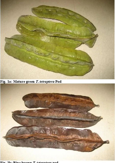
Fig. 1a: Mature green T. tetraptera Pod
Fig. 1b: Ripe brown T. tetrapterapod MATERIALS AND METHODS Samples collection and preparation
Triplicate samples of T. tetrapterafruit were collected at Idi-Ose village in Ibadan, Nigeria, at two phenologic stages of the fruit development, and were authenticated at the Department of Botany, University of Ibadan, Nigeria. The first stage was at the MG stage (seven months after fruiting, September, 2012), while the second was at the FRB stage (nine months after fruiting, November, 2012). The pods which were manually separated from seeds were later oven-dried at 50oC to a constant moisture content of about 5%. The
dried MG and RB pod samples (MGP and RBP, respectively) were subsequently milled to a fine particle size (0.5 mm)
packed in air-tight plastic vials and stored at 4°C until analysis.
All the chemicals used for analysis were of analytical grade.
Preparation of methanolic extract
Methanolic extract of the pod samples was prepared following the method of Chan et al.[18], by adding 25 ml of
methanol to 0.5g of sample contained in a 50 ml centrifuge tube, and shaking continuously for 1 h at room temperature. The mixture was centrifuged at 3,000 rpm for 10 min, and then the supernatant (subsequently referred to as methanolic extract) was collected and store at -4oC for further analysis.
Determination of total phenol content
The total phenol content of the methanolic extracts was determined according to the Folin–Ciocalteu method reported by Chan et al. [18]Briefly, 300 µl of extract was dispensed
into test tube (in triplicates). To this was added 1.5 ml of Folin–Ciocalteu reagent (diluted 10 times with distilled water), followed by 1.2 ml of Na2CO3 solution (7.5% w/v).
The reaction mixture was shaken, allowed to stand for 30 min at room temperature before the absorbance was measured at 765 nm against a blank prepared by dispensing 300µl of distilled instead of sample extract. TPC was expressed as gallic acid equivalent (GAE) in mg/g material.
Determination of tannin content
Tannin content of samples was determined according to the method of Padmaja [19] as follows. Sample (0.1g) was
extracted with 5 ml of acidified methanol (1% HCl in methanol) at room temperature for 15 minutes. The mixture was centrifuged at 3,000rpm for 20minutes. 0.1 ml of the supernatant was added to 7.5 ml of distilled water, followed by 0.5 ml of Folin-Denis reagent, 1 ml of 35% sodium carbonate solution and 0.9 ml of distilled water. The mixture was shaken, kept at room temperature for 30 min and absorbance was measured at 760 nm. Blank was prepared with water instead of the sample. Tannin content, expressed as tannic acid equivalent (TAE) in mg/g material, was subsequently calculated.
Determination of total flavonoid content
Total flavonoid content was determined using aluminum chloride method as reported by Kale et al. [20]. 0.5 ml of
methanolic extract was dispensed into test tube, followed by 1.5 ml of methanol, 0.1 ml of aluminum chloride (10%), 0.1 ml of 1M potassium acetate and 2.8 ml of distilled water. The reaction mixture was shaken, allowed to stand at room temperature for 30 minutes, before absorbance was read at 514 nm. TFC was expressed as quercetin equivalent (QE) in mg/g material.
Determination of total carotenoid content
Total carotenoid content of samples was determined according the method of Rodriguez-Amaya.[21]Briefly, 0.2 –
Determination of vitamin C content
The vitamin C content of the aqueous extract of samples was determined using the method reported by Benderitter et al.
[22] Briefly, to 300 µl of sample aqueous extract 100 µl of
13.3% trichloroacetic, 100 µl of distilled water and 75 µl DNPH reagent (2 g dinitrophenyl hydrazine, 230 mg thiourea and 270 mg CuSO4.5H2O in 100 ml of 5M H2SO4) were
added. The reaction mixture was subsequently incubated for 3 h at 37oC, after which 0.5 ml of 65% H
2SO4 (v/v) was
added to it and the absorbance was measured at 520 nm. Vitamin C content of the sample was subsequently calculated from a calibration curve prepared with ascorbic acid standard.
Estimation of DPPH free-radical-scavenging ability
The free-radical-scavenging ability of the methanolic extracts against 1,1-diphenyl-2-picrylhydrazyl (DPPH) free radical was estimated as described by Cervato et al.[23]with slight
modification. Briefly, appropriate dilutions of the extracts (0.024 to 0.096 mg/ml) totaling 1 ml was mixed with 3 ml of 60µM methanolic solution of DPPH radicals; the mixture was left in the dark for 30 min before the absorbance was taken at 517 nm. The decrease in absorbance of DPPH· on
addition of test samples in relation to the control was used to calculate the percentage inhibition (%Inh.) following the equation:
%Inhibition = [(A517control– A517sample) ÷ A517control] x 100.
The IC50 was calculated from the dose-inhibition linear
regression curve of each extract.
Estimation of ABTS* radical-scavenging ability
The 2,2-azinobis (3-ethyl-benzothiazoline-6-sulfonic acid) radical (ABTS*) scavenging ability of methanolic extract of samples were determined according to the method described by Sellappan and Akoh. [24] The ABTS* radical was
generated by incubating equal volume of a 7 mM ABTS aqueous solution with K2S2O8(2.45 mM) in the dark for 16 h
at room temperature and adjusting the absorbance at 734 nm to 0.7 ± 0.02 with 95% ethanol. Then 0.2 ml appropriate dilution of the extract was added to 2.0 ml ABTS* solution and the absorbance was measured at 734 nm after 15 min. The trolox equivalent antioxidant capacity (TEAC) was subsequently calculated.
Determination of ferric reducing antioxidant power (FRAP)
The reducing property of the methanolic extracts was determined by assessing the ability of the extract to reduce FeCl3solution as described by Oyaizu.[25]A 2.5 ml aliquot
was mixed with 2.5 ml of 200mM sodium phosphate buffer (pH 6.6) and 2.5 ml of 1% potassium ferricyanide. The mixture was incubated at 50oC for 20 min. and then 2.5 ml of 10% trichloroacetic acid was added. This mixture was centrifuged at 650 rpm for 10 min. Then 5 ml supernatant was mixed with an equal volume of water and 1 ml of 0.1% ferric chloride. The absorbance was measured at 700 nm. The ferric reducing antioxidant property was subsequently calculated.
Alpha-amylase inhibition assay
Alpha-amylase inhibition assay was conducted following the protocol described by Kwon et al. [26]Appropriate dilutions
(0.024 to 0.096 mg/ml) of methanolic extract amounting to 500μl, and 500μl of 0.02 M sodium phosphate buffer (pH 6.9 with 0.006 M sodium chloride) containing α-amylase solution (0.5 mg/ml) were incubated at 37°C for 10 min. After pre-incubation, 500μl of 1% starch solution in 0.02 M
sodium phosphate buffer was added. The reaction mixture was then incubated at 37°C for 15 min and the reaction was terminated with 1.0 ml of DNS acid color reagent (1% 3, 5-dinitrosalicylic acid and 12% sodium potassium tartrate in 0.4 M NaOH). The reaction mixture was then incubated in a boiling water bath for 5 min, cooled to room temperature, and diluted with 10 ml distilled water. The absorbance was measured at 540 nm.
Percentage α-amylase inhibition was calculated as using the formula:
%Inhibition = [(A540control– A540sample) ÷ A540control] x 100.
The IC50 was calculated from the dose-inhibition linear
regression curve of each extract.
Statistical Analysis
All assays were performed in three independent experiments and expressed as mean ± standard deviation (SD). Analysis of variance (ANOVA) was done by SPSS statistical software package version 17. The P values less than 0.05 were considered as statistically significant.
Table 1: Phenological variation in the antioxidant phytochemicals composition of T. tetraptera pod on dry weight basis.
Phytochemical MGP RBP
Total phenol (mg/g) 27.48 ± 0.16b 42.18 ± 0.16a
Tannin (mg/g) 32.54 ± 0.22b 46.99 ± 0.17a
Total flavonoid (mg/g) 0.18 ± 0.01b 0.44 ± 0.02a
Total carotenoid (µg/g) 5.03 ± 0.09a 1.31 ± 0.04b
Vitamin C (mg/100g) 3.91 ± 0.06a 2.86 ± 0.05b
Data represent the mean ± standard deviation of triplicate readings; values with the different lowercase superscript letter along the same row are significantly different (P >0.05).
Table 2: Phenological variation in the antioxidant activities and α -amylase IC50of methanolic extracts of T. tetraptera pod.
Antioxidant activity MGP RBP
TEAC (mM TE/g) 4.47 ± 0.02b 4.82 ± 0.03a
FRAP (µg GAE/g) 49.84 ± 0.43b 77.96 ± 0.85a
DPPH IC50 (mg/ml) 0.16 0.11
α-Amylase IC50 (mg/ml) 0.46 0.33
Data for FRAP and TEAC represent the mean ± standard deviation of triplicate readings; values with different lowercase superscript letter along the same row are significantly different (P >0.05).
RESULTS AND DISCUSSION
Fruits are known to undergo several changes in flavor, texture and color due to qualitative and quantitative variation in the composition of phytochemicals during the course of
maturity and development. [27] The antioxidant
phytochemicals results of T. tetraptera pod (on dry weight basis) at two different phenological stages of the fruit development (MGP and RBP) are presented in table 1. The results revealed that RBP had significantly higher (P < 0.05) total phenol, tannin and total flavonoid contents than MGP. This may be attributed to longer exposure of the RBP to sunlight (October to November) during which the pod became fully ripe and turned brown. According to Duenas et al. [28], phenolic compounds are synthesized in response to
light. Jaakola et al.[29]also reported that the expression of the
flavonoid biosynthetic genes is activated by enhanced light. While polyphenols are prominently known to exhibit antioxidant activity through a variety of mechanisms, including free radical scavenging, lipid peroxidation and chelating of metal ions [30], they also have many other
biological activities, such as anti-histamine [31],
anti-inflammatory and anticarcinogenic [32], antibacterial [33], and
In contrast to the phenolics results, MGP had significantly higher (P < 0.05) total carotenoid content than RBP. This may be due to the abundance of chlorophyll in the MGP as indicated by its greenness, than in the RBP. Gitelson et al.[35]
established a linear relationship between chlorophyll and carotenoids contents in higher plant leaves. In T. tetraptera fruit, the transition to ripening is accompanied by de-greening of the pod in which the photosynthetically active chloroplasts are differentiated to chromoplasts, resulting in its characteristic brown colour. Carotenoids are lipid-soluble antioxidant, which have been reported to have health benefits such as pro-vitamin A activity [36], antioxidant [37], anticancer
[38], and antiobesity effects.[39]
Similarly, MGP had significantly higher (P < 0.05) vitamin C content than RBP. This decrease could be attributed to the susceptibility of Vitamin C to oxidative destruction during ripening.[40]
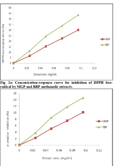
Fig. 2a: Concentration-response curve for inhibition of DPPH free-radical by MGP and RBP methanolic extracts.
Fig. 2b: Concentration-response curve for inhibition of porcine
pancreatic α-amylase activity by MGP and RBP methanolic extracts.
Vitamin C has powerful antioxidant properties and indirectly contributes to several key oxidative and reductive enzyme systems. It also has the ability to regenerate other biologically important antioxidants, such as glutathione and vitamin E, into their reduced state.[41]
Table 2 presents the antioxidant activities and porcine
pancreatic α-amylase IC50of methanolic extracts of MGP and
RBP. Antioxidants are substances which counteract free radicals, thus preventing oxidative damage to biomolecules such as lipids, proteins and DNA. [42-43] The ABTS*
scavenging ability reported as the Trolox equivalent antioxidant capacity (TEAC) showed that RBP had a
significantly higher (P < 0.05) scavenging ability than MGP. ABTS assay involves electron transfer process [32] and is
based on the discolouration of ABTS by antioxidant compounds, thus reflecting the amount of ABTS radicals that are scavenged within a fixed time period in relation to that of 6-hydroxy-2,5,7,8-tetramethylchroman-2-carboxylic acid (Trolox).
Similarly, the reducing power (from Fe3+to Fe2+) of MGP
and RBP reported as gallic acid equivalents, GAE (table 2) revealed that RBP had a significantly higher (P < 0.05) reducing power than MGP. FRAP assay is based on electron-transfer reactions, in which a ferric salt, potassium ferricyanide is used as an oxidant. The reaction mechanism involves the reduction of ferric 2,4,6-tripyridyl-s-triazine (TPTZ) to the coloured ferrous form.[44]
Free radical scavenging abilities of MGP and RBP methanolic extracts were further tested by the DPPH method at different concentrations (0.024 to 0.096 mg/ml). Both extracts exhibited very high DPPH radical-scavenging activity in a dose-dependent manner as depicted by their concentration-response curves (Figure 2a). Antioxidant reacts with DPPH, a stable free radical with characteristic deep
purple colour in solution, converting it to α,α-diphenyl-β -picryl hydrazine. On accepting proton from antioxidants, DPPH solution loses its characteristic deep purple, leading to absorption decrease (λmax 515–517 nm). So the degree of discoloration is an indication of the scavenging potentials of the antioxidant extract. Relative to MGP with IC50 of 0.16
mg/ml, RBP had IC50of 0.11 mg/ml. Since IC50 is a measure
of inhibitory concentration, a lower IC50value is a reflection
of greater antioxidant activity of the sample.[45]
MGP and RBP methanolic extracts displayed potent
inhibitory activity against porcine pancreatic α-amylase in a dose-dependent manner in-vitro(Figure 2b). The IC50values
showed that RBP (IC50 = 0.33 mg/ml) has a higher potency to inhibit the activity of porcine pancreatic α-amylase than MGP (IC50 = 0.46 mg/ml). The higher potency of RBP over MGP in inhibiting porcine pancreatic α-amylase may be attributed to its higher phenolic levels (total phenol, tannin and total flavonoid), which possibly were also responsible for the higher antioxidant activities observed in RBP. This finding is in agreement with earlier report of positive correlations between total phenolic content and antioxidant activity, and between antioxidant activity and α-amylase inhibition activity of plant extracts. [46] Tiwari and Rao [47]
reported that polyphenols inhibit alpha-amylase, sucrase, as well as the action of sodium glucose-transporter 1 (SGLUT-1) of the intestinal brush border, hence their antidiabetic action.
Inhibition of gastrointestinal carbohydrate hydrolyzing
enzymes such as the salivary and pancreatic α-amylase (EC 3.2.1.1), can significantly decrease the postprandial increase of blood glucose after a mixed carbohydrate diet and therefore is an important strategy in the management of postprandial blood glucose level in type 2 diabetic patients and borderline patients.[48]
and α-amylase inhibitory activity than MGP. Hence, for a more effective use of T.tetraptera pod for the management of oxidative stress and postprandial hyperglycemia in type 2 diabetes, it is recommended that the pod should be used at the ripe brown stage.
REFERENCES
1. Halliwell B, Gutteridge JMC. Free Radicals in Biology and Medicine, Edn 3, Oxford University Press, Inc., New York, 1999, pp. 105-245.
2. Baron AD. Postprandial hyperglycaemia and α-glucosidase inhibitors. Diabetes Res Clin Pract. 1998; 40:S51-55.
3. Kim JS, Kwon CS, Son KH. Inhibition of alpha-glucosidase and amylase by luteolin, a flavonoid. Biosci Biotechnol Biochem. 2000; 64(11): 2458-2461.
4. Tiwari AK, Rao JM. Diabetic mellitus and multiple therapeutic approaches of phytochemicals: Present status and future prospects. Curr Sci. 2002; 83(1): 30-37.
5. Iwu MM, Igboko AO, Onwubiko H, Ndu UE. Antisickling
properties of Cajanus cajan: Effect on hemoglobin gelation and oxygen affinity. Planta Medica. 1984; 24:431-432.
6. Kahkonen MP, Hopia AI, Vuorela HJ, Rauha JP, Pihlaja K, Kujala TS. Antioxidant activity of plant extracts containing phenolic compounds. J Agric Food Chem. 1999; 47:3954–3962.
7. Yen GC, Duh PD, Tsai HL. Antioxidant and pro-oxidant properties of ascorbic acid and gallic acid. Food Chem. 2002; 79:307–313. 8. Ozsoy N, Can A, Yanardag R, Akev N. Antioxidant activity of
Smilax excelsa leaf extracts. Food Chem. 2008; 110: 571-583. 9. Adetunji J, Aladesanmi J. Tetrapluera tetraptera: Molluscicidal
activity and chemical constituents. Afri J Trad Complement Altern Med. 2006; 4(Suppl 1): 23-36.
10. Opabode JT, Akinyemiju OA, Ayeni OO. Plant Regeneration via Somatic Embryogenesis from Immature Leaves in Tetrapleura tetraptera (Schum. & Thonn.) Taub. Arch Biol Sci Belgrade. 2011; 63(4): 1135-1145.
11. Essien EU, Izunwane BC, Aremu CY, Eka OU. Significance for humans of the nutrient contents of the dry fruit of Tetrapleura tetraptera. Plant Food Human Nutr. 1994; 45:47 -51.
12. Aladesanmi JA. Tetrapleura tetraptera: Molluscicidal activity and chemical constituents. Afr J Trad Complement Altern Med. 2007; 4:23-26.
13. Ojewole JAO, Adewunmi OC. Anti-inflammatory and
hypoglycaemic effects of Tetrapleura tetraptera. J
Ethnopharmacol. 2004; 95:177-182.
14. Jithendran KP. Clinical studies on coccidiosis in Angora rabbits. Blue Cross Book (Hoechst). 1997; 8:37-39.
15. Sahoo N, Manchikanti P, Dey S. Herbal drugs: Standards and regulation. Fitoterapia. 2010; 82:462–471.
16. Gobbo-Neto L, Lopes NP. Medicinal plants: Factors of influence on the content of secondary metabolites. Quimica Nova. 2007; 30:374-381.
17. Rathcke B, Lacey EP. Phenological patterns of terrestrial plants. Annu Rev Ecol Syst. 1985; 16:179-214.
18. Chan EWC, Lim YY, Chew YL. Antioxidant activity of Camellia sinensisleaves and tea from a lowland plantation in Malaysia. J Food Chem 2007; 102:1214-1222.
19. Padmaja G. Evaluation of techniques to reduce assayable tannin and cyanide in cassava leaves. J Agric food chem. 1989; 37:712-716.
20. Kale A, Gaikwad S, Mundhe K, Deshpande N, Salvekar J. Quantification of Phenolics and Flavonoids by Spectrophotometer from Juglans regia. Int J Pharm Biol Sci. 2010; 1: 1-4.
21. Rodriguez-Amaya DB. A guide to carotenoid analysis in foods. ILSI Press, Washington DC, 1999.
22. Benderitter M, Maupoil V, Vergely C, Dalloz F, Briot F, Rochette L. Studies by electron paramagnetic resonance of the importance of iron in the hydroxyl scavenging properties of ascorbic acid in plasma: Effects of iron chelators. Fundam Clin Pharmacol. 1998; 12: 510-16.
23. Cervato G, Carabelli M, Gervasio S, Cittera A, Cazzola R, Cestaro B. Antioxidant properties of oregano (Origanum vulgare) leaf extracts. J Food Biochem. 2000; 24:453-65.
24. Sellappan S, Akoh CC. Flavonoids and antioxidant capacity of Georgia-grown vidalia onions. J Agric Food Chem. 2002; 50:5338– 5342.
25. Oyaizu M. Studies on products of browning reaction: antioxidative activity of products of browning reaction prepared from glucosamine. Jpn J Nutr. 1986; 44:307-15.
26. Kwon YI, Vattem DA, Shetty K. Clonal herbs of Lamiaceae species against diabetes and hypertension. Asia Pac J Clin Nutr. 2006; 15:424-432.
27. Gull J, Sultana B, Anwar F, Naseer R, Ashraf M, Ashrafuzzaman M. Variation in Antioxidant Attributes at Three Ripening Stages of Guava (Psidium guajava L.) Fruit from Different Geographical
Regions of Pakistan. Molecules. 2012; 17:3165-3180;
doi:10.3390/molecules17033165.
28. Duenas M, Hernandez T, Estrella I. Assessment of in vitro antioxidant capacity of the seed coat and the cotyledon of legumes in relation to their phenolic contents. Food Chem. 2006; 98(1):95-103.
29. Jaakola, L, Maatta-Riihinen K, Karenlampi S, Hohtola A. Activation of flavonoid biosynthesis by solar radiation in bilberry (Vaccinium myrtillus L.) leaves. Planta. 2004; 218:721-728. 30. Shahidi F, Amarowicz R, He YH, Wettasinghe M. Antioxidant
activity of phenolic extracts of evening primrose (Oenothera biennis): A preliminary study. J Food Lip. 1997; 4:75-86.
31. Nitta Y, Kikuzaki H, Ueno H. Food components inhibiting recombinant human histidine decarboxylase activity. J Agric Food Chem. 2007; 55:299–304.
32. Srikanth G, Babu SM, Kavitha CHN, Rao MEB, Vijaykumar N, Pradeep CH. Studies on in-vitro antioxidant activities of Carica papayaaqueous leaf extract. Res J Pharm Biol Chem Sci. 2010; 1(2):59-65.
33. Romero C, Medina E, Vargas J, Brenes M, De Castro A. In vitro activity of olive oil polyphenols against Helicobacter pylori. J Agric Food Chem. 2007; 55:680–686.
34. Song JM, Lee KH, Seong BL. Antiviral effect of catechins in green tea on influenza virus. Antivir Res. 2005; 68:66–74.
35. Gitelson AA, Gritz Y, Merzlyak MN. Relationships between leaf chlorophyll content and spectral reflectance and algorithms for non-destructive chlorophyll assessment in higher plant leaves. J Plant Physiol. 2003; 160:271–282.
36. Rodriguez-Amaya DB. Carotenoids and Food Preparation: The Retention of Pro-vitamin A Carotenoids in Prepared, Processed, and Stored Foods. Department of Food Science Faculty of Food Engineering, State University of Campinas. Brazil. John Snow, Inc. /OMNI Project. 1997; pp 93.
37. Tian B, Xu Z, Sun Z, Lin J, Hua Y. Evaluation of the antioxidant effects of carotenoids from Deinococcus radiodurans through targeted mutagenesis, chemiluminescence, and DNA damage analyses. Biochem Biophys Acta. 2007; 1770:902-911.
38. Nishino H. Cancer prevention by carotenoids. Mutat Res. 1998; 402:159-163.
39. Jaswir I, Noviendri D, Hasrini RF, Octavianti F. Carotenoids: Sources, medicinal properties and their application in food and nutraceutical industry. J Med Plants Res. 2011; 5(33):7119-7131. 40. Aina JO. Physico-chemical changes in African Mango (Irvingia
gabogensis) during normal storage ripening. J. Food Chem. 1990; 36:205–212.
41. Jacob RA. The integrated antioxidant system. Nutr Res. 1995; 15:755-766.
42. Ratnam DV, Ankola DD, Bhardwaj V, Sahana DK, Kumar MNVR. Role of antioxidants in prophylaxis and therapy: A pharmeumaceutical perspective. J Control Release. 2006; 113:189-207.
43. Becker EM, Nissen LR, Skibsted LH. Antioxidant evaluation protocols: Food quality or health effects. Eur Food Res Technol. 2004; 219:561-571.
44. Ndhlala AR, Moyo M, Staden JV. Natural Antioxidants: Fascinating or Mythical Biomolecules? Molecules. 2010; 15:6905-6930; doi:10.3390/molecules15106905.
45. Irondi AE, Oboh G, Akintunde JK. Comparative and Synergistic Antioxidant properties of Carica papayaand Azadarichta indica Leaves. Int J Pharm Sci Res. 2012; 3(12): 4773-4779.
46. Manaharan T, Palanisamy UD, Ming CH. Tropical Plant Extracts as Potential Antihyperglycemic Agents. Molecules. 2012; 17:5915-5923.
47. Tiwari AK, Rao JM. Diabetic mellitus and multiple therapeutic approaches of phytochemicals: Present status and future prospects. Curr Sci. 2002; 83(1):30-37.