International Journal of Pharmaceutical Research&Allied Sciences, 2018, 7(3):81-90
Research Article
CODEN(USA) : IJPRPM
ISSN : 2277-3657
Vitamin D Receptor Gene Polymorphism as a Prognostic Marker in Acute
Myeloid Leukemia Patients
Amal Ahmed Zidan1, Fouad Mohammed Abu Taleb2, Alaa Abd El Moaty Omran1, Ola Elsayed Abdel Latif Elnaggar1*
1Clinical Pathology Department Faculty of Medicine, Zagazig University, Egypt, 2Medical Oncology Department Faculty of Medicine, Zagazig University, Egypt.
*Corresponding Author Email: olaelnaggar@gmail.com
ABSTRACT
Background : The purpose of this study was to evaluate the prognostic importance of vitamin D receptor start codon (FokI) polymorphism in acute myeloid leukemia. Patients and Methods : A total of 30 patients with acute myeloid leukemia were enrolled in this study. In addition 30 age and sex matched healthy volunteers were included as a control group. Five milliliters (ml) of venous blood were collected and poured into K3 ethylene di amine tetra acetic acid (EDTA), and genomic DNA was extracted from all the samples. A 260 bp fragment of VDR gene was amplified by PCR, and digested by restriction enzyme (FokI) to detect the polymorphism. Results : The results showed that wild genotype (FF) was significantly lower in the AML group than controls (83.3% VS 46.7%), while heterozygous genotype (Ff) was significantly higher in the AML group than controls, and homozygous genotype ff was found only in AML group (6.6%). There was a significant relation between patients’ FOKI genotypes and the occurrence of remission, FF genotype was the most common type in AML group who achieved CR (63.2%), while Ff genotype was 63.6% in those who failed to respond to induction therapy (NR), and finally all the patients with ff genotype (18.2%) did not show remission. Conclusion : The association of VDR FOKI polymorphism with increased risk of AML and poor response to therapy was observed, which was a potential indicator of prognosis in patients with AML.
Key words: VDR Start Codon Polymorphism, Acute Myeloid Leukemia.
INTRODUCTION
82
chromosome 12q12-q14, and several single nucleotide polymorphisms (SNPs) within the gene have been recognized that may affect cancer risk. Each polymorphism has been named according to the restriction site that was primarily used to recognize it [6]. Five well-known SNPs of human VDR, FokI (C/T), BsmI (A/G), ApaI (A/C), TaqI (T/C), and Cdx2 (A/G), were extensively studied previously for their association with cancer risk [7]. The FokI polymorphism is located at the first potential start site [6], and alters an ACG codon that results in the generation of an additional start codon [8]. Research studies conducted on the relationship between vitamin D receptor (VDR) start codon FokI Polymorphism and various kinds of cancers have revealed diverse results. An examination demonstrated considerable associations with VDR FokI polymorphism and prostate, breast, colon-rectum and skin cancers [9]. The pleitropic effect of VDR and its involvement in normal and malignant cells suggested that the VDR polymorphism might have a role in AML pathogenesis. This study aimed to evaluate the prognostic importance of vitamin D receptor start codon (FokI) polymorphism in acute myeloid leukemia.
PATIENTS AND METHODS:
This was a case control study carried out in Clinical Pathology and Medical Oncology Departments, Faculty of Medicine, Zagazig University Hospitals. A total of sixty subjects were included in this study; and they were classified into two groups as follows:
Control group: It included 30 apparently healthy adult volunteers. They were 17 males and 13 females with mean age of 43.36 years. They matched well with patients regarding age and sex.
Patient group: It included 30 adult patients with newly diagnosed AML. They were 19 males and 11 females with mean age of 46.33.
Sample collection
Five milliliters (ml) of venous blood were collected from each subject and poured into Ethylene di amine tetra acetic acid (EDTA) blood container.
DNA extraction and molecular analysis
Genomic DNA was extracted from all the samples by QIAamp DNA blood mini kit (QIAGEN). The reaction
mixture of 40 μl was prepared for each sample. It consisted of 6μl of genomic DNA, 1 μl of each forward primer AGCTGGCCCTGGCACTGACTCTGCTCT-3') and reverse primer (5'-ATGGAAACACCTTGCTTCTTCTCCCTC-3'), 10 μl master mix (MyTaq Red Mix) and 22 μl sterile distilled water.
DNA samples were amplified in Thermal Cycler Gene Amp, PCR system 2400 (Perkin Elmer, USA) with cycling conditions as follows: Denaturation at 95 o C for 1 min, 35 cycles, each consisted of 95o C for 15 seconds, 61 o C for 15 seconds and 72 o C for 10 seconds, and one final cycle of extension at 72oC for 7minutes.
The PCR product of 265 bp band was digested with 1 μl of Fok I restriction enzyme [Thermo Scientific FastDigest FOKI 100µL (for 100 rxns)], it is incubated at 37 o C for 5 minutes; after that, 5μL of the digested reaction mixture was loaded into 3% agrose gel containing ethidium bromide and visualized using gel documentation system (Syngene, Japan). 100 bp DNA ladder was loaded with each batch of samples. The digestion of the amplified 265 bp PCR product resulted in two fragments (196 bp and 69 bp). Depending on the digestion pattern, individuals were genotyped as (ff) when homozygous for the presence of the FokI site, (FF) when homozygous for the absence of the FokI site, or (Ff) in case of heterozygosity.
Statistical Analysis:
Data of this study was collected from patients’ medical files, and analyzed using Statistical Package for Social Sciences (SPSS). The frequencies of genotypes were calculated, and the correlation of genotypes with study groups was tested by Chi-square. The Hardy–Weinberg equilibrium was tested by a goodness-of-fit X2 test to compare the observed genotypic frequencies in normal individuals to the expected genotypic frequencies calculated from the observed allelic frequencies.
All P values were based on a 2-tailed distribution, and the corresponding P value: Non-significant (NS) difference if P > 0.05.
Significant (S) difference if P<0.05.
83
RESULTS:
A total of 30 patients with AML were enrolled in this study; 19 (63.3%) were males and 11(36.7%) were females. Further 30 healthy volunteers were included as a control group.
Table (1) shows the mean ± SD and the range of the age and sex of the AML and control groups.
Table 1: Demographic data among the studied groups:
Variable AML group
(No=30)
Control group (No=30)
Age: (years) Mean ± SD (Range)
46.33 ± 16.34 19 – 65
43.36 ± 10.71 26 - 63
No: % No: %
Sex: Male: Female:
19 11
63.3 36.7
17 13
56.7 43.3
SD: standard deviation No: number of subjects
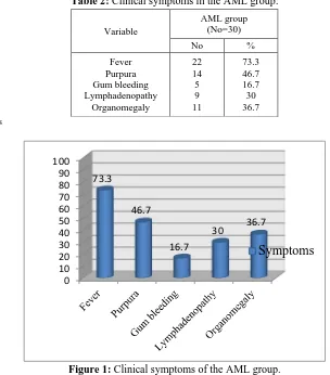
Table (2) and figure (1) show the clinical symptoms of AML group. Fever was the most common presenting symptom (73.3%), followed by purpura (46.7%), then organomegaly (36.7%). Almost one third of the patients experienced lymphadenopathy (30%), and gum bleeding was discovered only in 16.7% of patients.
Table 2: Clinical symptoms in the AML group:
Variable
AML group (No=30)
No %
Fever Purpura Gum bleeding Lymphadenopathy
Organomegaly
22 14 5 9 11
73.3 46.7 16.7 30 36.7
No: number of subjects
Figure 1: Clinical symptoms of the AML group.
Table (3) shows the hematological data of AML and the control groups. A triad of high WBCs count, anemia and thrombocytopenia was observed in the AML group by a single inspection of table (3).
0 10 20 30 40 50 60 70 80 90 100
73.3
46.7
16.7
30 36.7
84
Table 3: Hematological data of AML and the control groups:
Variable AML group
(No=30)
Control group (No=30) TLC (109/L)
Median (Range) 32.8 (2.5 -202) 6.80 (4 – 11) HB (gm/dl)
Median (Range) 8.4 (5.3 – 11.7) 13 (11.6 -15.3) Platelets (109/L)
Median (Range) 67.5 (6 – 259) 281 (167 – 423) BM blast (%)
Median (Range) 58.5 (15-93) ----
PB blast (%)
Median (Range) 45 (10-80) ----
ESR (mm/hr)
Median (Range) 47.5 (26 – 140) 4.5 (2 – 7)
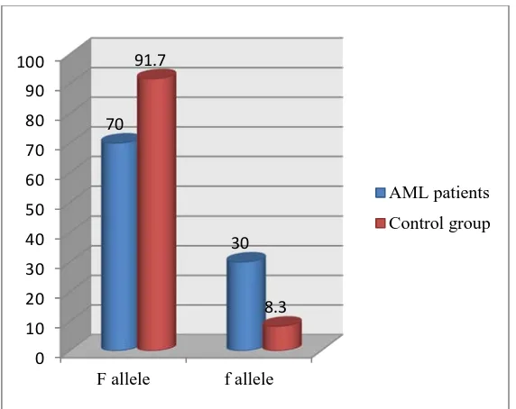
Table (4), figures (2) and (3) show the comparison of FOKI genotypes and alleles in AML and control groups. Wild genotype (FF) was significantly lower in the AML group than controls (83.3 VS 46.7), while heterozygous genotype (Ff) was significantly higher in the AML group than the controls. Of note homozygous genotype ff was found only in AML group (6.6%). Calculated risk estimation revealed that heterozygous genotype Ff conferred almost 5 fold increased risk of developing AML (OR=5,95% CI=1.48-16.8). The f allele was higher in AML group than controls (30% VS 8.3%), and the difference was statistically significant. The calculated risk estimation revealed that the f allele conferred almost a 4 fold increased risk of developing AML (OR=4.7, 95%CI=1.61-13.73).
Table 4: Comparison of FOKI genotypes and alleles in AML and control groups.
Variable
AML group (No=30)
Control group
(No=30) OR CI χ2 P
No % No %
Genotype: FF (wild) Ff (heterozygous) ff (homozygous) 14 14 2 46.7 46.7 6.6 25 5 0 83.3 16.7 0 5 7 1.4-16.8 1.8-9.4 8.71 6.13 2.03 0.003 (S) 0.01 (S) 0.153 Alleles: F allele (wild) f allele (mutant)
42 18 70 30 55 5 91.7
8.3 4.71 1.6-13.7 7.68
0.005 (S)
(S): significant difference. Ref.: reference. χ2: chi square
No: number of subjects OR: odds ratio P: p value
Figure 2: Frequency of FOKI genotypes among AML and control groups.
0 10 20 30 40 50 60 70 80 90 100
FF Ff ff
85
Figure 3: Frequency distribution of alleles in AML and control groups.
Table (5) and figure (4) show the response to induction therapy on day 28 in the AML group. After induction therapy, 63.3 % of the patients achieved complete remission, while no remission was encountered in 36.7%.
Table 5: Response to induction therapy of the AML group:
Variable AML group (No=30)
No %
Treatment: Complete remission
No remission
19 11
63.3 36.7
No: number of subjects
Figure 4: Response to induction therapy in AML group.
Table (6) and figure (5) show the relation between FOKI genotypes and alleles and response to induction therapy at day 28 in the AML group. There was a significant relation between the patients’ FOKI genotypes and the occurrence of remission. FF genotype was the most common type in AML group who achieved CR (63.2%), while Ff genotype was 63.6% in those who failed to respond to the induction therapy (NR). Of note, all the patients with ff genotype (18.2%) did not show remission.
Regarding FOKI alleles, there was a significant relation between the allele type and the occurrence of remission. Patients with F allele had significantly higher percentage of complete remission (81.6%) when compared to patients with f allele (18.4%).
0 10 20 30 40 50 60 70 80 90 100
F allele f allele
70
30 91.7
8.3
AML patients
Control group
63.3 36.7
Complete remission
86
Table 6: Relation between FOKI genotypes and alleles and response to therapy at day 28 in the AML group.
Variable
Complete remission (No=19)
No remission
(No=11) χ2 P
No % No %
Genotype: FF (wild) Ff (heterozygous)
ff (homozygous)
12 7 0
63.2 36.8 0
2 7 2
18.2 63.6 18.2
8.923 0.002 (S)
Alleles: F allele (wild) f allele (mutant)
31 7
81.6 18.4
11 11
50
50 8.06
0.004 (S)
S: significant difference. χ2: chi squa
Figure 5: Relation between FOKI genotypes and response to therapy.
Table 7: Logistic regression analysis of factors predicting prognosis of AML group:
Independent factors O.R (95%C.I ) P-value
Genotypes 7.714 (1.28 – 46.3) 0.02
TLC 1.5 (1.4 –5.4) 0.04
BM blasts 3.9 (1.5 – 8.9) 0.02
PB blasts 2.4 (1.6 – 5.5) 0.03
LDH 2.2 (1.8 – 7.1) 0.03
Significant at (P<0.05)
Table (12) shows Logistic regression analysis of factors predicting prognosis of AML group. Ff, ff genotypes, high TLC, LDH, BM and PB blasts were significantly related to bad prognosis of AML.
It has been assumed that all of these characters are predictors of bad prognosis.
DISCUSSION:
Acute myeloid leukemia (AML) is a heterogeneous disorder accompanied by clonal expansion of myeloid progenitors in the bone marrow and peripheral blood [10].
AML which is almost a third of all leukemias worldwide, has been considered as the most common form of leukemia in adults [11].
Despite the advancement in treatment options for AML, its prognosis has been very variable, ranging from survival of few days to cure. Many patients die either from the complications of intensive chemotherapy, and resistance to the current treatment options, or they experience relapse after initial response to the traditional chemotherapy [12]. Clinical outcome can be partly predicted by age, cytogenetic findings, and serum lactate dehydrogenase at the time of diagnosis [13]. However, the prognosisofanindividual AML patientcannotyetbeestimatedaccurately. Sothatadditional prognostic markers are required for more accurate estimation. Moreover, there is still a need for
0 10 20 30 40 50 60 70 80 90 100
FF Ff ff
63.2
36.8
0 18.2
63.6
18.2
Complete remission
87
simple, reliable, and easy measured factors which have impacts on the prognosis especially in areas where modern technology is not usually available.
Vitamin D is a fat-soluble steroid hormone precursor that is mainly produced in the skin by exposure to sunlight. Vitamin D is biologically inert, and must undergo hydroxylation steps to become active [14].
Just vitamin D3 can be synthesized in our body. Vitamin D2 enters our body with fortified food or by supplements. Physiologically, vitamin D3 and D2 are bound to vitamin D binding protein (VDBP) in plasma and moved to the liver to become 25-hydroxyvitamin D (OH)D. As serum (OH)D shows the major storage form, its blood concentration is used to evaluate the overall vitamin D status [15]. Moreover, it has been clarified that the receptors of vitamin D exist in various cells, and this hormone has biologic influences which go far beyond the control of the mineral metabolism. Its cellular differentiation, proliferation, apoptosis and angiogenesis lead it to have a role in the regulation of vital cellular processes [16].
The active form of vitamin D (1,25(OH)2D3) plays its vital biological role by VDR. 1,25(OH)2D3 joins with VDR, and combines retinoid X receptor (RXR) to form a dimeric complex, which binds to the target genes upstream to regulate downstream transcription of these target genes. These target genes are mostly cell cycle dependence kinase (CCDK) inhibitory protein P16, P21, P27, and so on, that hinder the activation of CCDK complexes and keep the cell in G0/G1 phase. So, the proliferation of tumor cells is hindered [17,18].
The VDR gene exists on the long arm of chromosome 12 (12q12-14) and contains almost 200 single nucleotide polymorphisms (SNPs). The most popular allelic scrutinized variants include a start codon polymorphism FokI (T/C) in exon II, BsmI (A/G) and ApaI (C/A) polymorphism in the intron between exon VII and IX and a TaqI (T/C) variant in exon IX [19].
Among the VDR polymorphisms, the FokI single nucleotide polymorphism of the translation start site has been the only one that results in a VDR protein with a different structure [20]. This polymorphism is identified by the existence of either two ATG start codons divided by six nucleotides in the long f-VDR, or just one start codon due to a T-to-C substitution in the most 50 ATG codons, forming a 3-aa shorter F-VDR protein (424 aa instead of 427 aa). Furthermore, it is the only polymorphism that is not connected to any of the other VDR polymorphisms [21], which gives it a unique role.
It has been reported that the longer VDR is less responsive to 1,25(OH)2D3, and has lower transcription activity [22] and may contribute to reduce immunity response, which lead to tumorigenesis.
Despite growing evidence for a relationship between VDR FokI Polymorphism, and the risk of solid tumor, far less has been known about the relationship between VDR FokI polymorphism and the risk of hematologic malignancy. The aim of this study was to evaluate the prognostic importance of vitamin D receptor start codon (FokI) polymorphism in adult acute myeloid leukemia patients, and correlate between the vitamin D receptor start codon (FokI) polymorphism and prognostic markers of AML. To achieve the aim, immunophenotyping, cytogenetic analysis and molecular detection of VDR FokI polymorphism were done on the patient group.
This study was conducted on 30 denovo AML patients. They were 19 males and 11 females. Their age ranged between 19 and 65 years with mean ± SD of 46.33± 16.34. Thirty age and gender matched healthy volunteers were included in the current study as a control group.
Among the newly diagnosed AML patients, fever was found in 22 patients (73.3%), followed by purpura which was found in 14 patients (46.7%) and organomegaly which was found in 11 patients (36.7%). Gum bleeding was found in 5 patients (16.7%), while lymphadenopathy was found in 9 patients (30%). These findings were in agreement with [23] who stated that fever was the most common initial clinical presentation. Symptoms related to AML were caused by the replacement of bone marrow and failure of normal hemopoiesis.
In the present study, the median value of WBC count in patients group at the time of diagnosis was 32.8 x10⁹/L and ranged from 2.5 to 202x 109/L. [24] and [25] reported that the median WBC count of the patients at the time of diagnosis was 11 and 6.6 x10⁹/L; respectively.
88
In this study, the median platelet count of the patient group was 67.5 x10⁹/L. [28] reported that the median platelet count was 40.25 x10⁹/L, and [26] reported that the median platelet count at the time of diagnosis was 42 x109/L. These findings were expected as these were directly attributable to the leukemic infiltration of bone marrow with resultant cytopenia [28].
In the present study, the median blasts percentage in the bone marrow was 58.5%, which was close to the study reported by [28], who found that the median BM blasts percentage was 41.5%.
In this study, the frequency of VDR start codon genotypic variants in egyptian AML patients with comparison to the control group was examined. The results showed that both wild FF and heterozygous Ff genotypes in the AML group were of the same percentage (46.7%), while heterozygous ff genotype in the same group was 6.6%. Regarding the control group, wild FF genotype was the most frequent (83.3%) followed by heterozygous Ff genotype (16.7%), while no homozygous ff genotype was detected. This was consistent with [29] who reported that wild FF genotype was significantly lower in AML group than controls, while heterozygous genotype Ff was significantly higher in AML group than the controls. The findings also disagreed with [30] who stated that wild FF genotype was lower in the controls than AML group, while heterozygous genotype Ff was higher in controls than AML group. This could be attributed to the different sample size.
This study revealed that those having the heterozygous genotype Ff conferred a five-fold increased risk of developing. [31] reported that the wild FF and the heterozygous Ff genotypes were associated with 1.4 and 1.6 fold increased risk for AML; respectively. [32] showed 0.2 fold increased risk of ALL in the individuals with the wild genotype FF and 4-fold increased risk in those with heterozygous genotype Ff. [29] observed a three-fold increased risk of CLL for those carrying the heterozygous genotype Ff. Another study by [33] reported that, the Ff genotype was associated with a two-fold increase in ovarian Cancer risk [34]. This was inconsistent with [33] who reported that there was a statistically significant correlation between CML and the VDR start codon ff genotype but not between the genotypes of FF and Ff.
This study showed that the mutant f allele was significantly higher in AML group than the controls (30% VS 8.3%). This was consistent with [29] who reported that the frequency of the f allele was 0.14 in the AML group and 0.08 in the control group.
After induction therapy, this study demonstrated a statistically significant relation between AML patients' FOKI genotypes and remission outcomes, as for the wild genotype FF, 63.2% of the patients achieved CR (complete remission), while 18.2% didn't achieve remission. Regarding the heterozygous Ff genotype, 36.8% of the patients showed CR, while 63.6% didn't achieve remission, and finally all the patients with the homozygous genotype ff didn't achieve remission who were 18.2%, so statistically FF genotype was the most common type to achieve CR, while the patients with ff genotype experienced no remission. The same finding was reported by [35] who found a significant association between better prognosis of the patients with EOC (Epithelial Ovarian Cancer) and the FOKI FF genotype. [36] has also reported that there was a significant association between the shorter progression-free survival time in patients with head and neck squamous cell carcinoma and FOKI ff genotype.
CONCLUSION:
The association of VDR FOKI polymorphism with the increased risk of AML and poor response to therapy was observed, which was a potential indicator of prognosis in patients with AML
REFERENCES
1. Thol F, Heuser M, Damm F, Klusmann J, et al. (2011) : DNMT3A mutations are rare in childhood acute myeloid leukemia. Haematologica ; 96 : 1238–1240.
2. Moore DD, Kato S, Xie W et al., (2006) : “International Union of Pharmacology. LXII. The NR1H and NR1I receptors : constitutive and rostane receptor, pregnene X receptor, farnesoid X receptor alpha, farnesoid X receptor beta, liver X receptor alpha, liver X receptor beta, and vitamin D receptor”. Pharmacol. Rev 58 : 742–59.
3. Haussler MR, Whitfield GK, Kaneko I, Haussler CA, Hsieh D, Hsieh JC, Jurutka PW : Molecular mechanisms of vitamin D action. Calcif Tissue Int. 2013 ; 92(2) :77-98.
89
5. Szpirer J, Szpirer C, Riviere M, Levan G, Marynen P, Cassiman JJ, Wiese R, DeLuca HF (September 1991). "The Sp1 transcription factor gene (SP1) and the 1, 25 dihydroxyvitamin D3 receptor gene (VDR) are co localized on human chromosome arm 12q and rat chromosome 7". Genomics 11 (1) : 168–73. 6. Ntais, C., Polycarpou, A. & Ioannidis, J. Vitamin d receptor gene polymorph isms and risk of prostate
cancer. Cancer Epidemiol Biomarkers Prev. 12, 1395 (2003).
7. Raimondi, S., Johansson, H., Maisonneuve, P. & andini, (2009) S. Review and meta-analysis on vitamin D receptor polymorphisms and cancer risk. Carcinogenesis. 30, 1170–1180
8. Kostner K, Denzer N, Muller CS, etal., (2009) : The relevance of vitamin D receptor (VDR) gene polymorphisms for cancer : A review of the literature. Anticancer Res. 2009 ;29 :3511–36.
9. Gandini S, Gnagnarella P, Serrano D et al., (2014). “Vitamin D receptor polymorphisms and cancer”. Adv Exp Med Biol 810 :69-105
10. Döhner H, Weisdorf DJ and Bloomfield CD (2015) : Acute myeloid leukemia. N. Engl. J. Med., 373, 1136–1152.
11. Deschler B and Lubbert M (2006) : Acute Myeloid Leukemia : Epidemiology and Etiology. Cancer ; 107(9) :2099-107.
12. Hwang K, Park CG and Jang S (2012) : Flow cytometric quantification and immunophenotyping of leukemic stem cells in acute myeloid leukemia. Ann. Hematol. :91 :1541–1546.
13. Estey EH, Müller-Tidow C, Berdel WE and Krug U (2001) : Prognostic factors in acute myeloid leukemia. Leukemia ;15 :670.
14. Holick MF (2007) : Vitamin D deficiency. N Engl J Med ;357 : 266– 281.
15. Hart GR, Furniss JL, Laurie D and Durham SK (2006) : Measurement of vitamin D Status : background, clinical use and methodologies. Clin Lab ; 52(7-8) : 335-343.
16. Moore DD, Kato S, Xie W, Mangelsdorf DJ, et al. (2006) : International Union of Pharmacology. Pharmacol Rev ;58 : 742–59.
17. Campbell MJ, Elstner E, Holden S, Uskokovic M, Koeffler HP (1997). Inhibition of proliferation of prostate cancer cells by a 19-norhexafluoride vitamin D3 analogue involves the induction of p21waf1, p27kip1 and E-cadherin. J Mol Endocrinol. ; 19(1) : 15–27.
18. Ylikomi T, Laaksi I, Lou YR, Martikainen P, Miettinen S, Pennanen P, et al. (2002) Antiproliferative action of vitamin D. Vitam Horm. ;64 : 357–406
19. Slatter ML, Yakumo K, Hoffman M and Neuhausen S (2001) : Variants of the VDR gene and risk of colon cancer (United States). Cancer Causes Control 12 : 359-364.
20. Arai, H., Miyamoto, K., Taketani, Y., Yamamoto, H., Iemori, Y., Morita, K., Tonai, T. et al., (1997) A vitamin D receptor gene polymorphism in the translation initiation codon : Effect on protein activity and relation to bone mineral density in Japanese women. J. Bone Miner. Res. 12 : 915–921.
21. Nejentsev, S., Godfrey, L., Snook, H., Rance, H., Nutland, S., Walker, N. M., Lam, A. C. et al., (2004) Comparative high-resolution analysis of linkage disequilibrium and tag single nucleotide polymorphisms between populations in the vitamin D receptor gene. Hum. Mol. Genet. 13 : 1633–1639.
22. Colin EM, Weel AE, Uitterlinden AG, Buurman CJ, Birkenhager JC, Pols HA, et al. (2000) Consequences of vitamin D receptor gene polymorphisms for growth inhibition of cultured human peripheral blood mononuclear cells by 1, 25-dihydroxyvitamin D3. Clin Endocrinol (Oxf). 52(2) :211–6.
23. Asif N and Hassan KH (2009) : Journal of Islamabad Medical & Dental College (JIMDC) ; 1211(1) :6-9. 24. Renneville A, Boissel N, Nibourel O, Berthon C, et al. (2012) : Prognostic significance of DNA
methyltransferase 3A mutations in cytogenetically normal acute myeloid leukemia : a study by the Acute Leukemia French Association. Leukemia ;26 :1247–1254.
25. Pezzi A, Moraes L, Valim V, Amorin B, et al. (2012) : DNMT3A Mutations in Patients with AcuteMyeloid Leukemia in South Brazil. Advances in Hematology ; 8 pages.
26. Hou H, Kuo Y, Liu C, Chou W, et al. (2011) : DNMT3A mutations in acute myeloid leukemia stability during disease evolution and the clinical implications. Blood ;23 : 541-549.
27. Mercadante V, Gebbia A and Marrazzo S (2000) : Filosto Anemia in cancer : pathophysiology and treatment Cancer Treat. Rev ;26 : 303–311.
90
29. Sara A. A. Rajab, Ibrahim K. Ibrahim, Enaam A. Abdelgader, Eldirdiri M. Abdel Rahman, Mahdi H. A. Abdalla. (2015) : Vitamin D receptor gene (FokI) polymorphism in Sudanese patients with chronic lymphocytic leukemia. American Journal of Research Communication ;3(7) : 71-78.
30. Esfahani, Z. Ghoreishi (2016) : Is there any association between vitamin D receptor polymorphisms and acute myeloid leukemia ? Annals of Oncology 27 (Supplement 6) : vi313–vi327.
31. Yasmine Ahmed Ibrahim, Elshazali Widaa Ali, Ebtihal Ahmed Babiker, Mohanad Altayeb Mohamed Ahmed (2015) : - Association of Vitamin D Receptor (VDR) Start Codon FokI Polymorphism with Acute Myelogenous Leukemia EUROPEAN ACADEMIC RESEARCH - Vol. II, Issue 12.
32. Ali, E.M., Ahmed, M. A. M., & Ibrahim, I. K. (2015) : association of vitamin-d receptor (vdr) gene start codon foki polymorphism with acute lymphoblastic leukemia. Journal of science Vol 5 / Issue 4 / 242-246. 33. Sheraz Fathi Saeed Algadal, Elshazali Widaa Ali, Hiba Abbas Elamin, Mohanad Altayeb Mohamed
Ahmed, Ebtihal Ahmed Babiker and Lubna babiker (2015) : Association of Vitamin D Receptor (VDR) Start Codon Fok-I Polymorphism with Chronic Myeloid Leukemia. IJAPBC – Vol. 4(1), ISSN : 2277 – 4688.
34. Mohapatra S, Saxena A, Gandhi G, Chandra B K, Chandra P R. (2013) : Vitamin D and VDR gene polymorphism (FokI) in epithelial ovarian cancer in Indian population. Journal of Ovarian Research 6 :37. 35. S Tamez, C Norizoe, K Ochiai, D Takahashi, A Shimojima and M Urashima (2009) : Vitamin D receptor
polymorphism and prognosis of patients with epithelial ovarian cancer. British journal of cancer ;101 :1957-1960.


