1
Original Article
CODEN: IJPNL6
Evaluation of Novel Ion Sensitive
In situ
Gelling Polymer in the Development of
Ophthalmic Drug Delivery System
Atish S Mundada
*,Deepak B Ghorpade
SNJB’s SSDJ College of Pharmacy, Neminagar, Chandwad, Nashik, India
*Corresponding author e-mail: [email protected]
Received on: 29-11-2017; Revised on: 02-12-2017; Accepted on: 12-12-2017
ABSTRACT
The attempt has been made in the present work to formulate an ion sensitive in situ gel of an antibacterial agent, Ciprofloxacin HCl
employing a novel in situ gelling low methoxy pectin (obtained from the heads of Helianthus annuus Linn) as bioadhesive polymer.
The drug loaded in situ gelling formulation was developed employing the cold method. The prepared formulations were evaluated for
appearance, clarity, pH, gelling capacity, viscosity, drug content, mucoadhesive force, gelling strength, in vitro drug release,
isotonicity study, sterility test and stability study. The results of various physicochemical evaluation shows that the formulation
comprising of 2.5% w/v low methoxy pectin to be considered as an optimized in terms of gelling capacity, viscosity, gelling strength,
the drug release study giving sustained release behavior over period of 11 h. Thus we claim that ion sensitive in situ gel of
Ciprofloxacin HCl was successfully formulated using low methoxy pectin obtained from heads of Helianthus annuus L as a natural
ion sensitive, bioadhesive polymer. Thus, it can be concluded that formulation containing low methoxy pectin (2.5% w/v) showed
better potential for sustained topical ocular delivery as supported by ex- vivo test studies.
Keywords: Ciprofloxacin HCl, Helianthus annuus L, in situ gel, Low methoxy pectin,Ophthalmic drug deliver
INTRODUTION
The most common route of ocular drug delivery is topical
delivery into cul-de-sac. To deliver drug in ocular therapeutics is a challenging task for the formulator and there are various
ocular diseases like glaucoma, conjunctivitis, dry eye syndrome, etc. that require frequent drug administration. The attainment of an optimum drug concentration at the site of
action is the major problem encountered in drug delivery to eyes [1]. Although external eye structures are readily accessible, the corneal epithelium, the outermost biological
barriers mainly limit ocular drug absorption. Consequently
after instillation of eye drop, typically less than 5% of an applied dose reaches to the intraocular tissues [2]. Rapid tear turnover, nonproductive absorption, rapid solution drainage by
gravity, induced lachrymation, blinking reflex, transient residence time in the cul-de-sac and the relative impermeability of the drugs to corneal epithelial membrane are the major
factors responsible for precorneal drug loss. Sometimes undesirable systemic side effects are observed due to systemic absorption of the drug through the nasolachrymal duct [3].
These factors force the clinician to recommend a frequent
International Journal of Pharmacy
2
dosing of ophthalmic products at an extremely high concentration and pulse-type dosing leads to several side effects. Some improvements are offered by novel ocular drugdelivery systems over a conventional liquid dosage form, however, blurring of vision (e.g., ointments) or lack of patient compliance (e.g., inserts), these formulations are not accepted
universally [3].
Problems associated with the above-mentioned formulations
suggest that a desirable ocular dosage form would be one that
could be delivered in the form of drops, creates little or no
problem for vision and needs to be dosed no more
frequently than once or twice daily [1]. In situ gel-forming
systems is such a drug delivery system that can these problems
as these formulations would be in the form of drops which on instillation into the eye undergo a sol-gel transition in the cul-de-sac. Different types of in situ gel-forming systems based on
different mechanisms (temperature, pH, or ion activated) using different polymers have been explored for sustained ocular drug delivery [4].
Low Methoxy Pectin (LMP) has an ability to undergo gelation
in the presence of divalent cations, usually calcium. In these
polymers gelation takes place due to the formation of
intermolecular junction zones between homogalacturonic
smooth regions of different chains [5]. The structure of such a
junction zone is generally described as 'egg box' binding
process, in this process, initial strong association of two
polymers into a dimer is followed by the formation of weak
interdimer aggregation, mainly governed by electrostatic
interactions. The ability of LMP to form gel increases with
decreasing degree of methylation. The LMP with a block wise
distribution of free carboxyl group is very sensitive to low
calcium levels [5].
The objective of the present work was to develop an ion
sensitive in situ gelling system of Ciprofloxacin HCl using
LMP obtained from heads of Helianthus annuus L. The LMP
acts as an in situ gelling polymer by undergoing sol-gel phase
transitions in response to physiological stimuli (calcium ions)
present in the tear fluid [6]. Thus the basic objective was to
develop an ophthalmic system that shows prolonged contact
time with corneal epithelium, simplicity and installation for
patient, non-irritable and comfortable form with appropriate
rheological considerations [7]. Gel formulations have found to
be successful in achieving much better drug product
effectiveness, reliability and safety [8]. Ciprofloxacin HCl
commonly used antibacterial drug in the treatment of ocular
infections was used as model drug. This drug shows its
Pharmacological action by Inhibition of DNA gyrase
(Topoisomerase II) which mediate the formation of supercoils
of DNA [9].
MATERIALS AND METHODS
The drug Ciprofloxacin HCl was procured from IPCA lab,
Silvasa, India. Low Methoxy Pectin (obtained from heads of
Helianthus Annuus L) was received as a gift sample from
Krishna Pectins, Jalgaon, Maharashtra, India. All other
chemicals used were of analytical grade.
Preparation of in-situ gelling system
The cold method as described by Bhoyar et al. was used to
prepare different formulations [8]. Known amount of LMP was
added slowly to the cold aqueous buffer solution with
continuous stirring. The dispersions were stored in a
refrigerator at 4°C overnight to get a clear solution. 0.3% of
Ciprofloxacin HCl (CFX) was added to the polymeric
solutions with continuous stirring until thoroughly mixed. To
the above solution, Benzalkonium chloride (BKC) was added
as preservative and Sodium Chloride (NaCl) as tonicity
modifier (Table 1). Finally volume was adjusted with aqueous
buffer solution and filled in amber colored vials, sealed with
aluminium cap and filled vials were terminally sterilized using
moist heat sterilization.
Table 1: Formulation table
Ingredients F1 F2 F3 F4 F5
Ciprofloxacin HCl (% w/v)
0.3 0.3 0.3 0.3 0.3
Low methoxy pectin (% w/v)
2.2 2.25 2.3 2.5 2.75
NaCl (% w/v) 0.9 0.9 0.9 0.9 0.9
Benzalkonium Chloride (% v/v)
0.01 0.01 0.01 0.01 0.01
Aqueous buffer vehicle
3
Physicochemical evaluations
The prepared formulations were evaluated for various
physicochemical properties like appearance, clarity, pH,
gelling capacity, viscosity, drug content, mucoadhesive force,
gelling strength, in vitro drug release, isotonicity, sterility and
stability.
Appearance and clarity
Visual inspection of the developed formulations was carried
out against black and white background to assess the color and
clarity and absence of the particulate matter if any [10].
pH
pH was measured by dissolving 1 ml formulation in 25 ml
distilled water using digital pH meter ( CL- 180 made by
Chemiline).
Gelling capacity
A drop of the each sample (about 100 µl) was put up into a test
tube containing 2 ml of simulated tear fluid (STF) equilibrated
at 35 ± 1°C to determine the gelling capacity of the
representative formulations. The composition of STF was
sodium chloride 0.67 g, sodium bicarbonate 0.2 g, dihydrate
calcium chloride 0.008 g and purified water added to 100 g.
The visual assessment of gel formation and dissolution with
time was recorded in triplicate [11].
Viscosity
Viscosity of formulation plays an important role in determining
residence time of drug in the ocular cavity. The determination
of viscosity of prepared formulations was carried out before
and after gelation using Brookfield’s Viscometer (GT-
21089-1512-T3, Brookfield) with spindle no.3. The developed sol
formulation was allowed to gel in the STF and then viscosity
was measured. Viscosity of samples was measured at different
angular velocities from 10 to 100 rpm. The hierarchy of
angular velocity was reversed (100 to 10 rpm) with similar
weight. The average of two readings (ascending and
descending angular velocity) was used to calculate the
viscosity [12].
Drug content
The vials containing formulation were shaken well to ensure
uniform distribution of the drug and then 1ml of the
formulation was transferred into volumetric flask using 1ml
calibrated pipette. 50 ml of STF was added to it. The
developed gel was then completely crushed with glass rod to
give clear solution. Final volume was adjusted up to 100 ml
with STF. Ciprofloxacin HCl concentration was determined at
272.4 nm by using UV spectrophotometer (Jasco, V-630) [13].
Mucoadhesive force
Excised goat cornea was used to determine the Mucoadhesive
force of all the batches. A section of corneal membrane
obtained from eye of a goat was instantly fixed using rubber
band with its mucosal side out on to each glass vial. The vial
with membrane was then connected to the balance in inverted
position whereas first vial was placed on a height adjustable
pan (modified balance method). Before applying the ocular gel
onto the ocular membrane, 150 μl of STF was evenly spread on
the surface of the test membrane. The height of the second vial
was then so adjusted that the mucosal surfaces of both vials
come in intimate contact and remain in that position for 2 min.
Then weights were added in ascending order on the other side
of pan until vials get detached. Mucoadhesive force was the
minimum weight required to detach two vials. The ocular
mucosa was changed for each measurement [12].
Detachment stress (dynes/cm2) = mg/A
Where, m is the weight added to the balance in grams,
g is the acceleration due to gravity,
A is the area of tissue exposed, i.e. 2.5 cm2.
Gelling strength
Formulated gels were placed in the test tubes in a thermostat at
37°C. The apparatus for measuring gel strength (Weight: 27
gm.) was then placed on to the gel as described by Patel et al.
[14]. The time taken by the apparatus to go downwards to a
depth of 5 cm through the prepared gel was measured for each
4
In vitro drug release: membrane less method
The test solution (2 ml) was taken in a circular plastic cup (2.5
cm internal diameter and 1.2 cm depth) and was placed on an
inverted USP basket kept inside a 250 ml beaker. Dissolution
medium (200 ml of STF) maintained at 37 ± 1°C was added
and stirred with a magnetic bead. At regular time intervals
samples (5 ml) were withdrawn and replaced with an equal
volume of pre warmed medium. The samples were analyzed by
UV Spectrophotometer at 274.2 nm [15].
In vitro drug release: (through dialysis membrane)
Dialysis membrane previously soaked overnight in STF was
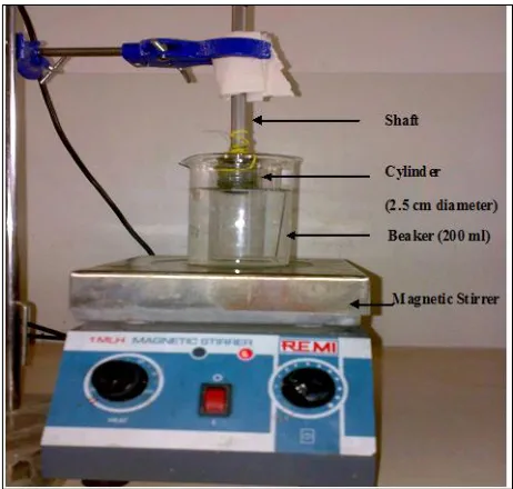
taken, washed and tied on to one end of the glass cylinder (2.5
cm diameter, open at both ends). One end of cylinder was
attached to the shaft of USP dissolution apparatus and then
suspended in 200 ml of dissolution medium in such a way that
membrane just touches the STF, maintained at 37 ± 1°C. The
dissolution medium was stirred using magnetic stirrer at 50
rpm. 1 ml of the formulation was added on the other side of the
cylinder which acts as a donor compartment. Aliquots were
withdrawn at intervals of 1 h and replaced by equal volumes of
dissolution medium. Aliquots were analyzed by UV
spectrophotometer at 274.2 nm [16].
Isotonicity study
Isotonicity is one of the important characteristics of the
ophthalmic formulations and it has to be maintained to prevent
tissue damage or irritation to the eye. Isotonicity testing of all
developed formulations was carried out as per the method
reported by Verma et al. [13]. The shape of red blood cell
(bulging or shrinkage) was compared with standard marketed
ophthalmic formulation containing Ciprofloxacin HCl.
Ex-vivo permeation study
Excised goat cornea was utilized for determining the
permeation across the corneal membrane. Whole eyeball of
goat was obtained from a slaughter house and transported to
laboratory in cold condition (4ºC) in normal saline. The cornea
was carefully removed along with a 5-6 mm of surrounding
scleral tissue and washed with cold saline. The washed cornea
was then kept in cold freshly prepared STF [3]. Corneal
membrane previously soaked overnight in STF was taken and
same procedure was followed for studying permeation of the
drug as that for dialysis membrane.
Sterility test
All ophthalmic preparations need to be sterile and therefore the
test for sterility is most important evaluation parameter. With
the help of a sterile pipette or with a sterile syringe or a needle
2 ml of the formulation was removed from test container and
was aseptically transferred to sterilized fluid thioglycolate
medium (20 ml). The inoculated media were then incubated for
not less than 14 days at 30 to 35°C and observed for bacterial
growth, if any [12].
In vitro antimicrobial efficacy
Antimicrobial efficacy study was carried out to ascertain the
biological activity of sol-to-gel systems against
micro-organisms. Agar diffusion test employing “cup plate technique” was used to determine antimicrobial efficacy.
Sterile solution of Ciprofloxacin HCl (as a standard) and the
developed formulations (test solutions) were poured into cups
bored into sterile nutrient agar gel previously seeded with test
organisms (Staphylococcus aureus). The plates were incubated
for 24 h at 37°C after allowing diffusion of solutions for 2 h.
The zone of inhibition (ZOI) measured around each cup and
was then compared with that of standard. Both positive and
negative controls were maintained throughout the study [17].
Stability study
The optimized formulations chosen based on the outcome of
various physicochemical evaluations was studied for stability
studies as per ICH guidelines. Formulations were kept at 40 ±
1ºC and 75% RH and evaluated at periodic intervals of 30, 60,
90 days for the drug content, clarity, pH, rheology, in vitro
drug release [10].
RESULTS AND DISCUSSION
The aim of the present work was to enhance the ocular
bioavailability of an antibacterial agent, Ciprofloxacin HCl by
formulating an in situ gelling solution based on LMP. The
developed formulations were characterized for the clarity, drug
content, viscosity, gelling capacity, mucoadhesive strength,
5
Drug excipients compatibility studies
From DSC spectrum of individual Ciprofloxacin HCl and LMP
it was observed that sharp endothermic peak was obtained at
319°C and 224°C. A sharp endothermic peak obtained at
170°C and 224°C in the DSC spectrum of combination
Ciprofloxacin HCl and LMP indicates thermal behavior of
drug (Ciprofloxacin HCl) and polymer (LMP). On comparing
individual DSC spectra with that of combination it was
observed that the drug and polymer are compatible with each
other as there was no interaction found between drug and
polymer.
Selection of buffer
Buffers play a crucial role in formulating ophthalmic drops.
Buffers not only contribute significantly to chemical stability
and clinical response but also influence the comfort and safety
of the product. The experiments done using various buffer
solutions indicated the drug was soluble in
acetate buffers of pH 4.6, 4.8 and 5.0 and in citrophosphate
buffer of pH 6.8 at the desired dosage level (0.3% w/v). The
solutions were stable to elevated temperatures and autoclaving.
However, their instability to light as evidenced by
discoloration of the exposed solutions necessitated their
packing in amber vials. The marketed eye drop was found to
have a pH of 6.6. Therefore we selected Citrophosphate buffer,
pH 6.8, as a vehicle for the development of eye drops.
Physicochemical evaluations
Appearance, clarity, pH, drug content
All the developed formulations were found to be clear and
were light yellow in color. Appearance was found to be
transparent for all formulations. Autoclaving done for terminal
sterilization had no deleterious effect on the physicochemical
properties of the formulations. The pH values for all the
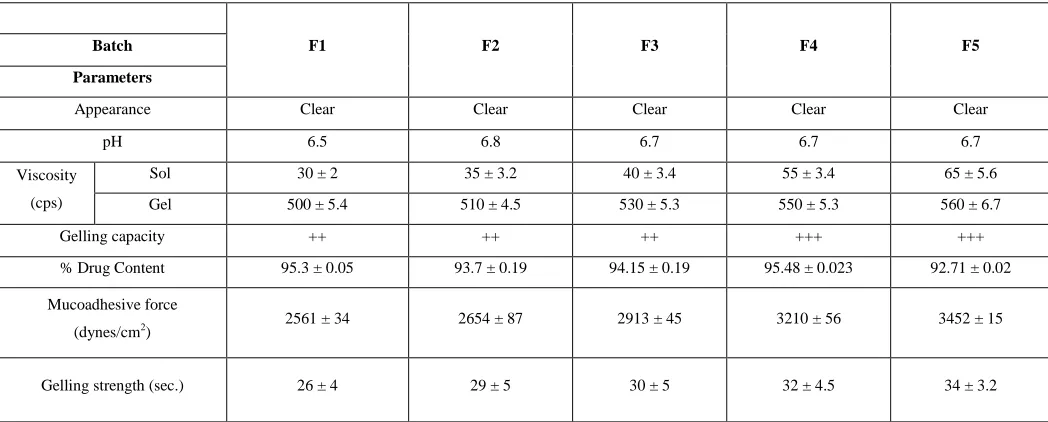
formulations have been depicted in Table 2.
Table 2: Physicochemical evaluation
F1 F2 F3 F4 F5
Batch Parameters
Appearance Clear Clear Clear Clear Clear
pH 6.5 6.8 6.7 6.7 6.7
Viscosity (cps)
Sol 30 ± 2 35 ± 3.2 40 ± 3.4 55 ± 3.4 65 ± 5.6
Gel 500 ± 5.4 510 ± 4.5 530 ± 5.3 550 ± 5.3 560 ± 6.7
Gelling capacity ++ ++ ++ +++ +++
% Drug Content 95.3 ± 0.05 93.7 ± 0.19 94.15 ± 0.19 95.48 ± 0.023 92.71 ± 0.02
Mucoadhesive force
(dynes/cm2) 2561 ± 34 2654 ± 87 2913 ± 45 3210 ± 56 3452 ± 15
Gelling strength (sec.) 26 ± 4 29 ± 5 30 ± 5 32 ± 4.5 34 ± 3.2
The pH was within acceptable range and hence would not
cause any irritation upon administration of the formulation.
Table 2 shows the result of percent drug content for all the
formulations. The drug content was also found to be in
acceptable range (92.00 - 96.00%) for all the formulations
6
formulation.Gelling capacity
LMP, the aqueous solution transforms into stiff gel in presence
of cations usually calcium ion was used as excipient in ion
sensitive in situ gelling system. However, the higher
concentration of LMP required for forming stiff gels results in
highly turbid solutions which are not clear for eye vision. A
selection of appropriate concentration of LMP without
disturbing the gelling capacity and rheological properties of the
delivery systems could be achieved between 2.2 to 2.75% w/v.
The formulation needs to have best possible viscosity that will
allow easy instillation into the eye as a liquid which would
then undergo a rapid sol-to-gel transition. Additionally, the gel
formed in situ should preserve its integrity without dissolving
or eroding for a prolonged period of time. In order to identify
the compositions suitable for use as in situ gelling, gelling
systems of various concentrations of LMP were prepared and
evaluated for gelling capacity. The outcomes of the gelling
capacity test of the developed formulations F1-F5 have been
depicted in Table 2 as dissolution time of the gel in STF.
Although all of them could form gel immediately as they make
contact with the STF equilibrated at 35 ± 1°C, the formulations
F4 and F5 were eroded by STF about 2 times slower than the
formulations F1, F2 and F3. As gelling is due to binding of
calcium ions (present in tear fluid) with LMP, the formulation
containing more LMP would possess stronger gel structure.
Since F4 showed ability to be retained as a gel for more than
few hours, it was selected as optimized formulation.
Viscosity
Formulations should have optimum viscosity which allows
easy instillation in to eye cavity, and should not hinder the
normal eye ball movement after instillation. The developed
formulations exhibited shear thinning or pseudoplastic
rheology. The viscosity was found to be directly dependent on
the polymeric content in each formulation. Table 2 shows the
viscosity values obtained before gelation and after gelation for
all the formulations using Brookfield’s Viscometer. No change
in the viscosity of the formulations as observed after
autoclaving. F5 showed the maximum viscosity of 65 Cps at
100 rpm whereas the minimum viscosity at 100 rpm was
shown by F1. Increased viscosity after gelation proves
excellent gelling ability of the formulations.
Mucoadhesive force
Ocular mucoadhesion depends upon the interaction between
the polymer and the mucin coat covering the conjunctiva and
corneal surfaces of the eye. Structurally, mucin consists of a
protein or polypeptide core with carbohydrate side chains
branching off the core. The polymer with many hydrophilic
functional groups (e.g. carboxyl group, hydroxyl group and
sulfate) can establish electrostatic and hydrophobic interactions
and hydrogen bond with the underlying surface. The
concentration of LMP has shown significant impact onto the
mucoadhesive force under physiological condition as depicted
in Table 2. The mucoadhesive force of 2.75% w/v LMP was
1.6 times greater than that of the 2.2% w/v. These can be
mainly attributed to the hydrogen bonds between the carboxyl
groups of the LMP molecule and the carboxyl groups of the
carbohydrate side chains of the mucin. Besides, electrostatic,
hydrophobic interactions and inter diffusion of the mucin and
the polymer also have some contribution. The mucoadhesive
force is an important physicochemical parameters for
prolonging ocular retention time and there by better therapeutic
effects. Detachment stress of F4 was found to be satisfactory in
comparison with other formulation. All the formulations
showed mucoadhesive force in increasing order i.e. highest
mucoadhesive force for F5 and lowest for F1. This could be
attributed to the concentration of LMP in the formulations.
Table 2 shows mucoadhesive forces of all formulations.
Gelling strength
For sustained release ability of polymer, measurement of
gelling strength is an important parameter. All the formulations
(F1 to F5) exhibited good gel strength (in the range of 25 to 50
sec).
In vitro drug release
The cumulative percent of Ciprofloxacin HCl released versus
time profiles for developed formulation is shown in Figures 1
7
Figure 1: in- vitro drug release, %CDR (membrane less method)
Figure 2: in - vitro permeation study, %CDR
Initially drug released study was carried out with
membrane-less method. F1 shown drug released around 100.29% in 8 h.
While formulations F2, F3, F4, F5 released 99.86%, 97.38%,
94.12%, and 93.22% of drug respectively in 8 h, which
indicated slow release of drug with increased concentration of
LMP. Further release study was carried out till 100% release of
drug through cellophane membrane (Figure 2) and it
demonstrated that the formulations F4 and F5 had relatively
better sustained-release effect as compared to other
formulations. F4 releases 99.84% of drug and F5 releases
98.08% of drug in 11 h. This was indicating that F4 can be
used as an ophthalmic sustained release drug delivery system.
Hence F4 was selected as optimized formulation on basis of %
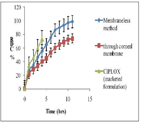
drug released. Finally optimized formulation shows 27.01% cumulative drug release after 1 h and 98.81% after 11 h by
membrane-less method as shown in Figure 3.
Figure 3: Modified USP dissolution apparatus
Drug release kinetics
In order to investigate drug release mechanism, the release data
was analyzed by PCP disso software fitted to models
representing Zero order, First order, Higuchi and Korsemeyer
Peppas equation. The examination of coefficient of correlation
(r2) values for the formulation F4 indicated that Korsemeyer
Peppas equation was more suitable fit to drug release
mechanism from developed ion activated in situ gelling
system.
Korsemeyer Peppas equation:
Mt/M∞ = Ktn
Log (Mt/M∞) = log K + n log t
Where Mt/M is the amount (%) of Ciprofloxacin HCl released
at time t,
n is the diffusional exponent,
K is the apparent release rate.
Our data shows that the release index (n) of the formulations
studied was 0.69 which suggest drug release taking place by
combination of diffusion and dissolution from the dosage form.
The release mechanism for semisolid vehicles containing
dissolved drug was found to be non-fickian or anomalous
involving both diffusion and polymer relaxation (0.5< n< 1).
The value of n suggest that Ciprofloxacin HCl release from
formulation was dependent on two simultaneous processes:
water migration into the in situ gelling system and drug
8
Isotonicity study
In order to be nonirritant to the ocular tissues, formulation
must be isotonic in nature. The shape and size of red blood cell
was found to be same or nearly same as that of blood cell
suspended in the solution of standard marketed formulation.
Hence it can be concluded that developed formulations were
isotonic.
Ex-vivo permeation study
Ex-vivo trans-corneal permeation studies of Ciprofloxacin HCl
formulations were conducted and higher and prolonged permeation across goat cornea was observed with LMP based formulation as compared to marketed eye drop. This might be
attributed to the gelling ability of LMP in presence of STF. Ex-vivo Trans-corneal permeation shows 22.2% cumulative drug
release after 1 h and 73.76% after 11 h as shown in Figure 4.
Figure 4: Drug releasestudy (%CDR): In-vitro drug release (membrane less method), Ex-vivo permeation study foroptimized
and marketed formulation
Ex-vivo Trans-corneal permeation study was also carried out
for marketed formulation (Ciplox) containing Ciprofloxacin HCl. It releases same % of drug in 4 h. This indicates that LMP based formulation increases the residence time of drug.
Sterility test
The preparations being examined for sterility were found to
pass the test for sterility as turbidity was not observed when all
the formulations were incubated for not less than 14 days at
30°C to 35°C in case of fluid thioglycolate medium. This was
clear evidence suggesting no microbial growth in the
formulations.
In vitro antimicrobial efficacy
The result of the antimicrobial efficacy tests shown that the
Zone of Inhibition (ZOI) values for the developed formulations
were approximately same as that of the ZOI values for the
standard antibacterial ophthalmic preparation. The satisfactory
ZOI values obtained for the formulations could be attributed to
the slow and prolonged diffusion of the drug from the
polymeric solution due to its higher viscosity. % inhibition for
F4 was found to be 96.10%.
Stability study
Stability studies were performed onto the developed
formulation at 40 ± 1°C, 75 ± 5% RH. Formulations were
screened at periodic interval for clarity, drug content, pH,
gelling capacity, viscosity and in vitro drug release. Every
formulation at the end of each month was found to be clear.
There was insignificant change in the pH of formulation for all three month’s batches. Drug content at the end of each month
was found to be 92-96% indicating drug content uniformity.
Gelling capacity and viscosity was in the acceptable range and
finally in-vitro drug release after 11 h was approximately
(97%) equal for all the batches. Hence optimized formulation
containing 2.5% w/v LMP was found to be stable in all
aspects.
CONCLUSION
Ciprofloxacin HCl was formulated successfully as in situ
gelling ocular drug delivery system using novel ion activated
bioadhesive polymer obtained from the heads of Helianthus
annuus L. Very simple and cost effective methodology was
adopted for the preparation of in situ gelling system was.
Polymer used in present research work proved its utility by
improving residence time and in turn bioavailability of the
Ciprofloxacin HCl by prolonging drug release. The developed
formulation could serve as best alternative to the conventional
ocular formulations by the virtue of its method of preparation,
decreased frequency of administration and better patient
9
formulation containing 2.5% w/v of the novel polymerexhibited better potential for sustained topical ocular drug
delivery.
ACKNOWLEDGMENTS
Authors would like to thank to Principal, SSDJ College of
Pharmacy, Chandwad for providing all necessary facilities to
carry out present work. We are also grateful to Krishna Pectins,
Jalgaon, India and IPCA laboratories for providing gift sample
of Low Methoxy Pectin and Ciprofloxacin hydrochloride
respectively.
REFERENCES
1. A. S. Mundada, J. G. Avari., Crit. Rev. Ther. Drug Car. Syst.2009, 26(1), 85-118.
2. K. Jarvinen, T. Jarvinen, A. Urtti., Adv. Dr. Del. Rev.1995, 16, 3-19.
3. H. Gupta, M. Aqil, R. Khar, A. Ali, A. Bhatnagar, G. Mittal, S. Jain., A. A. P. S. Pharm. Sci. Tech. 2009, 10, 540-546.
4. B. Nanjawade, F. Manvi, A. Manjappa., J. Cont. Rel. 2007, 122, 119-34.
5. B. Sharma, L. Naresh, N. Dhuldhoy, S. Merchant, U. Merchant., Tim. Fo. Proc. J. 2006, 11, 44-51.
6. C. Dollery., Therape. dru.1999, 230-232.
7. M. Iglesias, J. Lozano., J. Fo. Engg. 2004, 62, 215-223.
8. M. Bodhankar., Int. J. Pharm R. & D. 2011, 3, 212-222.
9. B. Bhoyar, V. Agnihotri, M. Bodhankar., Int. J. Res. Pharm. Chem. 2011, 1(3):591-600.
10. Srividya B, Cardoza R, P. Amin., J. Con. Rel. 2001, 73, 205-211.
11. R. Asasutjarita, S. Thanasanchokpibullb, A. Fuongfuchatc, S. Veeranondha., Int. J. Pharm.2011, 411, 128-135.
12. M. Reddy, S. Firoz, R. Rajalakshmi, C. Ashok., Int. J. Innov. Pharm. Res.2011, 2(2), 131-138.
13. L. Verma, M. Sakir, N. Singh, R. Gilhotra, S. Mehan., Int. J. Pharm. Profes. Res. 2010, 1, 137-144.
14. M. Patel, H. Thakkar, P. Kasture., Int. J. Pharm. Sci. 2010, 2, 116-120.
15. R. Gilhotra, M. Mahur, R. Saroot, N. Gilhotra, D. Mishra., Acta. Pharm. Sci. 2010, 52, 145-154.
16. S. Vodithala, S. Khatry, N. Shastri, M. Sadanandam., Int. J. Biopharm. 2010, 1(1), 39-45.