GENOMIC ORGANISATION AND TRANSCRIPT
IDENTIFICATION IN THE CLASS II REGION OF THE
HUMAN MAJOR HISTOCOMPATIBILITY COMPLEX
A thesis submitted for the degree of Doctor of Philosophy by
Amanda Jackson
Human Immunogenetics Laboratory
Imperial Cancer Research Fund
44 Lincoln's Inn Fields
London WC2A 3PX
and
Department of Genetics and Biometry
University College
Gower Street
London
ProQuest Number: 10106022
All rights reserved
INFORMATION TO ALL USERS
The quality of this reproduction is dependent upon the quality of the copy submitted.
In the unlikely event that the author did not send a complete manuscript and there are missing pages, these will be noted. Also, if material had to be removed,
a note will indicate the deletion.
uest.
ProQuest 10106022
Published by ProQuest LLC(2016). Copyright of the Dissertation is held by the Author.
All rights reserved.
This work is protected against unauthorized copying under Title 17, United States Code. Microform Edition © ProQuest LLC.
ProQuest LLC
789 East Eisenhower Parkway P.O. Box 1346
ABSTRACT
The aim of the work described in this thesis was to provide a complete picture
of the class II region of the human major histocompatibility complex (MHC),
identifying all the genes present, their genomic organisation and function.
Completion of the class II cosmid map was achieved by cloning two
remaining regions: 1) 40 kb of the DNA between the D M B and LM P2 genes
and 2) 24 kb of the DNA between the genes D P A l and D N A .. This map facilitated the localisation of a retinoid receptor (3 gene, hRXRp, 60 kb
centromeric of D P B 2 and eight cDNAs, one of which encoded a pseudogene
with homology to a rabbit muscle phosphatase inhibitor-2 (IPP-2) gene. Exon
amplification revealed two exons within 2.2 kb centromeric of D M B which
could be alternate splice products of the D M B gene.
Three cDNAs composed of two exons and one intron, mapping between
the D M B and L M P 2 genes, were isolated from cDNA libraries. The three
transcripts were not detectable on northern blots. Four unspliced transcripts contiguous with genomic DNA from within the class II region, were present in
cDNA libraries. All seven transcripts probably result from aberrant mRNA
transcription. Screening and partial sequencing of cosmids covering the class II
region identified no other transcripts between DPB2 and D R B l.
Experiments were designed to investigate the evolutionary origins of the
TAP and L M P genes. To this end, TM f-related genes on other chromosomes were investigated for linkage to TAP-related sequences. Three cDNAs were
identified, mapping within 12 kb of the LM P7-related gene, M B l, on
chromosome 14. No sequence homology between the cDNAs and TAP genes
was observed. The M B l gene consisted of three exons and two introns. This
organisation is very different from L M P 7which is composed o f six exons and
five introns. Hence, it is likely that M B l and L M P 7 are separated by
ACKNOWLEDGEMENTS
I would like to thank my friends and colleagues who have helped to make my
time at the ICRF such an enjoyable experience. Firstly, I gratefully
acknowledge my supervisor, John Trowsdale, for his guidance and support.
Next, I would like to thank members of the Human Immunogenetics
Laboratory, both past and present: Gordon Allan, M onica Belich, Ian
Campbell, Will Foulkes, Richard Glynne, Ulrike Griineberg, Jethro Herberg,
Adrian Kelly, Lesley-Anne Kerr, Pat Miller, Ian Mockridge, Beatrice
Plougastel, Stephen Powis, Jiannis Ragoussis, Derek McCusker, Sheetal Shah,
Frances Sanderson, Claire Thomas and Marieka van Ham. Particular thanks
must go to Louise Hosking for her friendship no matter how bad my moods
and to Philippe Sanséau who is, without a doubt. My Favourite Frenchman. I am also indebted to Stephan Beck, Fiona Francis, Gabriele Senger, Denise
Sheer and my external supervisor Yvonne Edwards. I am particularly grateful to
my parents, Roger and Edwina, and my sister Zoë for their continued support
and encouragement. Many thanks to my friends and flatmates Jill Amos, Robin
Dey, Wendy Douglass, Susan Kettle, Erinna Laffey, Robert Mettler, Ann
Smith and Ashley Vaughan for many memorable evenings. Last, but by no
means least, I would like to thank William Newell for his constant support and
C O N T E N T S
Chapter 1
The Human Major Histocompatibility Complex
1 .1 . Discovery and localisation of the MHC 10
1 . 1 .1 . Class I molecules 10
1 . 1 .2 . Class n molecules 13
1 . 1 .3 . Class i n molecules 14
1 .2 . Function of the MHC class I and class II loci 14 1 .3 . Structure of MHC class I and class E molecules 17
1 .4 . Antigen processing 23
1 . 4 .1 . The class I antigen processing pathway 23 1 . 4 .2 . Function of the transporters associated with antigen 25
processing (TAPs)
1 . 4 .3 . Alternative class I processing pathway 29 1 . 4 .4 . Involvement of the proteasome in class I antigen processing 30 1 . 4 .5 . Heat shock proteins and antigen processing 31 1 . 4 .6 . Antigen processing by the class E pathway 33 1 . 4 .7 . Function of the invariant chain (E) 33 1 .4 .8 . Involvement of DM in class E antigen processing 35
1 .5 . Polymorphism of the MHC 36
1 .6 . Molecular genetics of the MHC 38
1 . 6 .1 . Physical location of the MHC 38
1 . 6 .2 . Mapping the MHC region 40
1 .6 .3 . The class E region 43
1 . 6 .4 . The class I region 47
1 . 6 .5 . The class IE region 49
1 .7 . Evolution of the MHC 51
1 .8 . The MHC gene cluster 53
1 .9 . The MHC and disease associations 55
1 .1 0 . Approaches to finding novel genes 58
Chapter 2
M aterials and Methods
2 . 1 . Solutions 62
2 . 2 . Screening recombinant DNA libraries 65
2 . 2 . 1 . cDNA libraries 65
2 . 2 . 2 . Cosmid hbraries 66
2 . 3 . Preparation of DNA probes 67 2 . 3 .1 . Kinase reaction to label oligonucleotides 68
2 . 4 . Filter hybridisation 69
2.5. Transformation of bacterial cells with DNA by 70 electroporation
2 . 5 . 1 . Preparation of electro-competant E. coli cells 70
2 . 5 . 2 . Electroporation 7 0
2 . 6 . Preparation of DNA from transformed bacterial cells 71 2 . 6 . 1 . Small scale plasmid preparations (mini-preps) 71 2 . 6 . 2 . Large scale plasmid preparations (maxi-preps) 72
2 . 6 .3 . Small scale PI preparations 73
2 .7 . Restriction endonuclease digestion of DNA 7 3
2 . 8 . Electrophoresis of DNA 74
2 . 8 . 1 . Agarose gel electrophoresis 74
2 . 8 .2 . PFGE of PI digests 74
2 . 9 . Southern blotting 75
2 .1 0 . Sequencing of double stranded plasmid DNA 75
2 .1 1 . Shotgun sequencing 77
2 .1 1 .1 . Vector preparation 77
2 .1 1 .2 . Insert preparation 78
2 . 1 1 .3 . Ligation 78
2 .1 1 .4 . Preparation of competant cells 79
2 .1 1 .5 . Transformation 79
2 . 1 1 .6 . Template preparation 80
2 .1 2 . RNA preparation and manipulation 80
2 . 1 2 .1 . Isolation of mRNA 80
2 . 1 2 .2 . Isolation of poly (A)+ RNA 81
2 . 1 2 .3 . Electrophoresis of RNA 82
2 .1 2 .4 . Hybridisation of RNA blots 82
2 .1 3 . Exon amplification 83
2 .1 4 . PCR amplification of exon trapped products 83
2 .1 5 . Subcloning of PCR products 84
Chapter 3
Completion of an overlapping cosmid map of the human MHC class II region
3 . 1 . Introduction 85
3 . 1 . 1 . Cloning the DMB to LMP2 gap 86
3 . 2 . Results and discussion 89
between the genes DMB and LMP2
3 . 2 .2 . Construction of a cosmid library from the Y AC 11.2 89 3 . 2 .3 . Flow-sorted chromosome 6 cosmid library 93
3 . 2 .4 . PI genomic library 93
3 . 2 . 5 . Isolation of cosmid clones from the DMB to LMP2 94 region
3 . 2 . 6 . Isolation of a PI clone 96
3 . 2 . 7 . Cloning of the gap between DPAl and DNA 100
3 . 3 . Conclusions 103
Chapter 4
Genomic organisation and transcript identification in the class II region K E 3 to D M B
4 . 1 . Introduction 107
4 . 1 . 1 . Screening cDNA libraries with cosmid inserts to identify 107 novel genes
4 . 1 . 2 . Exon amplification of the MHC class II region 108
4 . 1 . 3 . Sequencing the class II region 109
4 . 1 . 4 . Localisation of a gene provisionally mapped to the MHC: 111 the human retinoid X receptor P gene (hRXRP)
4 . 2 . Results and discussion 112
4 . 2 . 1 . Exon amplification 112
4 . 2 . 2 . Exon amplification of the cosmid 0 19 A 114 4 . 2 . 3 . Exon amplification of the cosmid HA 14 115
4 . 2 . 4 . Localisation of the hRXRP gene 123
4 . 3 . Conclusions 127
Chapter 5
Screening for transcripts in the human class II region
D M B to D R B l
5 . 1 . Introduction 129
5 . 2 . Results and discussion 130
5 . 2 . 1 . Mapping a phosphatase inhibitor pseudogene to the 130 class II region
5 . 2 . 2 . Phosphatase inhibitors 130
5 . 2 . 3 . IPP-2 is a pseudogene 133
5 . 2 . 4 . Isolation of four cDNAs from the class II region 138
DMB to LMP2
5 . 2 .7 . Sequence analysis 151
5 . 3 . Conclusions 151
C h ap ter 6
Cloning and characterisation of genes linked to the -related proteasome gene, M B l
6 . 1 . Introduction 158
6 . 1 .1 . The proteasome 158
6 . 1 .2 . Involvement of the proteasome in the class I antigen 159 processing pathway
6 . 1 . 3 . LMP2 and LMP7: two proteasome subunits localised 160 within the MHC class II region
6 . 1 . 4 . Two proteasome components related to LMP2 and LMP7 162 6 . 1 .5 . Evolution of the LMP/TAP gene cluster: are the LMP-related 163
proteasome genes, M Bl and delta, linked to T4P-related genes?
6 . 2 . Results and discussion 164
6 . 2 . 1 . Isolation of cosmids encompassing the M Bl and delta 164 genes
6 . 2 . 2 . Isolation of cDNA clones with MB4 cosmid insert 164 6 . 2 . 3 . Partial nucleotide sequence of cDNA clones encoded 165
within the cosmid MB4
6 . 2 . 4 . Initial mapping of the cDNA clones M il, M14 and M15 166 6 . 2 . 5 . Genomic sequencing of cosmid MB4 166
6 . 2 . 6 . Genomic organisation of M Bl 172
6 . 3 . Conclusions 174
C h ap ter 7
Concluding discussion
7 . 1 . Genomic analysis of the human MHC class II region 178 7 . 2 . Isolation of genes linked to the LMP-related proteasome gene, MB 1 187 C h ap ter 8
R eferen ces
FIGURES
1 .1 . Molecular genetic map of the MHC 11
1 . 2 . Schematic representation of the MHC class I and class II 18
molecular structures
1 .3 . Diagrammatic representation of the three-dimensional 20 crystal structure of HLA-A2
1 . 4 . Diagrammatic representation of the three-dimensional 22
stmcture of class II HLA-DRl superimposed on class IHLA-A2
1 .5 . Schematic model of the class I antigen processing pathway 32 1 .6 . Schematic model of the class II antigen processing pathway 37
1 . 7 . Position of the MHC on chromosome 6 41
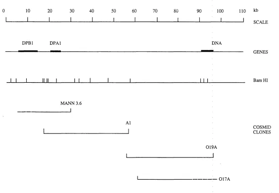
3 . 1 . Map of the class II region showing areas uncloned in cosmids 85
3 . 2 . Map of the MHC class II region showing deletion mutants 87
spanning the area
3 . 3 . PFGE mapping of the class II region 90 3 . 4 . Cosmid and PI map of the class II region between the genes 95
DMB and LMP2
3 . 5 . Flow-sorted chromosome 6 cosmid library hybridisation 97 3 . 6 . Southern blot analysis of cosmids A12 and A15 98 3 . 7 . FISH mapping of cosmids A12 and A15 99
3 . 8 . FISH mapping of PI clone, DPI 101
3 . 9 . Southern blot analysis of cosmids mapping to the region 102 telomeric of DPAl
3 .1 0 . Cosmid map of the class II region between DPBl and DNA 104 3 .1 1 . Complete YAC and cosmid map of the class II region 106
4 . 1 . Map of the class II region from KE3 to DMB 107
4 . 2 . Schematic representation of exon amphfication 113
4 . 3 . Nucleotide and amino acid sequence of exons HA 14-2 116
and HA 14-4
4 . 4 . Southern blot analysis of exons HA 14-2 and HA 14-4 118
4 . 5 . Determination of positions of sequences homologous 120
to exons HA 14-2 and HA 14-4 in the mouse MHC
4 . 6 . Zoo blot analysis of exon HA 14-4 122
4 . 7 . FISH mapping of cosmid H7 124
4 . 8 . Map of the class II region showing locahsation of 125
5 . 1 . Map of the class n region from DM5 to D/M 129 5 . 2 . Cosmid map of the class II region between DMB and LMP2 131 5 . 3 . Southern blot analysis of cDNA P I9 132 5 . 4 . Nucleotide and amino acid sequence of human IPP-2 134
cDNA P19
5 . 5 . Genomic Southern blot and Zooblot analysis of cDNA P I9 136 5 . 6 . Northern blot analysis of human IPP-2 137 5 . 7 . Amino acid sequence comparison of human and rabbit 139
IPP-2 cDNAs and human genomic IPP-2
5 . 8 . FISH mapping of human IPP-2 140
5 . 9 . Cosmid map of the class II region 142
5 .1 0 . Nucleotide and amino acid sequence of cDNA SI 143 5 .1 1 . Splicing patterns of cDNAs S 1, S2, S3 and S4 145 5 .1 2 . Nucleotide and amino acid sequence of cDNA S4 147 5 .1 3 . Nucleotide and amino acid sequence of class I- 148
homologous sequence from cosmid A15
5 .1 4 . Map of the class II region showing cosmid inserts 150 screened onto cDNA libraries
5 .1 5 . Expressed transcripts of genomic DNA, mapping 152 to the class II region, isolated from cDNA hbraries
5 .1 6 . Genomic sequence analysis of the class II region 153 6 . 1 . Southern blot analysis of cDNA clone M14 167 6 . 2 . Southern blot analysis of cDNA clone M 15 168
6 . 3 . Southern blot analysis of M Bl 169
6 . 4 . Partial nucleotide and amino acid sequence of cDNA 170 clone M 14
6 . 5 . Partial nucleotide and amino acid sequence of cDNA 171 clone M l5
CHAPTER 1: INTRODUCTION
1. The Human Major Histocompatibility Complex
The major histocompatibility complex (MHC) is one of the best characterised
regions of the human genome. Situated on chromosome 6, region p21.3, it spans
3.8 Mbp of DNA and contains over 100 genes (figure 1.1). These include
2^ class II genes at the centromeric end of the MHC and 34- class I-associated genes at the telomeric end. Between these two regions is a
heterogeneous collection of 45 class III genes including those for
complement components C2, C4 (C4A and C4B) and factor B together with three
members of the Hsp70 gene family, and the cytokines tumour necrosis factor (TNF) a and p.
Several features of the MHC have stimulated the enormous amount of work
on this gene cluster: it encodes molecules which play a central role in the
regulation of the immune response. Class I and II genes are highly polymorphic
and are associated with a large number of diseases, many of the autoimmune type.
The MHC represents 1/750th of human genetic material and can be considered as a model for the molecular organisation of the human genome. This chapter will
discuss the MHC in three sections: 1) general background including classical
studies of the MHC; 2) genomic organisation of the MHC and 3) outline of
research carried out during the course of this study.
1.1. Discovery and localisation of the MHC
1.1.1. Class I molecules
The MHC was originally discovered because of the role of its products in the
rejection of transplants made between incompatible individuals. The first
experiments identifying a phenotype controlled by the MHC were carried out by
Little and Tyzzer in 1916. It was shown that the growth of tumours transplanted
from one strain of inbred mouse to another were dependent on the strains selected
as donor and host (Little and Tyzzer, 1916; Klein, 1986). It was concluded that
the trait of susceptibility to a grafted tumour was genetically determined. In 1937
Gorer carried out a series of experiments to investigate the genetic basis of tumour
KE3
KE4
RING2 C0LIIA2 DPBl RING1\ KE5i DPA2|DPA1
i ' D P B 2 ' I I
10
□ DDO
RING9
DMA TAP1\LMP7 DQB2
RING3 I LMP2\ \iTAP2 IDQA2 DQB1
DNA I IdMB \ \ IiDOB I IDQB3 IDQA1 DRB1 I DRB3 DRBSl
DRB2 DRA
D D
D3I
0 0
□ □ □
C l a s s II rc !> io ii300 400 500 600 700 800 900
G16 G15
/ G14 / / G13
EDGD □
CYP
21 P Bf Hsp70 LTB
RDI G4 8 1 4 4 TNF
0 S G \ C 4 B i / C 4 A \ G10 G9 2 1 | G7b G6 G 5 | G 3 G l \ ' AB G1 2 ' A \ 1/ /g i i\I C 2 'G9a Ga\ 11 /G7a G7l BATS \ I I G2l Ml/ BAT1
1 cv
CYP 21
21 fXA'
i n i m f i n n o i
o h i o m d d
q
1 10 0 1200 1300 1400 1500 1600 1700 1800 1900
TEN LIKE IV-2 P5.3
OCT-3 X HSR1 VI
17 B
1 1
C
1
1 TUBB _ l
1
_|P5-1
II
lE
II
30 92
1 I I
PS-2 80 VII-3 \II-V
\
\
121
/70 16 P5-5 7/IV-3 I I 90
/ ' / P 5 - 4 |h Ig 175 F
1000
C l a s s III r o ^ i o n
2000
C l a s s I r e n i o n
2100 2200 2300 2400 2500 2600 2700 2800 2900 3000 3100 3200 3300 3400 3500 3600 3700 3800 3900 4000 ! ' c la ss II II a n d |J K tiics
I I cidla K cn g in c
p rciiL asu m c-lik c g e n e s A B L ir a n sp o r ie r gen es
IA l’2 l 2 1 O H g e n e s e iim p le -n ie m g e n e s l i s p ’ ll
o th e r g e n e s nt k n o w n iiin e iin n
various crosses. A transplantable tumour from an albino mouse was transferred to
all animals and its growth or regression monitored. The albino mice possessed an
antigen (called antigen II) in their erythrocytes, detectable with sera from
immunised rabbits, which was lacking in the blacks. Haemagglutination
experiments were carried out on the animals to test for the presence of antigen II.
A correlation was found between erythrocye agglutination by rabbit antiserum
containing antibody II and tumour rejection, leading to the conclusion that
"antigen II must be present in the tissues of the host, otherwise the tumour will
regress" (Gorer, 1937). The study of blood groups in man and animals had shown
that antigenic differences were determined by dominant genes and, in the light of
Gorer's experiments, it was suggested that the genes determining susceptibility to
tumour transplantation might be identical with those determining antigenic
differences. Snell (1948) suggested that the genes concerned should be called
histocompatibility genes, denoted by the symbol H. Since one of these genes was
defined by the blood group antigen n, (Gorer, 1937) the locus for the genes was
called H-2. Further genetic linkage and serological studies determined that the H-
2 genotype carried two components, defined by the series of antigens H-2K and
H-2D (Amos et al., 1955). These antigens also played a major role in determining
the success of tumour transplantation.
The presence of histocompatibility antigens in humans was shown by a
series of observations. First, multitransfused patients were found to raise
antibodies to the transfused leucocytes of some donors but not others (Dausset,
1958). Second, women who had undergone multiple pregnancies contained
leucocyte antigens in their blood which induced fetomaternal leucocyte
incom patibility (Payne and Rolfs, 1958). This was demonstrated by the
production of leukoagglutinins in maternal sera which were capable of
agglutinating the leucocytes of some of their children. Family studies showed that
these leucocyte antigens, detected by leukoagglutinins, were present in seven
families for two generations and one family for three generations. This suggested
that the leucocyte factors were genetically transmitted to the offspring either as
dominants or récessives.
Initial attempts to classify leucocyte antigens into allelic systems used
antisera obtained from multitransfused patients. These studies were unsuccessful,
largely because the reagents were multispecific. Leucocyte agglutinins from
women sensitised during pregnancy are less complex and were used in
and Van Leeuwen, 1963). This system was based on a statistical analysis of the
reactivity patterns of sera from multiparous women with large panels of donors
and defined a single locus. The existence of a further leucocyte antigen system,
defining a locus independent of Group 4, was described in 1964 using similar
methods (Payne et al., 1964). These two loci were named HLA-A and HLA-B for human leucocyte antigen locus A and human leucocyte antigen locus B. It was
soon demonstrated by family and population studies that these loci were closely
linked (Ceppellini et al., 1967). Later, a third locus, HLA-C, was described which was found to be closely linked to both the HLA-A and HLA-B loci (Svejgaard et al., 1973). Survival of human tissue grafts was found to be determined by
matching the HLA-A, -B and -C loci which suggested that the HLA antigens were
analogous to the mouse histocompatibility (H-2) loci. These molecules were
classified together as the classical transplantation or class I antigens.
1.1.2. Class II molecules
The development of a histocompatibility test in vitro promoted the discovery of a fourth human histocompatibility locus. When lymphocytes from unrelated individuals were cultured together in the same tube they underwent cell division
and morphological transformation, a phenomenon called the mixed lymphocyte
reaction (MLR: Bach and Hirschhom, 1964). It was discovered that the MLR
reaction occurred with leucocytes matched for products at the HLA-A, -B and -C loci (Yunis and Amos, 1971), leading to the proposal that the HLA and MLR loci
were closely linked but genetically separable identities. Interpretation of data from
a number of sources, including MLR and antibody typing led to the concept of the
class II region, HLA-D. Further antibody typing and serological studies in HLA-
D matched individuals led to the discovery that the HLA-D region could be
subdivided into three loci known as the HLA-DP, -DQ and -DR loci (Tosi et al., 1978; Shaw et al., 1980). Combining these results led to the concept of the major
histocompatibility complex: a gene cluster involved in the survival of tissue
transplants.
The development of typing assays in the mouse provided evidence for the
existence of the mouse MHC class II region. Synthetic polypeptides were injected
into different inbred strains of mice and their ability to mount an immune
response tested. This response was found to be controlled by 'inunune response'
loci. These two genes {1-A and I-E) were found to be correspond to the human
HLA-DQ and -DR loci respectively, thus defining the mouse MHC class II region. 1.1.3. Class III molecules
In 1963 a mouse serum protein was described which mapped to the H-2 complex,
midway between the H-2K and H-2D loci (Shreffler and Owen, 1963). This was later identified, immunochemically, as being the complement component C4
(Meo et al., 1975). Similarly, in humans, two genes coding for C4 were found to
be closely linked to the HLA-B locus (O'Neill et al., 1978). Analysis of a family with complement component C2 deficiency revealed evidence for close linkage
between the C2 defect and the HLA loci (Fu et al., 1974). The factor B (Bf) locus
was assigned to a position between HLA-B and H L A -D based on family recombinant studies (Lamm et al., 1976). Further studies showed that the C4, C2
and B f loci were tightly linked and mapped to a region which later became known
as the class m region (Weitkamp and Lamm, 1982).
1.2. Function of the MHC class I and class H loci
A series of key experiments contributing to an understanding of the function of
class I and class II molecules was carried out by Zinkemagel and Doherty (1975;
Doherty et al., 1976). Different strains of mice were infected intracerebrally with
lymphocytic choriomeningitis virus (LCMV) and killed one week later. Their
cytotoxic T lymphocytes (CTLs) were tested for their ability to lyse target cells
(LCMV-infected fibroblasts) from mice with varying H-2 haplotypes. It was
discovered that the H-2*^ restricted target cells were lysed only by H-2*^
compatible virus CTLs. All the mouse strains produced CTLs in response to the
viral infection but in each case these lymphocytes were only able to lyse target
cells of the same MHC haplotype. This led to the suggestion that viral and H-2
components were present in a common complex which was recognised by T cells.
Studies investigating helper T cell activation arrived at similar conclusions
showing that T cell activation by antigens could only occur in an MHC-
compatible environment (Sprent, 1978). Purified mouse helper T cells were
injected into irradiated syntenic mice together with sheep erythrocytes to act as
donor and host mouse shared H-2 determinants. It was later determined that CTLs
recognise and kill infected target cells that share class I molecules of the MHC
with the host in which the CTL developed (reviewed by Swain and Dutton, 1980).
The products of the class II region were essential for accessory cells such as
macrophages to present antigens to helper T cells (Heber-Katz et al., 1983). Thus,
the concept of MHC-restricted antigen presentation was evolved in which
cytotoxic T cells and helper T cells recognise foreign antigen only in association
with self class I or class II molecules respectively.
At first it was not known whether the T cells recognised the antigen and
MHC molecule separately with different receptors or together with the same
receptor. Over subsequent years it became apparent that T cells had a single
receptor which recognised a single complex of MHC molecule in the form of a
peptide (Davis and Bjorkman, 1988). Initial experiments carried out on class II
molecules showed that an immunogenic peptide derived from chicken ovalbumin
(residues 323-329) showed high binding affinity for these molecules (Buss et al.,
1986). This observation was complemented by Babbit and coworkers who showed
that a 10 amino acid stretch of the hen-egg lysozyme protein could be presented in
H-2K mice to T cells, creating the structural determinant recognised by helper T
cells (Babbitt et al., 1985). Over subsequent years it became apparent that MHC
class II molecules bound peptides of 12-25 amino acids in length (Chicz et al.,
1992).
Similar binding studies were carried out on MHC class I molecules. It was
demonstrated that epitopes of influenza A viral nucleoprotein, recognised by
CTLs in association with MHC class I molecules, could be defined by short
synthetic peptides derived from the nucleoprotein sequence (Townsend et al.,
1986). MHC class I molecules are now known to bind peptides of 8-10 amino
acids in length (Saper et al., 1991).
The interaction between T cells and antigen involves the CD4 and CD8
glycoproteins found on the surfaces of T cells. The majority of CD4+ T cells are
of the helper phenotype, whereas most CD8+ cells are cytotoxic (Littman, 1987).
In general, class I molecules present peptides derived from endogenous proteins
to CD8+ cytotoxic T lymphocytes while class II molecules present peptides
derived from exogenous proteins to CD4+ helper T lymphocytes. The interaction
between MHC/antigen complex and the T cell receptor results in T cell activation.
production by B cells and assist in the activation of CTLs. Stimulated CTLs lyse
the cell presenting the foreign antigen (Klein, 1986).
A second mechanism by which class I and class II molecules play an
important role in determining the specificity of an individuals immune response is
through the selection of the T cell repertoire during development. Classical
studies on the control of the immune response in mice had indicated that the MHC
was responsible for determining whether or not an immune response could be
mounted against a given peptide (Benacerraf, 1981). It was shown that the ability
to mount an immune response was directly correlated with the affinity of purified
MHC molecules for that peptide (Babbitt et al., 1985; Buus et al., 1986). Thus
MHC molecules dictate, by their ability to selectively bind peptides from a protein
antigen, whether a T cell response can be generated against a given protein
antigen. The entire family of antigen-specific receptors on lymphocytes is
required to discriminate between antigens that are self and antigens that are
foreign. Recognition of non-self antigens activates the immune response to
eliminate the foreign molecules. The immune system learns to differentiate
between self and non-self antigens during development. This is accomplished by providing each lymphocyte with a unique receptor then selectively deleting or
inactivating the population of cells bearing anti-self receptors. This process, the
acquisition of self-tolerance, occurs by positive and negative selection of T cells.
Selection of T cells appropriate for antigenic peptides in association with
self-MHC, is developed in the thymus gland. Double positive CD4+CD8+ T cells,
bearing receptors which recognise self-MHC molecules (on epithelial cells) are
positively selected for differentiation into CD4+8" or CD4"8+ single positive cells.
Thus, CD4+8+ cells only differentiate into CD4+ or CD8+ mature T cells if they
come into contact with thymic epithelial cells of the MHC class II or class I
haplotype that are recognised by their receptor. This is known as positive
selection (Schwartz, 1989; Marrack and Kappler, 1988).
Negative selection also occurs in the thymus and is responsible for the
deletion of T cells with T cell receptors (TCRs) specific for peptides derived from
self-proteins bound to self-MHC molecules. These T cells would otherwise be
autoreactive (Schwartz, 1989). This event occurs at the CD4+CD8+ stage of T cell
development and involves participation of the CD4 or CD8 molecules in the
recognition o f MHC class I or II molecules, respectively. Evidence for this
a defect in the Ea genes possessed mature T cells utilising V pi7a, whereas strains which expressed 1-E normally, selectively deleted their V p i7 a positive T cells
(Schwartz, 1989). Negative selection is thought to be the major mechanism for
establishing immunologic self tolerance. However, such clonal deletion does not
remove all autoreactive T cells.
Many self-T cell epitopes, including all housekeeping gene products,
ubiquitous cell surface molecules and constituents of lymphoid and epithelial cells
are present within the thymus and can induce negative selection. However,
autoreactive T cells not expressed in the thymus, such as tissue specific antigens,
are not removed by clonal deletion. It has been postulated that self-antigens with
highly specific tissue distribution are controlled in the periphery (reviewed in
Nossal, 1994). Transfer of such self-antigens to local lymphatic tissue is thought
to enable induction of anergy, a phenomenon whereby lymphocytes can be
functionally silenced without being killed. The anti-self cells may receive a
nonlethal down-regulatory signal and be induced into a state of anergy. Such
anergic cells may remain susceptible to further signals on renewed contact with
antigen and may lead to eventual deletion. As yet, this process has not been fully
elucidated.
1.3. Structure of MHC class I and class II molecules
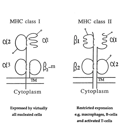
Class I and class II molecules were initially characterised in 1973 (Cresswell et al., 1973). They are cell surface glycoproteins composed of a|3 polypeptide chain
heterodimers (figure 1.2). Both molecules have four extracellular domains, each
approximately 90 amino acids long and encoded on separate exons (Malissen et
al., 1982). In class I molecules, three of these domains are contained within the 44kD a-chain ( a l , a2 and oc3 domains) which is encoded within the MHC and is
often referred to as the heavy chain. The fourth domain is composed of the 12kD, non-covalently bound light chain, P-2 microglobulin (p-2m). p-2m is encoded on
human chromosome 15. The a3 domain and p-2m are relatively conserved and
show amino acid sequence homology to immunoglobulin constant domains (Orr et al., 1979). The polymorphic a l and a 2 domains show no significant homology
to immunoglobulin constant or variable domains, but have been reported to show
weak sequence homology to each other (Orr at al., 1979). In the case of class II molecules, two domains are contained within the 34kD a-chain and two within
MHC class I
MHC class II
TM
Cytoplasm
I
s
TM
C ytoplasm
Expressed by virtually
all nucleated cells
Restricted expression
e.g. macrophages, B-cells
and activated T-cells
F igure 1.2. Schematic illustration of the structure of class I and class II
molecules. TM, transmembrane region. Glycosylation sites are indicated
domains are furthest away from the cell surface and are highly polymorphic whereas the a 2 and p2 domains are membrane proximal and less polymorphic.
Class I molecules are expressed by virtually all nucleated cells. Class II molecules
are expressed primarily by B lymphocytes, macrophages, dendritic cells and
activated T lymphocytes (Cresswell, 1987).
Recently, the crystal structures of both class I and class II molecules have
been determined (HLA-A2: Bjorkman et al., 1987a/b; HLA-Aw68: Garrett et al., 1989; HLA-B27: Madden et al., 1991; HLA-DRl: Brown et al., 1993). HLA-A2
consists of two pairs of structurally similar domains (figure 1.3). The a 3 and p2-m
domains form the base of the molecule and are both P-sandwich structures
composed of two anti-parallel P-pleated sheets. The a l and a 2 domains each
consist of an anti-parallel P-pleated sheet spanned by a long a-helical region. A
deep groove was observed on the top surface of the HLA-A2 molecule composed
of eight strands of anti-parallel P-sheets as a floor and two a-helical regions as the
sides. This was proposed to form the peptide binding groove of the class I
molecule. Additional electron density was observed in the groove, suggesting that
a peptide or mixture of peptides had copurified and cocrystallised with HLA-A2.
A polyalanine 9-mer was later modelled into the groove (Saper et al., 1991)
providing a model for peptide binding. It was suggested that the peptide in the
cleft was a nonamer in a largely extended conformation, with side chains protruding in a roughly alternating pattern.
The crystal structure of H LA-Aw68 also showed uninterpreted electron density (Garrett et al., 1989), which seemed to be different to that seen in HLA-A2. Comparison of the three-dimensional structures of HLA-A2 and HLA-Aw68
demonstrated how different pockets within the class I cleft were produced by
amino acid differences between the two molecules. This polymorphism resulted in
limited local structural changes, the backbone structure generally remaining
unperturbed. These structural changes provide a basis for the observed allelic
specificity in peptide binding. Since the affinity of antigenic peptides for
individual MHC class I molecules could depend on only a few of the peptide's
side chains fitting into the binding groove pockets, a wide range of peptides could
be accommodated by various combinations of filled and empty pockets.
Peptide binding specificity was further explored by the crystallisation of
Figure 1.3. D ia g ra m m a tic re p re s e n ta tio n o f the th re e -d im e n sio n a l crystal structure o f H LA -A 2. (a) Side on view show ing the a l and a 2 dom ains and the antigen binding groove, located betw een them , on the top surface o f the m olecule. T he a l and a 2 dom ains form a platfo rm w ith a single eig h t-stran d ed p -p leated sheet co vered by a helices, (b) T he antigen binding groove view ed from above (from B jorkm an et al.,
motif, since these side chains were bound in pockets in the bottom and sides of
the binding groove. A TCR could interact directly with the accessible peptide side
chains (PI, P4, P5, P6, P8).
Acid elution and sequencing of peptides bound to class I molecules revealed
specific consensus sequences. For HLA-B27, the most restricted peptide position was P2 (arginine) followed by PI and P9 (charged amino acids). These well
conserved positions are termed "anchor" positions. Further constraints were found
at positions P3 (hydrophobic) and P6 (nonpolar residues). The remaining
positions were unrestricted. Other anchor residues occur at different positions
dependent on the class I molecule (for example asparagine at position 5 in H-2D*^
(Falk et al., 1991).
Determination of the class II H LA -D R l crystal structure revealed overall similarity with that of class I molecules (figure 1.4; Brown et al., 1993). The two a-chain domains, a l and a2, of D R l superimpose closely on the corresponding a l and ^2 - ^ 1 subunit of HLA class I. The p i domain of D R l superimposes on the
a l domain of the HLA class I molecule, and the P2 domain less closely on the class I a 3 domain. The class II peptide binding groove has the same eight
stranded anti-parallel p-sheet floor and a-helical sides as that of the class I
groove. However, there are detailed changes in the class II structure so that the
peptide binding site is open at both ends. Electron density in the binding groove
indicates that long peptides, at least 15 residues, are bound in an extended
conformation, projecting out of both ends of the site. This is consistent with the
observation that class II peptides vary in length from 12-24 amino acids (Chicz et
al., 1992). This contrasts with peptides bound to class I molecules where octamers
and nonamers bind with extended conformations, and the N- and C-termini are bound in conserved, peptide terminal binding sites. A dimer of the class H a p
heterodimer was discovered in the three H L A -D R l crystals studied. It was suggested that class II molecules dimerised during their recognition by helper T
cells. This dimer of dimers could lead to an increased affinity for the CD4
receptor and to the crosslinking of T cell receptors to initiate cytoplasmic
signalling pathways.
Recently, binding of a decamer peptide to the class I molecule H IA -A 2 has been reported showing the carboxy terminal residue positioned outside the peptide
binding site (Collins et al., 1994). Rearrangement of several protein side chains at
the C-terminus created an opening through which the peptide was able to extend.
N-Figure 1.4. D iagram m atic representation o f the three dim ensional crystal structure o f class II H L A -D R l (blue) superim posed on class I H LA-A2
' ' t ' t w V'A
RED-#K T
terminal 10 amino acids of the 17 amino acid signal sequence of calreticulin. It is found bound to HLA-A2, in vivo, whereas the nonamer (MLLSVPLLL) is not. These observations suggest that peptides could extend out of the class I binding
site. It will be interesting to see if the novel surfaces presented by peptides
extending out of the ends of class I molecules are recognised by T cells.
1.4. Antigen processing
Antigen processing can be defined as the structural modification and trafficking of
protein antigens that enable the determinants recognised by T cells to interact with
MHC molecules. These antigenic determinants are then transported to the cell
surface for T cell recognition (Yewdell and Bennink, 1990). Two distinct
pathways are used to process antigens: the endogenous and the exogenous
pathways. Viruses, bacteria and protozoan parasites, such as those that cause
malaria and sleeping sickness establish their infections inside the host cells, where
antibodies cannot reach them. In infected cells, class I MHC molecules bind to
and display peptides derived from the parasite. The complexes of parasite peptides
and host class I molecules form antigens which can be recognised by cyotoxic T
lymphocytes (Engelhard, 1994). In this way, T lymphocytes can identify and kill
infected cells, selectively sparing healthy cells. Since class I-associated peptides
are produced from proteins that originate from the cytoplasm, this pathway
became known as the endogenous or class I pathway.
Specialised antigen processing cells (APCs), such as macrophages, roam the
body ingesting extracellular materials, degrading them to produce peptides and
presenting the peptides as antigens complexed with MHC class II molecules.
When helper T lymphocytes recognise a class E-peptide complex on these antigen
presenting cells, they secrete lymphokines that promote differentiation of immune
system cells. Class II molecules associate with peptides either derived from
proteins in the extra-cellular medium or from proteins located on the outer
membrane. This pathway of antigen recognition is known as the exogenous or
class n pathway.
1.4.1. The class I antigen processing pathway
The data summarised above led to the suggestion that the proteins recognised by
derived from them were subsequently transported to the cell surface in association
with class I molecules of the MHC (Towsend and Bodmer, 1989). Association of
class I molecules with peptide was proposed to occur in the endoplasmic
reticulum (ER). Supportive evidence came from a number of sources. Class I
antigen processing is blocked by brefeldin A which inhibits exocytosis by
interfering with normal vesicular traffic between the ER and Golgi (Yewdell and
Bennink, 1989). Similar effects were observed in antigen presenting cells infected
with a recombinant vaccinia virus expressing the adenovirus E19 glycoprotein
(Cox et al., 1990). E19 specifically binds class I molecules and retains them in the
ER. The effects of brefeldin A and E19 on antigen presentation indicate that
peptides enter the secretory pathway in a pre-Golgi compartment, presumably the
ER.
Association of class I molecules with antigen in the ER is supported by
studies using the mutant mouse cell line RMA-S (Townsend et al., 1989). These
cells express only 5% of the wild type level of class I molecules on their cell surface, even though heavy chains and P2-IÏI are synthesised in the normal
amounts. This results from a deficiency in the assembly of class I heavy chains with P2-m, the chains remaining trapped in the ER. Incubation of these cells with
high concentrations of the appropriate peptides enhances class I assembly and the
cells express more normal levels of class I molecules on the cell surface. Thus,
peptides stabilise the assembly of class I molecules, a process which occurs in the
ER.
During assembly, class I molecules are associated with an 88kD molecular
chaperone, calnexin, that is released on peptide binding (Jackson et al., 1994). The
ability of calnexin to influence the transport of incompletely assembled class I
molecules in Drosophila melanogaster cells was assessed. Stably transfected
Drosophila cell lines were prepared that expressed murine K^, D^, or heavy chains with or without p2-m in the presence or absence of calnexin. Expression of
calnexin was shown to slow intracellular transport of both peptide-deficient heavy chain-P2-ni heterodimers and free heavy chains. Calnexin also impeded the rapid
intracellular degradation of free heavy chains. Thus, calnexin might act to
facilitate assembly of class I complexes by retaining or protecting assembly
1.4.2. Function of the transporters associated with antigen processing {TAPs)
Since class I assembly occurs in the ER, there must be specific mechanisms by
which peptides are generated in the cytoplasm and transported across the ER
membrane. Insight into these mechanisms came from studies on cell lines with
defective class I presentation including the human mutant lymphoblastoid cell line
(LCL) 721.174 and the murine mutant RMA-S. These cell lines were derived in a similar manner by y-irradiation (LCL 721.174) or ethylmethane sulfonate (EMS)
mutagenesis (RMA-S) followed by several rounds of selection with anti-MHC
antibody plus complement (DeMars et al., 1984; Ljunggren et al., 1989). The
mutant cell lines show similar phenotypes. First, they retain most of their class I
MHC molecules in the ER in an incompletely assembled state (Salter and Cresswell, 1986; Cerundolo et al., 1990). Treatment of these cell lines with y-
interferon does not result in increased expression of class I MHC molecules.
Second, the mutants are unable to present intact protein antigens to class I-
restricted CTLs but can present exogenously supplied peptides. This leads to increased cell surface expression of stable class I molecules on the cell surface
suggesting that their class I molecules are fully functional (Hosken and Bevan, 1990). Third, the genetic defect in these cell lines is not due to defects in either the class I heavy chains or (Salter et al., 1985; Ohlen et al., 1990). These
observations have led to the hypothesis that these cell lines are defective in the
ability to transport antigenic peptides into the lumen of the ER.
A similar phenotype to those defined by the LCL 721.174 and RMA-S
mutants, called the d m (class I modification) phenotype, was observed in the rat (Livingstone et al., 1989; Livingstone et al., 1991). Natural alleles at the d m locus modify the specificity of class I antigen presentation and the time of residence of
newly synthesised class I molecules in the ER. It was therefore suggested that the
d m locus product was also involved in peptide loading or assembly of class I antigens.
The genetic location of the defects in LCL 721.174 was mapped to the class
n region of the human MHC on chromosome 6 (Cerundolo et al., 1990). LCL
721.174 has a deletion sparming from the DPB2 gene in the class II region to the com plem ent gene cluster in the class III region, thus mapping the gene
responsible for the antigen presentation defect within this interval. Similarly, the
genetic defect responsible for the RMA-S phenotype was mapped to the murine
locus was mapped to the rat MHC between the human DP homologue, R T l.H ,
and the DQA homologue RTLB^, (Deverson et al., 1990).
Two genes mapping to the region of the MHC known to encompass the
gene(s) responsible for the antigen presentation defect were discovered
simultaneously in the mouse, rat and human. These are now known as TAPI and
TAP2 for transporter associated with antigen processing. Initially they were known as HAM 1/2, PSFl/1 or RING 4/11, and M TPl/2 in the mouse, human and rat respectively (Deverson et al., 1990; Monaco et al., 1990; Trowsdale et al.,
1990; Powis et al., 1992; Spies et al., 1990). These genes showed protein
sequence homology to the ATP-binding cassette (ABC) superfam ily of transporters which includes the human and mouse multidrug-resistance proteins
(MDR: Hyde et al., 1990). Typically, eukaryotic ABC transporters consist of two
hydrophobic, membrane spanning domains and two ATP-binding domains. All
known eukaryotic members of this family (MDR, cystic fibrosis gene, and the
yeast STE-6 gene) contain all four domains within a single polypeptide. Thus, the products of the TAPI and TAP2 genes were the first members of the family that contained only two domains. It therefore seemed likely that the TAPI and TAP2
gene products functioned as a heterodimer (Monaco et al., 1990). The ABC
transporter superfamily includes over 30 members, each specific for a different
substrate. Transported substrates include sugars, inorganic ions, amino acids,
peptides and proteins (McGrath and Varshavsky, 1989; Felmlee et al., 1985).
TAPI and TAP2 were strong candidates for a specialised transport system that passed the peptide products of proteolysis from the cytosol into the ER (Trowsdale et al., 1990).
Evidence for such a function came from a number of sources. Transfection
of TAPI cDNA into mutant LCL 721.134 cells restored normal levels of surface
HLA-A2 and -B5 expression (Spies and DeMars, 1991). However, transfection of the same cDNA could not restore the defect in LCL 721.174 cells. Both 721.174
and 721.134 cell lines contain class I genes HLA-B5 and HLA-A2 of one MHC haplotype (the other haplotype having been deleted). However, surface HLA-B5
levels are not detectable in the mutants and surface HLA-A2 levels are only 50- 60% of that o f the parental line, as determined by fluorescence activated cell
sorting (FACS) analysis. 721.174 has a further deletion over the class II region
and 721.134 contains a point mutation in TA P I. Hence, transfection of TAPI
restores class I surface expression in 721.134 but not in 721.174 since both TAPI
to restore the antigen processing defect. Similarly, the defect in RMA-S could be
restored by transfection of either a rat or mouse TAP2 cDNA, leading to increased class I assembly, surface expression and antigen presentation (Attaya et al., 1992;
Powis et al., 1991).
An antiserum raised against a peptide representing the C-terminal 15 amino
acids of the TAPI protein sequence immunoprecipitated both TAPI and TAP2
proteins from human cells. This suggested that the two TAP genes encoded proteins that associated with each other, possibly as a heterodimeric complex
(Kelly et al., 1992). The same antiserum was used to precisely locate the TAPI
protein by high resolution immuno-electron microscopy on ultrathin frozen cell
sections. Immunogold particles were found specifically on the membranes of the
ER and cw-Golgi. Since the antibody was raised against a peptide from the C-
terminus of the T A P I ATP-binding domain and labelling occurred on the cytosolic side of the membrane, it was suggested that the TAPI protein was oriented in the ER and Golgi membranes with its ATP-binding domain in the
cytosol (Kleijmeer et al., 1992). Transfection of TAPI and TAP2 cDNAs in the cell line T2, which is derived from 721.174 and lacks both transporter genes, is
both necessary and sufficient for the rescue of class I surface expression
(Momberg et al., 1992).
The development of a TAP 1-1- knockout mouse has allowed investigation of the involvement of the TAP complex in selecting the T cell repertoire (Van Kaer et al., 1992). A deletion was introduced into the TAPI gene of embryonic stem cells by gene targeting, and mice homozygous for this mutation were produced. Cell lines from such mice lacked cell surface class I expression and were unable
to present endogenously synthesised proteins to CTLs. Exogenously added
peptide could, however, be presented. An analysis of the T cells from various
organs showed that CD4+CD8+ and CD4+ cells were present at wild type levels.
However, CD8+ T cells were not found in the blood, spleen or lymph nodes of the
mutant mice. This is consistent with a role for class I in the positive selection of
CD8+ cells in the thymus.
Reconstitution of cell surface class I expression using TAP genes in mutant cell lines, immunoprécipitation and localisation experiments strongly suggest a
An assay was developed to study the mechanism of peptide transport across
the ER membrane into microsomal vesicles (Koppelman et al., 1992). Peptides
were synthesised containing N-linked glycosylation acceptor sequences, which
serve as glycosylation substrates. Their transport into microsomes results in the
depletion of the pool of dolichol high-mannose oligosaccharides in the lumen of
microsomal vesicles. Glycosylation of a marker protein present in the microsomal
vesicles would then be inhibited. This assay showed no effect of ATP depletion,
nor inhibition of transport by an ATPase inhibitor, oligomycin.
In vitro translation experiments also provided evidence suggesting that peptide transport was ATP-independent (Bijlmakers et al., 1993). In vitro
translation of mouse class I heavy chains and p2-ni was performed in rabbit
reticulocyte lysate containing dog pancreas microsomes and radio-iodinated
peptide. Microsomes were recovered by centrifugation, and their contents
analysed on SDS gels. Radio-iodinated peptides, found in association with
microsomes, displayed reduced electrophoretic mobility when compared to
peptides recovered from the microsomal supernatant. This shift was found to be
due to the attachment of an endoglycosidase H-sensitive carbohydrate chain to the
peptides. Since the enzyme responsible for carbohydrate attachment was found
only in the microsome lumen and the process occurred in an isotonic buffer, it
was concluded that peptide translocation was ATP-independent. Furthermore,
ATP depletion did not inhibit glycosylation of radio-iodinated peptide.
In both of these experiments the microsome preparation would be of great
importance in interpreting the results. If, for example, the microsomes were leaky
for small peptides this would mask any ATP- and hence TAP-dependent transport.
Additional evidence for TAP-dependent transport has recently been described.
Peptide translocation was assessed in the TAP-deficient cell line T2 and in
T2 transfected with rat TAPI and TAP2 (NeeQes et al., 1993). Permeabilised cells were incubated with an 11-mer peptide containing an N-linked glycosylation
sequence. N-linked glycosylation of the peptide was used to measure peptide
translocation. Glycosylated peptide could be recovered from T2ITAP1+2 cells but not from T2 cells. This glycosylation depended on ATP being present.
Nonhydrolysable ATP analogues did not yield glycosylated peptide, suggesting
that ATP hydrolysis is required for TAPT+2-dependent peptide translocation into
the ER.
A further assay demonstrating TAP-dependent transport was carried out on
TAP+/+ and TAP-/- mice could both assemble exogenously added peptide. When
the reaction temperature was lowered from 37°C to 23°C peptide translocation into TAP-I-/+ microsomes was 4 fold higher than in TAP-1- microsomes. This difference in rates was ATP-dependent.
These results seem to provide strong evidence for TAP-dependent transport
of peptides across the ER membrane. However, conclusive evidence will only be
provided by the purification of recombinant TAPs, their incorporation into a synthetic lipid bilayer, and reconstitution of peptide transport in vitro.
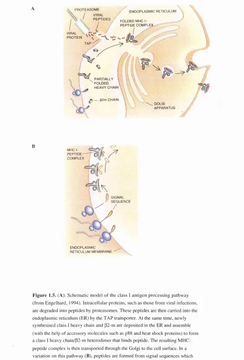
1.4.3. Alternative class I antigen processing pathway
Evidence for an alternative class I antigen processing pathway has also been
demonstrated (Henderson et al., 1992). Normal levels of H L A -A 2 class I molecules, complexed with peptides, were found to be expressed on the surface of
mutant T2 cells which lack both the peptide transporters TAPI and TAP2. Peptide sequencing revealed that the peptides were derived from the signal sequences of
cellular proteins.
Signal sequences are features of the amino terminal ends of newly
synthesised proteins that are heading to the cell surface or other internal cellular
compartments (reviewed in Sabatini et al., 1982). When ribosomes synthesise such proteins, the signal sequences ensure that the ribosomes will attach to the ER
before the protein is completed, directing the proteins to their final destination. As
the protein extrudes into the ER, an enzyme clips the signal sequence from the
proteins, providing a source of peptides that could associate with class I MHC
molecules.
Signal sequences are now known to be presented on the surface of normal
cells in conjunction with HLA-A2. Further work (Wei and Cresswell, 1992) has confirmed these results and shows that most signal-derived peptides bound to
HLA-A2 are longer than the nine residue peptides found in HLA-A2 isolated from non-mutant cells. The majority of HLA-A2 molecules isolated from T2 cells are unstable which may be due to the longer length of bound peptides. Trimming of
peptides may occur by specialised enzymes resident in the ER (Falk et al., 1990).
Peptides of more than nine residues in length may enter the ER and be trimmed to
the stable binding length by such enzymes. Trimming may occur in both the TAP-
dependent and signal-dependent pathways but may not be efficient if peptides
conclusive evidence exists to support this hypothesis. It has been suggested that
most peptides are processed by the established path. However, this new pathway
could provide an alternative processing route for specific pathogens.
1.4.4. Involvement of the proteasome in class I antigen processing
A pathway for antigen processing was proposed in 1989 (Townsend and Bodmer,
1989) suggesting that antigenic proteins were degraded in the cytoplasm into
peptides which were transported through the ER. These peptides then bound to
MHC class 1 molecules and travelled to the cell surface for presentation to T cells.
Biochemical evidence (described in section 1.4.2) has shown that the TAPI and
TAP2 genes are responsible for the transport of peptides through the ER and recently strong evidence has been produced to support the hypothesis that the
proteasome is responsible for peptide production in this pathway.
The discovery of the proteasome and its involvement in the class 1 antigen
processing pathway will be discussed in detail in chapter 6 and only a brief
outline of its function will be described in this chapter.
The eukaryotic proteasome is composed of 14-17 different polypeptide
subunits and has a sedimentation coefficient of 20S. It exhibits multiple peptidase
activities which cleave specifically after basic, hydrophobic and acidic amino acid
residues (Wilk and Orlowski, 1983). cDNAs encoding all the proteasome subunits
have been cloned and sequence comparisons show that they can be divided into two classes: a and p. The 20S proteasome forms the proteolytic core of a larger
protein complex, the 26S proteasome, which is responsible for protein
degradation in the ubiquitin pathway (Peters et al., 1993).
In 1982 a group of 16 proteins were precipitated from mouse cell lines, using
anti-MHC alloantisera, which were called Low Molecular Weight Proteins, or
LMPs (Monaco and McDevitt, 1982). The LMP and proteasome complexes were
subsequently shown to be similar in that they share most, but not all, subunits.
Two genes encoding shared subunits, LMP2 and LMP7, have been localised to the class n region of the MHC and are closely associated with the TAP peptide transporter (Glynne et al., 1991; Martinez and Monaco, 1991; Kelly et al., 1991b).
Incorporation of LMP2 and LMP7 into the proteasome enhances its capacity to cleave oligopeptides after hydrophobic and basic residues, and suppresses
cleavage after acidic residues (Gaczynska et al., 1993). This favours the
molecules. The proteasome is now thought to be responsible for the generation of
most peptides presented on MHC class I molecules, confirming its major role in
the class I antigen processing pathway (Rock et al., 1994).
1.4.5. Heat shock proteins and antigen processing
Recently, it has been suggested that heat shock proteins (HSPs) have a role in
antigen processing and presentation (Manara et al., 1993). Studies carried out on
normal antigen presenting cells demonstrated that the determinant recognised by
the anti-heatshock protein 72/73 monoclonal antibody (MAb) was constitutively
expressed on the cell surface of monocytes as well as of B cells. The ability of
monocytes to present an antigen to T cells was significantly decreased when
preincubated with an anti-HSP 72/73 MAb. This provides evidence for a role of
HSP72/73 in antigen processing. It has been suggested that HSPs constitute a
relay line in which the peptides, after generation in the cytosol by proteasomes,
are transferred from one HSP to another, until they are accepted by MHC class I
molecules in the ER (Srivastava et al., 1994).
Some HSPs are known to bind to a diverse array of molecules, including
peptides (Ang et al., 1991). It has been observed that immunisation of mice with the HSP gp96, isolated from a given virus-infected/transformed cell line, elicits an
antigen-specific MHC class I-restricted CTL response (Blachere et al., 1993).
This response is not elicited by gp96 itself but by virtue of the peptide that it
chaperones. It has been proposed that peptides are transported into the lumen of
the ER where they bind to gp96. Gp96 then transfers the peptides to MHC class I
molecules for antigen presentation to T cells (Li and Srivastava, 1993). As yet the
involvement of HSPs in the antigen presentation pathway has not been fully
elucidated but by combining all the data discussed in section 1.4 it is possible to
provide a model for class I restricted antigen processing (figure 1.5).
Antigenic proteins are degraded in the cytoplasm by the 20S proteasome.
Peptide products then traverse the ER membrane via the TAP complex. At least one TAP-independent processing route has been described which achieves
translocation of signal sequences across the ER membrane. Within the ER lumen
the peptides may then be bound to the HSP gp96 which passes them to the assembled class I heavy chain/p2-m complex. This complex passes to the cell
PR O T E A SO M E / VIRAL
' P E P T ID E S
/
EN D O PLA SM IC RETICULUM FOLDED MHC I-
PE PT ID E C O M PL EX VIRAL
PR O TE IN - ^ TA P
• PARTIALLY FO LD ED v ’\ HEAVY CHAIN
^ p2 m CHAIN
GOLGI A PPA R A TU S
B
MHC IP E IP T I D E -COM PLEX
1 SIGNAL / S E Q U E N C E
END O PLA SM IC RETICULUM M EM BRANE