REACTIVE OXYGEN SPECIES AND
THE ROLE OF ANTIOXIDANT
THERAPY IN INFLAMMATORY
BOWEL DISEASE
Andrew David Millar BSc MRCP
Submitted for the degree of Doctor of Medicine to the Faculty of
Medicine, University of London
October 1996
Gastrointestinal Science Research Unit
St Bartholomew’s and the Royal London School of Medicine and Dentistry
26 Ashfield Street, London E l 2AJ
ProQuest Number: 10046168
All rights reserved
INFORMATION TO ALL USERS
The quality of this reproduction is dependent upon the quality of the copy submitted.
In the unlikely event that the author did not send a complete manuscript and there are missing pages, these will be noted. Also, if material had to be removed,
a note will indicate the deletion.
uest.
ProQuest 10046168
Published by ProQuest LLC(2016). Copyright of the Dissertation is held by the Author.
All rights reserved.
This work is protected against unauthorized copying under Title 17, United States Code. Microform Edition © ProQuest LLC.
ProQuest LLC
789 East Eisenhower Parkway P.O. Box 1346
ABSTRACT
A proven role for antioxidant therapy in inflammatory bowel disease (IBD) would help establish the pathogenic importance of reactive oxygen species (ROS) and may provide new therapies with low toxicity. The major findings of the work for this
thesis were as follows:
• ROS production, as measured by the chemiluminescence response, of colonic biopsies from acetic acid-induced colitis in rats correlated with the macroscopic and microscopic scores of inflammation.
• A method for assessing the antioxidant actions o f novel IBD therapy was established by demonstrating that conventional antioxidants, and standard therapies for IBD, alter the chemiluminescence responses o f acetic acid-induced
colitis biopsies similarly to ulcerative colitis (UC) biopsies (previously published data). The novel compounds, amflutizole and LY231617 were potent
antioxidants.
• The chemiluminescence response of UC biopsies correlated with clinical disease activity and sigmoidoscopic scores, and with mucosal neutrophil infiltration.
• Recombinant human recombinant manganese superoxide dismutase (rh-MnSOD) was not an effective antioxidant using acetic acid-induced colitis biopsies. Pre treatment of rats with acetic acid-induced colitis with intraluminal rh-MnSOD did not alter the m acroscopic or m icroscopic scores o f inflam m ation nor chemiluminescence: Rh-MnSOD is probably not, therefore, a suitable agent for topical therapy in IBD.
• The iron chelators, desferrioxamine and 1,10-phenanthroline, reduced ROS production by inflamed biopsies from UC.
• A pilot trial of antioxidant nutrients, selenium, p-carotene, ascorbate, a-tocopherol and methionine, in active UC demonstrated no adverse events, remission in 4/10 patients, and significant improvements in stool frequency, rectal bleeding and sigmoidoscopic score, but not rectal mucosal histology, ROS production, or
plasma thiobarbituric acid reactive substances.
CONTENTS
Abstract i
Contents ii
Abbreviations xi
Index of tables xiii
Index of figures xvi
Index of colour plates XX
Acknowledgements xxi
Statement of originality xxii
CHAPTER 1 INTRODUCTION 1
Inflammatory bowel disease 1
Historical perspective 2
Epidemiology 2
Aetiology and pathophysiology 3
Genetic factors 3
Infectious agents 4
Mucin abnormalities 4
Vascular abnormalities 4
Platelets and thromboembolism 5
Mucosal permeability 5
Psychological factors 6
The immune response in inflammatory bowel disease 6 B-lymphocytes and immunoglobulin production 6
Influence of autoantibodies 7
Presence of immune complexes 7
Complement activation 8
T-lympbocytes and cell-mediated immunity 8
Neutrophils 8
Monocytes and macrophages 9
Eosinophils, basophils and mast cells 9
Adhesion molecules 9
Eicosanoids 9
Cytokines 11
Neuropeptides 11
Management of inflammatory bowel disease 13
Pharmacological therapy 13
Reactive oxygen species 14
The chemistry of free radicals in biology 14
Superoxide 15
Hydrogen peroxide 15
The hydroxyl radical 16
Fenton chemistry and the Haber-Weiss reaction as sources of the
Hypochlorous acid 16
Non-oxygen-centred free radicals 17
Sources of reactive oxygen species in biological systems 17 Reactive oxygen species in cellular respiration 17 Reactive oxygen species production by inflammatory cells 18
Superoxide production by polymorphonuclear leukocytes - the
‘respiratory burst’ 18
Action of neutrophil myeloperoxidase 19 Production of ROS by other inflammatory cells 20
The role of iron in biological tissues 20
Ischaemia-reperfusion injury and the role of xanthine oxidase 21
Arachidonic acid metabolism 21
Nitric oxide 22
Defences against damage from reactive oxygen species 24
Direct ROS scavengers 24
Ascorbate (vitamin C) 24
a-Tocopherol 26
p-Carotene 26
Glutathione 26
Selenium 27
Other endogenous non-enzymatic antioxidants 27
Enzymatic defences 27
Superoxide dismutase 27
Catalase 28
Glutathione peroxidase 28
Transition metal chelation 28
Pro-inflammatory consequences of excess reactive oxygen metabolite
production 29
Lipid peroxidation 29
Increased mucosal permeability 30
Increased intestinal secretion 30
Neutrophil adhesion and up-regulation of adhesion molecules 31
Interference with cell metabolism 31
Activation of the complement pathway 31
Release of pro-inflammatory mediators 32
Inactivation of antioxidant defence mechanisms 32 Activation of nuclear transcription factors 32 Beneficial effects of free radicals in biological systems 34
Antimicrobial defence 34
Maintenance of vascular tone 34
Reactive oxygen species in inflammatory bowel disease 34 Systemic reactive oxygen species in inflammatory bowel disease 35
Peripheral blood neutrophils 35
Peripheral blood monocytes 37
Reactive oxygen metabolites in the gastrointestinal tract in
Isolated intestinal phagocytes 37
Mucosal tissue in vitro 37
ROS in the intestinal lumen 38
Nitric oxide in inflammatory bowel disease 38 Antioxidant defence in inflammatory bowel disease 39 Antioxidant activity of drug therapy in inflammatory bowel disease 40
Aims of this thesis 42
Chapter 2 - Analytical methods and materials 42 Chapter 3 - Establishment of the acetic acid-induced model of colitis 42 Chapter 4 - Evaluation of antioxidant compounds using colonic
biopsies from the acetic acid-induced model of colitis in vitro 42 Chapter 5 - Chemiluminescence of human colorectal mucosal biopsies 43 Chapter 6 - The antioxidant and anti-inflammatory efficacy of human
recombinant manganese superoxide dismutase 43 Chapter 7 - The role of iron chelation as antioxidant therapy in
inflammatory bowel disease 43
Chapter 8 - Antioxidant nutrient therapy in active ulcerative colitis 43
Chapter 9 - General discussion 43
CHAPTER 2 METHODS AND MATERIALS 44
Statistical analysis 44
Computer-aided analysis 44
Graphing methods 44
Comparison of unpaired groups with non-parametric data 44 Comparison of paired groups with non-parametric data 44
Variability of assays 45
Calculation of the intra- and inter-observer repeatability of scoring
systems and intra- and inter-assay repeatability 45 Correlation between assay measurements and scoring 45
Multiple linear regression analysis 45
Acetic acid-induced colitis 45
Induction of acetic acid-induced colitis in rats 46
Histology 46
Light microscopy 46
Haematoxylin and eosin staining procedure 46
Measurement of reactive oxygen species in vitro 46
Chemiluminescence techniques 47
Luminol- and lucigenin-amplified chemiluminescence 47 The effect of physical conditions on amplified-chemiluminescence 51 Choice of chemiluminescence to measure ROS in the mucosa 51
Reagents 51
Cell-free systems 52
Chemiluminescence using colonic biopsies from acetic acid-induced
Choice of luminol and lucigenin concentration 52 Chemiluminescence using colorectal biopsies from humans 54
Chemiluminescence assay validation 54
Thiobarbituric acid reactive substances 55
Normal levels of thiobarbituric acid reactive substances (TEARS) 55
Adjustment by serum lipids 56
Systemic TEARS in inflammatory disorders 56 Choice of TEARS to assess circulating ROS 56
Reagents 57
Solutions 57
Samples 57
Assay procedure 57
Measurement of fluorescence 58
Calculation of the standard curve 58
Variability of the TEARS assay 58
CHAPTER 3 THE ACETIC ACID-INDUCED MODEL OF
COLITIS 61
Introduction 61
Spontaneous colitis in animals 61
Spontaneous colitis in C3H/HeJEir mice 64 Experimental models of intestinal inflammation 64
Transfection models 64
Ischaemic models 64
Immunologic models 65
Trinitrobenzene sulphonic acid 66
Eacterial cell wall products 66
Carrageenan 67
Genetic models 67
Acetic acid-induced colitis 67
Role of free radicals and use of antioxidant therapy in experimental
models of colitis 70
ROS and antioxidant therapy in animal models of colitis 70
Free radicalmediated colitis 71
Summary 71
Choice of an animal model of colitis 72
Hypothesis 72
Aims 72
Methods 73
Animals 73
Materials 73
Macroscopic assessment of acetic acid-induced colitis 73 Inter-observer coefficient of repeatability of the macroscopic score...74 Histological assessment of acetic acid-induced colitis 77
Fibrin deposition 78
Submucosal neutrophil margination 78
Submucosal oedema 78
Epithelial necrosis 78
Epithelial ulceration 78
Intra- and inter-observer coefficient o f repeatability o f the
histological score... 83 Establishment of method for acetic acid-induced model of colitis 84
Instillation of acetic acid into the colon 84 Choice of tube and assessment of tube position... 84 Assessment o f acetic acid volume...84 Assessment o f speed of injection of acetic acid into the colon... 85 Assessment of necessity to withdraw excess acetic acid after
exposure... 85 Assessment of acetic acid concentration... 86 Final protocol for acetic acid-induced colitis 87 Establishment of method of sampling the colonic mucosa 87 Chemiluminescence response of full-thickness biopsies from acetic
acid-induced colitis 88
Calculations and statistics 89
Results 89
Macroscopic changes 89
Variability of the macroscopic score 90
Histological score 90
Variability of the histological score 91 Chemiluminescence response in acetic acid-induced colitis and
controls 91
Variability of the initial chemiluminescence response 93 Chemiluminescence compared to the grade of inflammation 93
Luminol-amplified chemiluminescence compared with macroscopic
score 94
Lucigenin-amplified chemiluminescence compared with macroscopic
score 95
Luminol-amplified chemiluminescence compared with the
histological score 95
Multiple linear regression analysis of luminol-amplified
chemiluminescence and the histology score 96 Lucigenin-amplified chemiluminescence compared with the
histological score 96
Summary of main findings 98
Discussion 98
CHAPTER 4 THE ANTIOXIDANT POTENTIAL OF THERAPEUTIC AGENTS FOR
INFLAMMATORY BOWEL DISEASE 100
Introduction 100
Use o f Colorectal Biopsies and in Vitro Techniques for Detection of
Choice of Compounds to Validate this Model of Assessing the Antioxidant Potential of Novel Therapies for Inflammatory Bowel
Disease 101
Potential New Therapeutic Agents for Inflammatory Bowel Disease 101
LY231617 101
Amflutizole 101
Hypothesis 102
Aims 102
Materials and methods 102
Reagents 102
Induction of Experimental Colitis in Rats 105 Assessment of Antioxidant Activity of Test Compounds Using Colonic
Biopsies from Acetic Acid-Induced Colitis 105
Water-soluble Compounds 105
Water-insoluble Compounds 105
Calculations And Statistics 106
Effect of Water-Soluble Compounds 106
Effect of Water-Insoluble Compounds 106
Results 110
Variability of the Chemiluminescence Assay 110 Variability of the change in chemiluminescence after addition of
vehicle 110
Variability of the change in chemiluminescence after incubation of
acetic acid biopsies in 10% acacia 110 Comparison of Response of Biopsies from Acetic Acid-Induced colitis
and UC to Conventional Antioxidants 110
Comparison of Response of Biopsies from Acetic Acid-induced Colitis
and UC to Standard Therapies 112
Response of Biopsies from Acetic Acid-Induced Colitis to Potential
New Therapies for Inflammatory Bowel Disease 113
Summary of main findings 113
Discussion 114
CHAPTER 5 CHEMILUMINESCENCE OF HUMAN
COLORECTAL MUCOSAL BIOPSIES 115
Introduction 115
Hypothesis 115
Aims 115
Materials and methods 116
Clinical scoring of disease activity in ulcerative colitis 116
Sigmoidoscopic scoring 116
Histological scoring 117
Variability of the histological score of colorectal biopsies from
patients with ulcerative colitis 118
Patients 118
Calculation and statistics 119
Results 120
Variability of the luminol-amplified chemiluminescence response in
colorectal biopsies 120
Clinical score compared with the sigmoidoscopic score 120 Luminol-amplified chemiluminescence according to the
sigmoidoscopic score 121
Luminol-amplified chemiluminescence according to clinical score 122 Multiple linear regression analysis of luminol-amplified
chemiluminescence and clinical and sigmoidoscopic scores 123 Luminol-amplified chemiluminescence according to the histological
score of adjacent biopsies 124
Multiple linear regression analysis of luminol-amplified
chemiluminescence and elements of the histology score 124 Luminol-amplified chemiluminescence according to therapy 125 Luminol-amplified chemiluminescence according to disease extent 125 Luminol-amplified chemiluminescence compared with age 125
Summary of main findings 127
Discussion 128
CHAPTER 6 RECOMBINANT HUMAN MANGANESE
SUPEROXIDE DISMUTASE 130
Introduction 130
Hypothesis 131
Aims 131
Methods and materials 132
Materials 132
Methods 132
Cell-free systems 132
Acetic acid-induced colitis in vitro 132 Acetic acid-induced colitis in vivo 132
Statistics 134
Results 134
Rh-MnSOD in a cell free system 134
Effect of rh-MnSOD on ROS production of in vitro colonic biopsies
from the acetic acid-induced colitis model in rats 135 Effects of pre-treatment with intraluminal rh-MnSOD in vivo in acetic
acid colitis 136
Macroscopic score of colonic biopsies from acetic acid-induced
colitis treated with rh-MnSOD and controls 136 Microscopic score of colonic biopsies from acetic acid-induced
colitis treated with rh-MnSOD and controls 137 Chemiluminescence of colonic biopsies from acetic acid-induced
Summary of main findings 141
Discussion 141
CHAPTER 7 IRON CHELATING AGENTS IN ULCERATIVE
COLITIS 143
Introduction 143
Iron as a pro-oxidant in inflammatory bowel disease 144 Potential sources for increased mucosal free iron in inflammatory
bowel disease 145
Iron chelating properties of current therapy 146 Iron therapy in inflammatory bowel disease 146 Evidence for an anti-inflammatory role for iron chelation 146 Iron chelators used in the present study 148
Hypothesis 148
Aims 148
Methods and materials 148
Materials 148
Desferrioxamine and 1,10-phenanthroline 148
Ferric citrate 149
In vitro studies using rectal biopsies from patients with ulcerative
colitis and acetic acid-induced colitis 149
Acetic acid-induced colitis 149
Ulcerative colitis 149
Calculations and statistics 150
Results 150
Chemiluminescence response to iron chelators and iron in acetic acid-
induced colitis 150
Chemiluminescence response to iron chelators in ulcerative colitis 150 Chemiluminescence response to ferrous citrate in ulcerative colitis 151
Summary of main findings 152
Discussion 152
CHAPTER 8 ANTIOXIDANT NUTRIENT THERAPY IN
ACTIVE ULCERATIVE COLITIS 154
Introduction 154
Antioxidant nutrient therapy 154
p-Carotene 155
a-Tocopherol 155
Ascorbate 157
Selenium 157
Methionine 158
Advantages o f combination therapy 158
Potential risks with selenium-pCE/methionine therapy 158
Aims 160
Methods 160
General study design 160
Patients 162
Inclusion/exclusion criteria 162
Exclusion criteria 162
Patient withdrawals 162
Study methods 162
Main outcome measures 165
Rectal biopsy ROS assay 165
Serum oxidant activity 165
Statistics 166
Results 166
Patients recruited 166
Course of disease, achievement of remission and subsequent therapy 167 Number o f liquid or soft stools passed per day 168 Number of bloody stools passed per day 168
Number of solid stools passed/day 168
Clinical score 168
Sigmoidoscopic score 169
Histological score 169
Blood markers of disease activity - erythrocyte sedimentation rate,
haemoglobin and albumin 169
Thiobarbituric acid reactive substances 169
Rectal mucosal chemiluminescence 169
Adverse effects 181
Patients compliance 181
Summary of main findings 181
Discussion 181
CHAPTER 9 GENERAL DISCUSSION AND FUTURE WORK 184
REFERENCES 188
APPENDIX 236
ABBREVIATIONS
1,1,3,3-Tetramethoxypropane (TMP) 1 -Chloro-2,4-dinitrobenzene (DNCB)
2,2’-azo-bisamininopropane (AAPP) 2,4-Dinitrochlorobenzene (DNCB) 5-Aminosalicylic acid (5-ASA) Adenosine triphosphate (ATP)
Anti-neutrophil cytoplasmic antibodies (ANCA) Butyrate hydroxytoluene (BHT)
C-reactive protein (GRP)
Coefficient of repeatability (CR) Coefficient o f variation (CV) Colony stimulating factor (CSF)
Copper zinc superoxide dismutase (CuZnSOD) Crohn’s disease (CD)
Cyclic adenosine monophosphate (cAMP) Dextran sulphate sodium (DSS)
Diethylenetriaminopenta-acetic acid (DTPA) Dimethyl sulphoxide (DMSO)
Dulbecco’s phosphate buffered saline (D-PBS)
Endothelial leukocyte adhesion molecule-1 (ELAM-1) Erythrocyte sedimentation rate (ESR)
Ethylenediaminetetra-acetic acid (EDTA) FormyImethionine (fMet)
Glutathione (GSH)
Gut-associated lymphoid tissue (GALT)
H eat-in activ ated copper zinc superoxide dism utase (HI-CuZnSOD)
Heat-inactivated recombinant human recombinant manganese superoxide dismutase (HI-rh-MnSOD)
High performance liquid chromatography (HPLC) Hydroxy-6,8,11,14-eicosatetraenoic acid (HETE) Inflammatory bowel disease (IBD)
Inhibitory factor kappa-B (IkB )
Inter-cellular adhesion molecule-1 (ICAM-1) Interleukin (IE)
Interleukin-2 receptor (IL-2R) Intraepithélial lymphocytes (lEL) Leukotriene (LT)
Lipopolysaccharide (LPS)
Major histocompatability complex (MHC) Malondialdehyde (MDA)
Manganese superoxide dismutase (MnSOD) Monocyte chemoattractant protein-1 (MCP-I)
Myeloperoxidase (MPO)
N-formyl-methionyl-leucyl-phenylalanine (fMLP) Nicotinamide adenosine dinucleotide (NAD)
Nicotinamide adenosine dinucleotide phosphate (NADPH)
Nitric oxide (NO)
Non-steroidal anti-inflammatory drugs (NSAIDs) Nuclear factor kappa-B (NFkB)
N^^-nitro-D-methy 1-arginine methyl ester (D-NAME) N^-nitro-L-methyl-arginine methyl ester (L-NAME) Oxidised glutathione (GSSH)
Rara-aminosalicylic (4-ASA)
Peptidoglycan-polysaccharide polymers (PG-PS) Peripheral blood mononuclear cells (PBMC) Phorbol-12-myristate-13 -acetate (PMA) Phospholipase A] (PLA2)
Platelet activating factor (PAF)
Polymorphonuclear leukocytes (PMN)
Polyunsaturated fatty acids (PUVA) Prostaglandin (PG)
Reactive oxygen species (ROS)
Recombinant human recombinant manganese superoxide
dismutase (rh-MnSOD) Sulphasalazine (SSP)
Superoxide dismutase (SOD) T Helper (TH)
Thiobarbituric acid (TBA)
Thiobarbituric acid-reactive substances (TBARS) Thromboxane (TX)
INDEX OF Table 1.1 Table 1.2 Table 1.3 Table 1.4 Table 1.5 Table 1.6 Table 1.7 Table 1.8 Table 1.9 Table 1.10 Table 1.11 Table 2.1 Table 2.2 Table 2.3 Table 3.1 Table 3.2 Table 3.3 Table 3.4 Table 3.5 Table 3.6 Table 3.7 Table 3.8
Table 3.9
TABLES
The epidemiology of ulcerative colitis and Crohn's disease...3
Eicosanoids in inflammatory bowel disease...10
Cytokines in inflammatory bowel disease... 12
Mechanisms of action of sulphasalazine and 5-ASA... 13
Biologically important ROS... 15
Important sources of ROS in biology...18
Biomolecular inducers of neutrophil superoxide production... 20
The effects of nitric oxide on the gastrointestinal tract... 23
Primary antioxidant defences in eukaryotic cells... 25
Oxidative metabolism of neutrophils in ulcerative colitis... 36
Sources of nitric oxide in inflammatory bowel disease...38
Some of the techniques for detection o f ROS in biological systems... 48
Coefficient of repeatability of fluorescence readings from the same sample of serum in the TBARS assay...59
Inter-assay coefficient of repeatability of TBARS... 60
Spontaneous enterocolitis in animals...62
Animal models of intestinal inflammation for investigation of novel therapies...63
Similarities between acetic acid-induced colitis and ulcerative colitis... 68
Drugs compounds with a beneficial effect in acetic acid-induced colitis... 69
The ideal model of inflammatory bowel disease... 72
Scoring system for the macroscopic appearances of acetic acid- induced colitis...74
Coefficient of repeatability of macroscopic scoring of acetic acid- induced colitis between 2 observers, A and B... 75
Histological assessment of full-thickness biopsies from acetic acid-induced colitis in rats...77
Table 3.10 Colonie volumes after injection o f normal saline to the point at which serosal markings are lost (taken as the limit of acceptable
distension)... 85
Table 3.11 Macroscopic score in acetic acid-induced colitis at varying concentrations of acetic acid instilled for 30secs...87
Table 3.12 M ultiple linear regression analysis o f lum inol-am plified chemiluminescence (co-variable) against elements o f the histological score in acetic acid-induced colitis...97
Table 4.1 Study compounds - mechanism of antioxidant activity... 103
Table 4.2 Study compounds and experimental conditions... 104
Table 5.1 Clinical scoring system for disease activity in ulcerative colitis... 116
Table 5.2 Scoring system for sigmoidoscopic appearances in UC... 117
Table 5.3 Histological assessment of colorectal biopsies from patients with ulcerative colitis...117
Table 5.4 Coefficient o f repeatability in histological scoring o f colorectal biopsies from patients with ulcerative colitis... 118
Table 5.5 Patient demography... 119
Table 5.6 M ultiple linear regression analysis o f lum inol-am plified chemiluminescence (co-variable) against the sigmoidoscopic and clinical scores in patients with ulcerative colitis... 123
Table 5.7 M ultiple linear regression analysis o f lum inol-am plified chemiluminescence (co-variable) against elements o f the hisstological score for ulcerative colitis...125
Table 7.1 Disorders in which iron-mediated free radical release may be pathogenic... 144
Table 7.2 Animal models on inflammation in which desferrioxamine has been used... 147
Table 8.1 Daily doses in antioxidant nutrient trial in active ulcerative colitis and safety limits...159
Table 8.2 Details of patients recruited into the study...166
Table 8.3 Study period, remission and post-study therapy in 10 patients with active UC treated with Se-pCE/Met for 2-8 weeks...167
Table 8.4 Average number of liquid stools/day in 10 patients with active UC treated with Se-j3CE/Met for 2-8 weeks...170
Table 8.5 Average number of bloody stools/day in 10 patients with active UC treated with Se-pCE/Met for 2-8 weeks...171
Table 8.7 Clinical score in 10 patients with active UC treated with
Se-pCE/Met for 2-8 weeks...173
Table 8.8 Sigmoidoscopic score in 10 patients with active UC treated with
SepCE/Met for 2-8 weeks... 174
Table 8.9 Histological score in 10 patients with active UC treated with
Se-pCE/Met for 2-8 weeks... 175
Table 8.10 ESR (mm/hr) in 10 patients with active UC treated with
Se-pCE/Met for 2-8 weeks... 176
Table 8.11 Haemoglobin (g/dl) in 10 patients with active ulcerative colitis
treated with antioxidant nutrient therapy... 177
Table 8.12 Serum albumin (g/1) in 10 patients with active UC treated with
Se-pCE/Met for 2-8 weeks...177
Table 8.13 TBARS (nmol/1 /mmol serum lipid) in 10 patients with active UC
treated with Se-pCE/Met for 2-8 weeks...178
Table 8.14 Uncorrected TBARS (pmol/1) in 10 patients with active UC
treated with Se-pCE/Met for 2-8 weeks...179
Table 8.15 Mean chemiluminescence response (photons/min/mg wet weight of tissue) of two biopsies taken from 10 patients with active UC
INDEX OF FIGURES
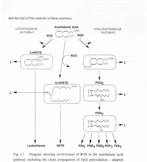
Fig 1.1 Diagram showing involvement of ROS in the arachidonic acid
pathway including the chain propagation of lipid peroxidation... 22
Fig 1.2 The potential pro-inflammatory effects of ROS in inflammatory
bowel disease...29
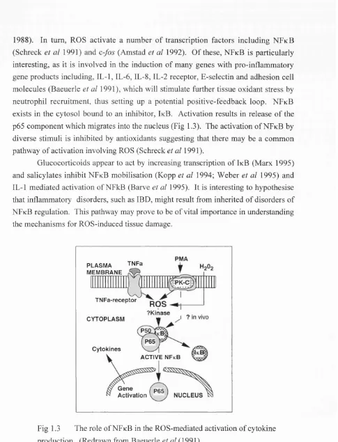
Fig 1.3 The role of NFkB in the ROS-mediated activation of cytokine
production... 33
Fig 1.4 The main sources of ROS in IBD, showing the important enzyme systems involved in ROS production and antioxidant
defence... 34
Fig 1.5 The main antioxidant defences in inflammatory bowel disease...39
Fig 2.1 The reaction of luminol with ROS to produce light... 50
Fig 2.2 Schematic representation of a colorectal biopsy placed in a solution containing the chemiluminescence amplifier which react with ROS to release light detected by the photomultiplier and photon detector comprising the liquid scintillation counter
analyser... 50
Fig 2.3 Method for assessing the amplified chemiluminescence response produced by inflamed colorectal biopsies from rats
and human subjects... 53
Fig 2.4 Percentage increase in chemiluminescence with increasing concentrations of luminol and lucigenin in acetic acid-induced
colitis... 54
Fig 2.5 The reaction of malondialdehyde and thiobarbituric acid to give
a chromogen which absorbs light at 532nm... 55
Fig 2.6 Concentration gradient o f TMP fluorescence against rodamine to assess malondialdehyde levels in serum from patients with
IBD and controls...58
Fig 3.1 Relationship of the macroscopic score of acetic acid-induced colitis rats at 24 hours to the time taken for spontaneous expulsion of acetic acid to occur after instillation o f 3% acetic
acid... 86
Fig 3.2 Method of obtaining and storing full-thickness biopsies from
the rat colon prior to experiments... 88
Fig 3.3 Macroscopic scores of inflammatory damage in rats pre-treated with 3% intra-colonic acetic acid or 0.9% saline, assessed at 24
hours and in untreated control animals...90
Fig 3.4 Histological scores of inflammatory damage in rats pre-treated with 3% intra-colonic acetic acid or 0.9% saline, assessed at 24
Fig 3.5 Logio-transform ed luminol-amplified chemiluminescence counts in full-thickness colonic biopsies from rats with acetic
acid-induced colitis, saline controls and untreated controls... 92
Fig 3.6 Logio-transformed luciginen-amplifîed chemiluminescence counts in full-thickness colonic biopsies from rats with acetic acid-induced colitis, saline-perfused controls and untreated
controls... 93
Fig 3.7 Relationship of luminol-amplified chemiluminescence to the macroscopic score in colons from rats pre-treated with 3% acetic acid-induced colitis at 24 hours, saline-perfused rats and
untreated controls... 94
Fig 3.8 Relationship of lucigenin-amplified chemiluminescence to the macroscopic score in colons from rats pre-treated with 3% acetic acid-induced colitis at 24 hours, saline-perfused rats and
untreated controls... 95
Fig 3.9 Relationship of luminol-amplified chemiluminescence to the histology score in biopsies from rats pre-treated with 3% acetic acid-induced colitis at 24 hours, saline-perfused controls and
untreated controls... 96
Fig 3.10 Relationship of lucigenin-amplified chemiluminescence to the histology score in biopsies from rats pre-treated with 3% acetic
acid-induced colitis at 24 hours and untreated controls... 97
Fig 4.1 The chemical structures of Amflutizole and LY231617... 102
Fig 4.2 M ethod for assessing the effect o f w ater-soluble test compounds on the amplified chemiluminescence produced by
inflamed colonic biopsies from acetic acid-induced colitis... 107
Fig 4.3 M ethod for assessing the effect o f water-insoluble test compounds on the amplified chemiluminescence produced by
inflamed colorectal biopsies from acetic acid-induced colitis... 108
Fig 4.4 The percentage change in chemiluminescence response o f inflam ed biopsies from acetic acid-induced colitis to conventional antioxidants compared with results using mucosal biopsies from active UC (data redravm from (Simmonds et al
1992a; Simmonds 1992b; Simmonds et al 1992c)... 110
Fig 4.5 The p ercen tag e change in the lu m in o l-am p lified chemiluminescence response of inflamed biopsies from acetic acid-induced colitis to standard treatments in IBD compared with results using mucosal biopsies from active UC (data redrawn from (Simmonds et al 1992a; Simmonds 1992b;
Fig 4.6 M ean p e rc e n ta g e ch an g e in lu m in o l-a m p lifie d chemiluminescence response of inflamed biopsies from acetic acid-induced colitis to proposed new therapies, LY231617,
amflutizole, and to 5-ASA... 112
Fig 5.1 Clinical Score in patients with UC and controls according to the
sigmoidoscopic score...121
Fig 5.2 Logio-transformed luminol-amplified chemiluminescence in patients with UC and controls according to the sigmoidoscopic
score... 122
Fig 5.3 Logio-transformed luminol-amplified chemiluminescence of biopsies from patients with UC according to their clinical
score...123
Fig 5.4 Logio-transformed luminol-amplified chemiluminescence of biopsies from patients with UC according to the histological
score of an adjacent biopsy... 124
Fig 5.5 Logio-transformed luminol-amplified chemiluminescence in
patients with UC sub-divided by treatment... 126
Fig 5.6 Logio-transformed luminol-amplified chemiluminescence in
patients with UC sub-divided by the extent of the disease...126
Fig 5.7 Logio-transform ed luminol-amplified chemiluminescence
compared with age in patients with UC... 127
Fig 6.1 Effect of human recombinant manganese superoxide dismutase (rh-M nSOD) and copper zinc superoxide dism utase (CuZnSOD) on lucigenin-am plified chem ilum inescence
produced by the xanthine-xanthine oxidase... 135
Fig 6.2 The percentage change in the lucig en in -am p lified chemiluminescence response of inflamed biopsies from acetic
acid-induced colitis to CuZnSOD and rh-MnSOD... 136
Fig 6.3 The macroscopic score from acetic acid-induced colitis treated with rh-MnSOD in 0.2mls hydrogel administered by gavage intra-rectally 1 hour prior to induction of colitis and appropriate
control groups... 138
Fig 6.4 The histological score from acetic acid-induced colitis treated with rh-MnSOD in 0.2mls hydrogel administered by gavage intra-rectally 1 hour prior to induction of colitis and appropriate
control groups... 139
Fig 6.5 The chemiluminescence response by two full-thickness colonic sections from each animal from acetic acid-induced colitis treated with rh-MnSOD in 0.2mls hydrogel administered by gavage intra-rectally 1 hour prior to induction of colitis and
Fig 7.1 The percen tag e change in the lu m in o l-am p lifie d chemiluminescence response o f inflamed biopsies from rats with acetic acid-induced colitis to desferrioxamine and ferrous
citrate... 150
Fig 7.2 The percen tag e change in the lu m in o l-am p lified chemiluminescence response of inflamed biopsies from patients w ith UC and normal controls to the iron chelators,
desferrioxamine and 1,10-phenanthroline and to ferrous citrate... 151
Fig 8.1 Study design for open-label study o f antioxidant nutrient therapy (Selenium pCE and Methionine (Sej3CE/Met)) in active
ulcerative colitis...161
Fig 8.2 Diary card for patients to record symptoms... 164
Fig 8.3 The average number of liquid or soft stools per day in 10 patients with active UC treated with Se-pCE/Met for 2-8
weeks... 170
Fig 8.4 Average number of bloody stools/day in 10 patients with active
UC treated with Se-pCE/Met for 2-8 weeks...171
Fig 8.5 Average number of solid stools/day in 10 patients with active
UC treated with Se-pCE/Met for 2-8 weeks...172
Fig 8.6 Clinical score in 10 patients with active UC treated with
Se-pCE/Met for 2-8 weeks... 173
Fig 8.7 Sigmoidoscopic score in 10 patients with active UC treated
with Se-pCE/Met for 2-8 weeks... 174
Fig 8.8 Histological score in 10 patients with active UC treated with
Se-pCE/Met for 2-8 weeks...175
Fig 8.9 ESR in 10 patients with active UC treated with Se-pCE/Met for
2-8 weeks...176
Fig 8.10 Lipid-adjusted TBARS in 10 patients with active UC treated
with Se-pCE/Met for 2-8 weeks... 178
Fig 8.11 Uncorrected TBARS in 10 patients with active UC treated with
Se-pCE/Met for 2-8 weeks...179
Fig 8.12 L o g 1 0 o f the m ean o f the lu m in o l-a m p lifie d
chemiluminescence response from two biopsies from 10 patients with active UC treated with Se-pCE/Met for 2-8
INDEX OF COLOUR PLATES
Plate 2.1 Infant feeding tube, shown with syringe attached, used to instil
acetic acid or saline into the colon of rats... 49
Plate 2.2 Scintillation counter used for chemiluminescence experiments...49
Plate 3.1 Macroscopic appearances o f colons 2-8cm from the rectum removed and opened longitudinally from acetic acid-induced
colitis and 0.9% saline-perfused controls... 75
Plate 3.2 Macroscopic appearances of acetic acid-induced colitis...76
Plate 3.3 Microscopic appearances of normal rat colon... 79
Plate 3.4 M icroscopic appearances o f a saline-perfused rat. The histological appearances are normal except for mild, superficial
oedema of the lamina propria... 79
Plate 3.5 Microscopic appearances o f acetic acid-induced colitis. The inflammation is mild and demonstrates the presence of confluent
submucosal oedema...80
Plate 3.6 Microscopic appearances of acetic acid-induced colitis. Mild inflammation with confluent submucosal oedema and a mild- moderate neutrophil infiltrate in the lamina propria, submucosa
and serosa... 80
Plate 3.7 Microscopic appearances of acetic acid-induced colitis. Marked infiltrate of neutrophils in the lamina propria and particularly in the submucosa, focal necrosis o f the mucosa and submucosal
fibrin deposition...81
Plate 3.8 Microscopic appearances of acetic acid-induced colitis. There is a marked neutrophil infiltrate in the submucosa and submucosal oedema. Fibrin deposition is present in the perivascular space in the lamina propria and in the submucosa. There is marked
epithelial necrosis... 81
Plate 3.9 M icroscopic appearances o f acetic acid-induced colitis. Submucosal neutrophil margination in a vessel and shows fibrin
deposition which characteristically is peri-vascular... 82
Plate 3.10 M icroscopic appearances o f acetic acid-induced colitis.
Moderate neutrophil infiltrate in the muscularis propria...82
Plate 8.1 Study medication for trial of Selenium pCE and Methionine in
ACKNOWLEDGEMENTS
This research work was undertaken with the support and advice o f many colleagues in the Gastrointestinal Science and Bone and Joint Research Units at the St. Bartholomew’s and Royal London School of Medicine and Dentistry. I am very grateful to David Rampton, my supervisor, for his intellectual drive, constant availability and unerring help in stimulating ideas for projects and reviewing the results with discussion and constructive criticism.
I am also grateful to David Wingate and David Evans for supplying the facilities and equipment for this research, to Tito Castillo for advice on computer use,
to Chaman Chander and Andrew Claxson for help with animal experiments, to Selina Blades and Adam Coumbe for their help with histology, and to Martin Grootveld, Cliff Stevens and Eva Dowling for advice on the experiments herein. Professor David Blake was instrumental in initiating this project and stimulating ideas; he also provided access to laboratory facilities, without which these studies would not have been possible. Much o f this thesis was written whilst in the employ of Dr Colin Ainley, who gave considerable support towards its completion.
Dr Philomena Pereira, of the Department of Epidemiology and Statistics, and Dr Julian Holmes, o f the Hill Centre, Queen Mary and Westfield College were particularly helpful with advice on statistics and data analysis.
STATEMENT OF ORIGINALITY
All experimental work presented in this thesis was designed, performed and analysed by the author apart from the following exceptions:
Some of the histological processing and staining was carried out by Selina Blades. Histological processing of biopsy material for the open study of antioxidant nutrients in active ulcerative colitis was undertaken in the routine hospital laboratory.
Scoring of histological material was done in conjunction with Dr Adam
1
IN T R O D U C T IO N
The term inflammatory bowel disease (IBD) defines a group o f chronic, relapsing inflammatory disorders of the intestine o f unknown aetiology. There have been few changes in the therapy of IBD for two decades and the most frequently used agents, aminosalicylates and glucocorticoids, have non-specific anti-inflammatory
actions, giving few clues as to the important pathogenic mediators. There has therefore been much research effort to establish the aetiology and pathogenesis of these diseases with the aim o f developing more specific, effective, and less toxic, therapies.
Oxygen free radicals and their associated metabolites, collectively termed reactive oxygen species (ROS), are highly reactive and pro-inflammatory. They are produced in excess by the inflamed mucosa in IBD (Ahnfelt-Ronne et al 1990; Simmonds et al 1992a; Keshavarzian et al 1992b), but their exact role in pathogenesis remains poorly understood. This thesis examines the potential role o f ROS and in particular antioxidant therapy, in IBD. A proven role for specific antioxidant therapy would help to establish the pathogenic importance of ROS and potentially lead to novel therapies with low toxicity.
The first part of this introduction presents an overview of the history, epidemiology, aetiology and pathophysiology, clinical features and therapy o f inflammatory bowel disease. The second part presents the chemistry and biological origins of ROS. In the third section, evidence for a role for ROS in IBD is examined. The final part sets out the aims of this thesis.
INFLAMMATORY BOWEL DISEASE
The term IBD encompasses ulcerative colitis (UC), Crohn’s disease (CD), collagenous colitis and microscopic colitis. At least 5% o f patients cannot be classified into one of these categories. Only UC and CD will be specifically considered further in this thesis.
UC is a chronic, relapsing, inflammatory disease affecting a variable extent of
steroidal anti-inflammatory drugs (NSAIDs) and antibiotics), emotional stress and intestinal infection. Patients with pancolitis are at particular risk o f developing malignancy late in the course of the disease. Proximal spread occurs in up to 30% of patients with proctitis alone at diagnosis (Ekbom et al 1991). Extraintestinal manifestations are common, including arthropathy, uveitis, pyoderma] gangrenosum, erythema nodosum and hepatobiliary disorders.
CD is a chronic, relapsing, inflammatory condition of the intestine which can affect any part o f the gastrointestinal tract from the mouth to the anus. The most frequently affected parts are the terminal ileum (30%), ileum and colon (50%), and
colon alone (20%) and perineum (Campieri et al 1993). The inflammation is usually transmural and classically produces discrete affected areas ('skip-lesions'). It is characterised by stricture formation and fistulae to other viscera and to skin. Similarities to UC include the range o f extraintestinal manifestations and an increased, though less well recognised, risk o f colorectal carcinoma (Gillen et al
1994).
Historical Perspective
Idiopathic, extensive ulceration of the colon was first described in the late 19th Century (Wilks et al 1875; Hale-White 1888), and the term ulcerative colitis became established in the early part of this century (Hawkins 1909). Previously described as regional ileitis, the detailed description by Crohn et al (Crohn et al 1932) led to the eponym commonly used today.
Epidemiology
The incidence of IBD varies with geographical location, race, age, and
possibly sex (Table 1.1). UC is commoner in developed countries, particularly in Northern Europe, and in urban areas (Ekbom et al 1992). Jews have a higher incidence of IBD (Ekbom et al 1992), which varies according to their geographical origin. In Israel, the incidence is lower in Jews of Asian or African origin than those from America or Europe (Stenson et al 1991). Conversely, IBD is less common in black groups than among Caucasians in both America (Calkins et al 1984) and South Africa (Wright et al 1986).
UC is probably marginally commoner in women; the reported degree o f the difference varying between a factor of 30% and equal incidence (Stenson et a l\9 9 \) .
UC is very uncommon before the age of 10, and its highest incidence is at 15-25 years, with a second peak at 55-65. However, UC that presents for the first time in those over 50 is often less severe and has a lower incidence o f relapse (Tysk et al
1992b).
between UC and CD, being much less common in UC, and more common in CD, than in the general population (Stenson et al 1991). In addition, UC is more likely to occur
after giving up smoking, and patients with UC who continue to smoke experience improvements in symptoms (Tysk et al 1992a). Recently, a controlled clinical trial in
active UC has shown some benefit from nicotine patches (Pullan et al 1994). Conversely, CD runs an unfavourable course in those who continue to smoke (Lindberg et al 1992).
Table 1.1 The epidemiology of ulcerative colitis and Crohn's disease.
Factor Ulcerative Colitis Crohn’s Disease
Incidence (per 100,000/year) 2-10 1-6
Prevalence (per 100,000) 35-100 10-100
Racial Incidence whites, jbiacks in USA whites, jbiacks in USA
Ethnic incidence Jews Jews
Sex (Probably similar) (Probabiy simiiar)
Age 15-35* 15-35*
(55-65)
Smoking Decreased increased
* Peak at this age does not occur in low-risk populations. ( ) no consensus.
Adapted from (Stenson et al 1991).
Aetiology and Pathophysiology
Although the aetiology of IBD is unknown, a number of pathogenic factors have been proposed.
Genetic Factors
The influence of inheritance in IBD is suggested by the inter-racial differences in disease frequency (Samuels et al 1974), the increased frequency in monozygotic compared with dizygotic twins (Tysk et al 1988) and the occurrence o f disease
aggregation within families (Orholm et al 1991). CD shows greater concordance rates for monozygotic twins (53%) than UC (6%) (Tysk et al 1988). For first degree relatives o f patients with IBD, there is a 15-fold risk for UC and 3.5-fold for CD (Langman 1979). In addition, genetic diseases, such as Turner's syndrome (Weinrieb
et al 1976) and Hermansky-Pudlak syndrome (Schinella et al 1980), are associated
with IBD.
families in which several members had CD, two loci on chromosome 16 were linked to disease susceptibility (Hugot et al 1996). It has been suggested that genetic factors may affect immunological control (Yang et al 1992).
Infectious Agents
The colonic lumen contains a vast reservoir of potentially pathogenic micro organisms. Abnormal host responses to commensal bacteria may be responsible for IBD (Phillips 1988), as suggested by the increased adhesion of E. colt to the mucosa in UC (Burke et al 1988). Alternatively, induction o f an autoimmune response via molecular mimicry with commensal organisms may lead to persistent inflammation.
It has been suggested that exogenous agents such as Mycobacteria (Wall et al
1993), the measles virus (Levin et al 1995), and elementary forms o f bacteria (L- forms) (Belsheim et al 1983; Ibbotson et al 1987), may be directly responsible for IBD. However, these studies largely rely on demonstrating the putative agent within the gut wall and could be explained by increased mucosal permeability (Wyatt et al
1993). The evidence for an infectious aetiology for IBD thus remains weak, especially given the clinical response to potent immunosuppressive agents.
An alternative possibility is that structural components o f microbes, rather than the viable organism, might cause mucosal inflammation. This is suggested by the presence o f circulating antibodies to lipopolysaccharide (LPS) in IBD (Kruis et al
1984) and the correlation of endotoxin plasma levels with disease activity in CD (Wellman et al 1986). Animal models in which colitis is induced by exposure o f the intestinal wall to bacterial cell wall components, such as peptidoglycan- polysaccharide (PG-PS) polymers and N-formyl-methionyl-leucyl-phenylalanine (fMLP) (Chester et al 1985; Sartor et al 1985), lends further support to this concept.
Mucin Abnormalities
Abnormal gastrointestinal mucus may reduce mucosal protection from the potentially deleterious luminal contents and thus provoke inflammation. A number of abnormalities have been described including the loss of goblet cell mucin in UC, possibly due to abnormal secretion (Cope et al 1988), structural abnormalities of the mucin layer (Podolsky et al 1984), decreased mucus sulphation (Raouf et al 1992) or acétylation of glucosamine (Burton et al 1983).
Vascular Abnormalities
It has been proposed that the microvascular changes found in CD (Wakefield
et al 1989) and UC (Fairburn 1973) may cause ischaemic injury to the mucosa
(Knutson et al 1968; Geller et al 1983; Wakefield et al 1991). Paradoxically, however, mucosal and sub-mucosal blood flow is increased in IBD (Hulten et al
(Bousen et al 1966) and preliminary evidence that the length of the colonic marginal artery in UC matches the extent of well-demarcated disease in UC (Hamilton et al
1995). It remains unclear, however, whether these abnormalities are o f aetiological importance or are secondary to the release o f vasoactive mediators. The vasoconstrictor, endothelin-1, is found in the submucosa in CD (Murch et al 1992)
and nitric oxide, a potent vasodilator, is produced in excess by the mucosa in UC (Boughton-Smith et al 1993; Middleton et al 1993). The hypothesis that these changes are secondary to inflammation is supported by evidence from animal studies in which abnormal mesenteric blood flow accompanies induction of inflammation in both dinitrochlorobenzene- and immune-complex-mediated colitis in rabbits (Hauser
et al 1988).
Platelets and Thromboembolism
There is good evidence that platelets may have a pathogenic role in IBD. IBD is a risk factor for thromboembolism (Talbot et al 1986; Novotny et al 1992), exacerbations o f IBD are associated with thrombocytosis (Harris et al 1983) and platelet thrombi are seen in the microvasculature in CD (Wakefield et al 1989). Furthermore, platelets circulate in an activated state in both UC and CD (Collins et al
1994). The release of inflammatory mediators, such as IL- 8 (Kaplanski et al 1993),
12-hydroxy eicosatetraenoic acid, platelet factor 4 (Page 1989) and ROS (Amieson et al 1985) by activated platelets suggests that they are pro-inflammatory. Reports of a beneficial therapeutic response to heparin in UC provide further supporting evidence for the pathogenic importance o f thromboembolism (Gaffney et al 1991; Dwarakanath et al 1995; Gaffney et al 1995).
Mucosal Permeability
Luminal pathogens and pro-inflammatory molecules may gain access to the mucosa if mucosal permeability is increased. Permeability to polyethylene glycol (PEG) is increased in the colon in active UC (Aimer et al 1993) and permeability to
PEG and 99mTc-labelled diethylenetriaminopenta-acetic acid (DTPA) is increased in the small bowel in CD (Casallas et al 1986; Teahon et al 1992).
Some workers have shown that permeability to lactulose and mannitol is only abnormal in CD (Casallas et al 1986) and others, using PEG, lactulose and mannitol, have found no differences between IBD and controls (Ruttenberg et al 1992; Munkholm et al 1994). Alterations in permeability are most likely to be secondary to
permeability to lactulose and mannitol in quiescent CD predicts subsequent relapse (Wyatt et al 1993).
Psychological Factors
Psychological stress is associated with disease activity in IBD (Porcelli et al
1994) and a link has been shown between exacerbations of disease and prior
psychological stress (Drossman 1988). The stress of captivity appears to increase the incidence of colitis in cotton-top tamarin monkeys (Kirkwood et al 1986). These observations may be explained by the influence of the brain-gut axis on the release of
pro-inflammatory neurotransmitters in the intestine (Foreman 1987). Naturally, the psychosocial status of patients with IBD must be taken into account in their management (Ramchandani et al 1994).
The Immune Response in Inflammatory Bowel Disease
CD is characterised by transmural inflammation with a predominantly chronic inflammatory infiltrate and granuloma formation, whereas in UC there is a mixed neutrophil and lymphoid infiltrate confined to the mucosa. The inflammatory cells comprise lymphocytes of the gut-associated lymphoid tissue (GALT) together with neutrophils, lymphocytes and macrophages recruited from the mesenteric vasculature. Immune protection is also provided by the physico-chemical barrier of intestinal mucus, the inter-cellular tight junctions, epithelial cells and intra-epithelial lymphocytes, which are predominantly T cells (Hirata et al 1986). Collections o f mucosal lymphoid cells comprise Peyer’s patches, which lie below specialised epithelial cells (M-cells) and are intimately involved in the uptake and processing of luminal antigens. A subset of B-cells enter the systemic circulation and later return to the GALT, thus exchanging immunological information with the rest of the immune system (Mayer 1992). Several abnormalities of the immune defence system in IBD
have been described.
B-lymphocytes and Immunoglobulin Production
IBD is associated with hypergammaglobulinaemia (Hodgson et al 1978a), and increases in immunoglobulin G (IgG) subtypes have a disease-specific tendency. Serum levels of IgGl predominate in UC and IgG2 in CD (MacDermott et al 1989).
Antibodies to Baker's yeast {Saccharomyces cerevisiae) have been described in CD (Giaffer et al 1992; Lindberg et al 1992), although not in UC (McKenzie et al
1990). However, it is not currently thought that antibodies to exogenous antigens are important in the pathogenesis of IBD.
Lymphocytes are markedly increased in the lamina propria in IBD and these intra-mucosal lymphocytes differ phenotypically from circulating forms (James et al
the lamina propria is reduced by a concomitant increase in the population o f IgG- secreting cells, particularly in CD, where they concentrate near Peyer’s patches and
granulomata (Baklien et al 1975; Baklien et al 1976). IgG production by endoscopic biopsies is disease specific, as it is in the serum (see above); IgG2 secretion predominates in CD and IgGl in UC (Ruthlein et al 1992).
Intra-mucosal lymphocytes in IBD also secrete an increased amount o f IgM
(Jewell et al 1990), and IgA, particularly IgAl (Kett et al 1987). These findings may impair the mucosal immune response as, unlike IgA2, IgAl is susceptible to bacterial proteases, and the resulting Fab and Fc fragments are unable to induce bacterial agglutination or prevent bacterial adherence (Plaut 1983). Further evidence for qualitative defects of immunoglobulin secretion in IBD is the overall decrease in
J-chain production and binding capacity of the cytoplasmic secretory component by mucosal lymphocytes (Brandtzaeg et al 1984), and the production of abnormal light chains by peripheral blood lymphocytes (Ginsberg et al 1981).
Influence of Autoantibodies
The pathophysiological similarity o f IBD to other autoimmune diseases suggests a role for autoantibodies. Autoantibodies directed against colonic antigens (Das et al 1978) have been described in UC, although their target epitope(s) and role in pathogenicity remain to be established.
Anti-neutrophil cytoplasmic antibodies (ANCA), may also be involved in IBD, given their pathogenicity in the vasculitis of Wegener’s granulomatosis. ANC A
are found in 30-80% of patients with UC, and 2-25% of CD (Saxon et al 1990; Cambridge et al 1992; Seibold et al 1992). They have neither the cytoplasmic staining (cANCA) pattern of Wegener's granulomatosis, nor the perinuclear pattern
(pANCA) typical of polyarteritis nodosa, but have a mixed diffuse and peri-nuclear pattern. Serum levels of cANCA are a marker of disease activity in W egener’s granulomatosis (Lai et al 1990), but not in UC (Deusch et al 1993; Lee et al 1995), and persist after colectomy (Saxon et al 1990), though others have shown reduced titres with long-term steroid therapy and after total colectomy (Rump et al 1993).
Relatives o f patients with IBD have increased levels o f ANC A (Shanahan et al
1992a), and thus ANCA may be useful as a genetic marker of disease susceptibility.
Presence of Immune Complexes
Circulating immune complexes are raised in IBD (Doe et al 1973; Jewell et al
1973) and correlate with disease activity (Nielson et al 1978), though this has been disputed by other workers using alternative methods of detection (Lurhuma et al
Complement Activation
Complement is activated by many o f the immune mediators that are o f proposed pathogenicity in IBD. Circulating levels o f complement components are normal, though there is some evidence for activation of the classical pathway (Jewell
et al 1990). Tissue deposition of the terminal complement complex has been found in
submucosal vessels in both UC and CD (Halstensen et al 1989) and in the epithelium
in UC (Halstensen et al 1990). Furthermore, complement components are released into the lumen in CD (Ahrenstedt et al 1990) and it has been suggested that there is defective degradation of the C3b fragment in IBD (MacDermott et al 1988). Others, however, have failed to detect complement deposition in the inflamed mucosa (Keren
et al 1984) and thus the pathogenic importance o f complement activation in IBD
remains to be established.
T-lymphocytes and Cell-Mediated Immunity
There is good evidence that there is marked mucosal T-cell activation in both UC and CD (Shanahan et al 1992b), and activation o f peripheral blood lymphocytes in CD (Pallone et al 1987). In contrast to normal bowel, epithelial immune activation in CD is associated with a predominance of T-helper (CD4+) cells over T-suppressor (CD8+) cells (Mayer et al 1990) and suppressor cell activity is suppressed in CD
(Goodacre et al 1982). However, the proportion of CD4+ to CD8+ cells in UC is not
different to controls (Neil et al 1994). It has been suggested that these alterations might reflect a primary defect of feedback inhibition of T-cell activation, leading to persistent release of pro-inflammatory mediators, such as cytokines, eicosanoids and
superoxide (Chang et al 1990). Isolated lamina propria T-cell lymphocytes demonstrate a polyclonal response in IBD, as assessed by analysis of the T-cell receptor genes (Kaulfersch et al 1988), suggesting that multiple antigens are involved in immune stimulation in IBD.
HLA class II antigens are over-expressed on the epithelial cell surface o f both diseased, and non-diseased intestine, in CD (Mayer et al 1991), probably under the influence o f interferon-y (Salomon et al 1991), indicating a role for antigen- presentation in the inflamed bowel.
Neutrophils
Mucosal infiltration by neutrophils is a cardinal feature o f IBD. There are at
least 50 cytopathic toxins (divided into those localised to the cell membrane, and those confined to intracellular granules) which are released by the activated neutrophil (Weiss 1989), as well as other pro-inflammatory mediators, such as elastase, collagenase and gelatinase.
dinucleotide phosphate (NADPH) oxidase, and the release o f the intra-cellular enzyme, myeloperoxidase, in the extracellular release o f ROS, is discussed in more detail below. Neutrophil-mediated tissue destruction normally halts when the
initiating antigen is destroyed (Clark 1983), but one might hypothesise that a defect in this autoregulation would result in the persistent inflammation seen in IBD. The role
o f neutrophil-mediated release of ROS and their role in inflammation will be discussed in more detail below.
Monocytes and Macrophages
M acrophages, derived from circulating monocytes, m igrate into the gastrointestinal wall in IBD and produce greater amounts of interleukin-1 (IL-1) and colony-stimulating factor (CSF) than those from control tissue (Pullman et al 1992). Monocytes and macrophages are involved in antigen presentation in IBD (Mahida et
al 1989b), and there is evidence of increased numbers o f a sub-population o f macrophages, ‘veiled cells’, which are particularly active in antigen presentation (Wilders et al 1984).
Eosinophils, Basophils and Mast Cells
Mast cells are often seen in the inflamed mucosa in IBD (Dvorak et al 1980; Bakazs et al 1989) and secrete pro-inflammatory mediators. This may explain the
therapeutic benefit of mast cell stabilisers, such as sodium cromoglycate, in some patients with UC (Grace et al 1987).
Adhesion Molecules
Adhesion molecules play a critical role in the migration, activation and differentiation of lymphoid cells, and in the adhesion of leukocytes to the vascular
endothelium and diapedesis through it. Their importance in IBD is demonstrated by the marked increase in the expression of, for example, endothelial leukocyte adhesion molecule-1 (ELAM-1) (Koizumi et al 1992) and inter-cellular adhesion molecule-1 (ICAM-1) by the colonic vascular endothelium (Malizia et al 1991), and the release of E-selectin by intestinal macrophages in IBD (Pooley et al 1995). However,
expression of vascular cell adhesion molecule-1 (VCAM-1) is normal in IBD .
Eicosanoids
Eicosanoids are a group of lipid mediators derived from essential fatty acids, o f which the most important is arachidonic acid. Arachidonic acid is released from cellular phospholipids and metabolised to leukotrienes via the lipoxygenase pathway, and prostaglandins and thromboxanes via the cyclo-oxygenase pathway (Table 1.2). Platelet activating factor (PAF) is formed by the action o f acetyl transferase.
membrane phospholipid, is increased in the mucosa in IBD (Minami et al 1994). Eicosanoids are predominantly produced by inflammatory cells in response to inflammatory stimuli. Arachidonic metabolism is both stimulated by ROS and induces ROS production (Chakraborti et al 1989; Riendeau et al 1989; Harris et al
1992). Prostaglandins are important in the maintenance o f mucosal integrity, and
leukotrienes, particularly leukotriene-B4 (LTB4), mediate increased microvascular
permeability and the recruitment of polymorphonuclear leukocytes.
Table 1.2 Eicosanoids in inflammatory bowel disease.
Eicosanoid Ulcerative Crohn’s Main Pathophysiological
Colitis Disease Effects
M
sM
S
LeukotrienesLTB4
LTC4/D4
5-, 12-, 15-HETE
r
ff
ft
ft ft* ft
ft
ft
Potent stimulant of PMN chemotaxis, adherence and secretion, activates platelets Increases vascular tone and permeability, increases intestinal smooth muscle contractility
P rostaglandins
PGE2
ft*
N
ft* ft* Increase epithelial chloride andmucus secretion, decrease activation of infiammatory cells
PGI2
ft
-ft
PG p2a ft - — —
O thers - — —
PAF
TXB2
ft
ft’ N
ft
ft ft
Increases PMN chemotaxis, adherence, increases vascular tone and permeability,
activates platelets
Activates inflammatory cells, platelets, increases vascuiar tone (metabolite of short-lived TXA2)
M - Mucosal/Rectal dialysis, S - Serum levels or production by peripheral blood cells. N = Normal or conflicting data, * Correlates with disease activity, fPresent in inactive disease. - Not known.
LT-n - leukotriene-n, n-HETE - n-Hydroxy-6,8,11,14-eicosatetraenoic acid, PG - prostaglandin, PAF - platelet activating factor, XX - thromboxane.
(Lauritsen et al 1988) (Table 1.2). Similar increases in eicosanoids have been found in acetic acid-induced colitis (Sharon et al 1984; Eliakim et al 1992) and trinitrobenzene sulphonic acid (TNBS)-induced colitis (Rachmilewitz et al 1989). However, specific LTB4 antagonists have only limited efficacy in UC (Rask-Madsen
et al 1992). Furthermore, prostaglandin synthesis inhibition by NSAIDs, which
inhibit cyclo-oxygenase, is associated with disease relapse (Rampton et al 1981).
Cytokines
Cytokines are glycosylated polypeptides, with a molecular weight ranging from 10,000 to 30,000kDa, that modulate inflam m ation by intercellular communication (Fiocchi 1992) and act as the hormones o f the immune system
(Brynskov et al 1994). The role of cytokines in IBD is summarised in Table 1.3. Mucosal and serum levels of the pro-inflammatory cytokines IL-ip (Ligumsky
et al 1990) and IL- 8 (Mahida et al 1992), and mucosal levels o f IL- 6 (Isaacs et al
1992), are raised in IBD. Circulating levels of IL- 6 are raised in CD, but rarely in UC
(Mahida et al 1991b). Serum levels of IL-2 are decreased in IBD (Fiocchi et al 1984), though the IL-2 receptor (IL-2R) is increased (Matsuura et al 1993), which may explain the efficacy of the IL-2 antagonist, cyclosporin A in active UC (Lichtiger et al
1994). Conversely IL-4, which normally down-regulates the production of IL-1, fails to do so in IBD (Schreiber et al 1995), and levels o f another regulatory cytokine, IL-10, are increased in both the serum (Kucharzik et al 1995) and mucosa in IBD (Niessner et al 1995). Further evidence of the importance o f interleukins in colonic inflammation is the development of spontaneous colitis in IL-2 and IL-10 knockout mice (Kuhn et al 1993; Sadlack et al 1993).
Tumour necrosis factor-a (TNFa) is increased in the mucosa (MacDonald et al 1990), serum (Murch et al 1991) and stool (Braegger et al 1992; Nicholls et al
1993) o f children with IBD, and may contribute to persistent inflammation by a local effect on the mucosa, though mucosal levels in adults are normal (Stevens et al 1992).
Preliminary data suggest that a single parenteral dose of a monoclonal antibody against TNFa antibody is of therapeutic benefit in CD (van Dullemen et al 1995).
Neuropeptides
The gut neurogenic peptides, vasointestinal peptide (VIP), substance P and somatostatin may mediate inflammation by altering immune function (Foreman 1987; Shanahan et al 1988). Increased levels of VIP are found in the inflamed mucosa and submucosa in CD (Bishop et al 1980), and in the inflamed mucosa in UC (O’Morain
et al 1984), though others have disputed these findings (Kubota et al 1992). Plasma
levels o f VIP increase in parallel with the degree of inflammation in both IBD (Duffy